


Abstract
Objectives: To determine the effect of age and gender on diameters and lengths of the renal arteries.
Methods: This is a retrospective study. A total of 50 asymptomatic volunteers were selected randomly and scanned by multi-detector CT to assess the diameters and lengths of the renal arteries. The study conducted at King Abdulaziz Specialized Hospital (KAASH) and King Faisal Hospitals, Taif, Saudi Arabia between October 2017 and March 2018. The lengths and diameters of the main arteries were measured and compared to age and gender of the participants.
Results: The mean length of right renal artery was significantly longer than the left one (4.47±.70 versus 3.714±.68 cm, p<0.001). Length of right and left renal arteries were significantly higher in males than females (p=0.02 and p =0.03). Diameters of both left and right renal arteries were higher in males than females (5.482±1.37 versus 5.288±1.09 cm, and 5.544±1.14 versus 5.188±1.05 cm). The diameters of renal arteries varied significantly with age, specifically in elders (p=0.001).
Conclusion: The mean length and mean diameter were significantly different between females and males, and between left and right main renal arteries. Age and gender have a significant impact on the length and diameters of main renal arteries.
The renal arteries are the suppliers of kidneys with oxygenated blood. They play an effective role in the circulatory system since they convey a significant amount of oxygenated blood supply to the kidneys.1 The most common lesion that affects the renal arteries (RA) was stenosis which is the common cause of hypertension. The RAs might also be involved by atherosclerotic changes and aneurysm, which significantly affect the diameters.2 Computed tomography (CT) is reliable and accurate for demonstrating the morphology of the renal arteries. Computed tomography angiography has provided a unique diagnosis of cardiovascular diseases and management.3,4 Computed tomography angiography was utilized to assess several diseases of the renal vasculature.5 Aging was considered one of the most significant risk factor for many RA abnormalities. It was associated with vascular compliance and elevated vascular rigidity.6 It was reported that aging has a strong influence on narrowing of RA diameter.7
The ultimate aim of this study is to determine the impact of age and gender on the average diameter and length of the renal arteries using CT.
Methods
This study was a retrospective and analytical type conducted at King Abdulaziz Specialized Hospital (KAASH) and King Faisal Hospitals, city, Saudi Arabia for 50 asymptomatic participants, between 2015 and 2016. The participants were investigated by CT angiography to assess the RAs. Data collection sheet was designed to gather the demographic data such as age and gender. The participants are selected from the donor in the renal transplantation process.
An expert radiologist interprets the CT images and analyzed all measurements and findings of the RAs. Patients with abnormal renal findings or previous renal surgery were excluded from the study. Oral consent was taken from the patients. This study was approved by the ethics committee of Taif University, Taif, Saudi Arabia.
Statistical analysis
The data has been analyzed using the software Statistical Package for Social Sciences Version 23 (Version 22.0. Armonk, NY: IBM Corp.). Descriptive and quantitative data were displayed as mean±standard deviation. Continuous variables were analyzed using the student independent t-test, and one-way Analysis of Variance tests to compare diameters and lengths of RAs with males and females and among age groups. P-value lesser than 0.05 was considered to be significant.
Results
Out of the 50 participants, 34 (68%) were males and 16 (32%) were females. The comparison of length and diametes between right and left RAs are shown in Table 1.
Comparison of length and diameters between right and left renal arteries.
In males, the mean diameter of the right RA was 5.482±1.37mm, and left RA was 5.288±1.09 mm. The diameters were higher in males than females without significant difference. In males, the mean length of right RA was 4.618±.64mm and of left RA was 4.156± .69 mm. A significant difference of mean lengths of both left and right RAs was observed between females and males (Table 2).
Comparison of mean length and diameters of renal arteries in males and females
Table 3 summarizes the influence of age in diameters and lengths of RAs. There are significant differences among the age groups (p<0.001). It was observed that the diameters increased in the age group of 36-55 years and decreased in 56-75 years old and 76 to 90 years.
The impact of age on diameters and lengths of the renal arteries
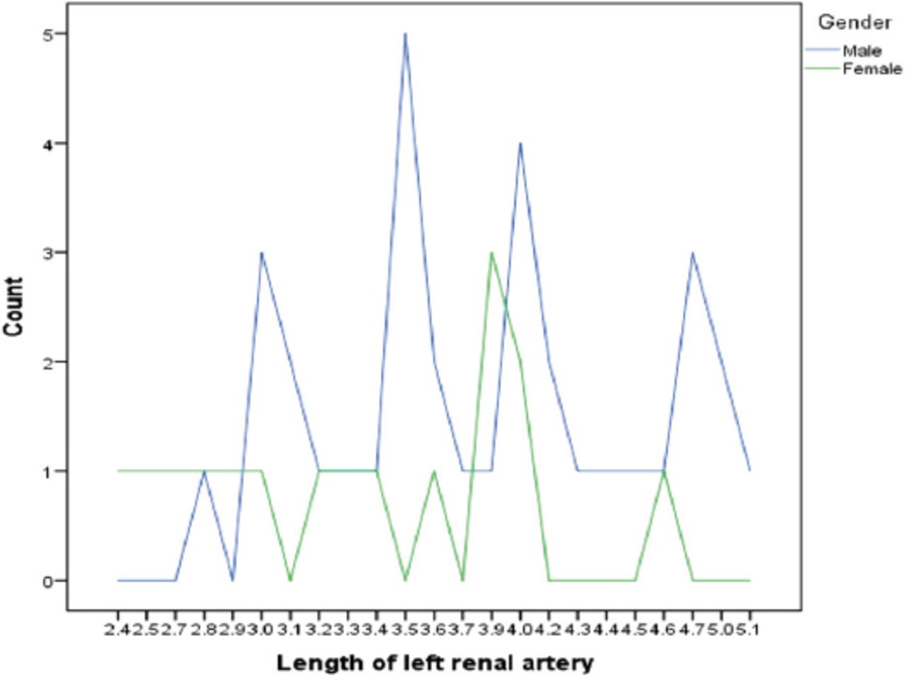
Figure 1 and Figure 2 showed the variation of length of RAs and gender differences.

Measurement of the length of right renal arteries in males and females.

Measurement of the length of left renal arteries in males and females.
Discussion
Aging was a risk factor that influences the diameter of RA. Advanced CT plays a useful role for delineating anatomical structures, specifically the morphology the RA that helps in clinical and diagnosis decision. In this study, we determined the impact of age and gender on length and diameters of the RAs.
The current study revealed that there was a significant difference in length and diameters between left and right RAs. It was observed that the width of the right RA was significantly lesser than that of the left. Our results are in agreement with the findings reported by Saldarriaga et al8 that the right RA diameter was significantly lesser than the left RA. A study performed by Tarzamni et al, who used a multi-slice CT scan reported that the diameter of right RA was smaller than the left RA (p=0.35).9 It was noted that the mean length of the right RA in the current study was significantly longer than the mean length of the left RA (p<0.001) (Table 1). In a previous study, it was reported that the mean length of the right RA was longer than the left RA (p=0.002).10 This is attributed to the location of the abdominal aorta (AA) to the left median sagittal plane in the abdominal cavity and the long path of the RA on the right side, as reported by previous studies.8,10,11
The present study revealed that gender differences significantly influenced the lengths and diameters of RAs. It was observed that the length of the right and left RAs were significantly longer in males than females (p=0.03 and p=0.02). The diameters of RAs were higher in males than females. The results of the study were consistent with previous studies7 that show that the lengths and widths of RAs were smaller in males than males. The gender variation in renal measurements was attributed to the relatively large body habitus of males compared to females.13
Aging is established to cause vascular compliance and increased vascular rigidity.14 It was observed that mean diameters of both renal arteries was significantly reduced in advanced age groups (56-75 and 76-90 years old) (p<0.001), while it increased in the age groups between 36 and 55 years. Many authors studied the influence of age on diameters of RA. A study stated that a strong correlation was found between age and narrowing of renal RAs diameters.15 The increase in diameter in adults is attributed to increased physical activity and consequently elevated cardiac output.16 The reduction in luminal diameter of RA with advancing age is probably due to the progressive thickening of the internal intima layer increase in the collagen matrix in the renal arterial wall.7
Study limitation
Limitation of this study was the small sample size, and the authors could not follow-up all the abnormalities that affect the morphology of the RAs. Further studies are recommended with a larger sample size to confirm the initial results of this study.
In conclusion, there was a significant difference of length and diameter between left and right RAs. Widths and lengths both arteries were significantly different between males and females. Significant reduction of diameters of RAs was attributed to aging.
Acknowledgment
I would like to express my gratitude too all my colleague who helped me during the study period. Also to Dr. Hamid Osman, for his encouraging words to carry out this study. And also I would like to acknowledged King Abdulaziz Hospital Specialist, Taif, Saudi Arabia for their collaboration during data collection. We would also like to thank Scribendi for the English Language editing of the manuscript.
Footnotes
Disclosure. Authors have no conflict of interests, and the work was not supported or funded by any drug company.
- Received March 23, 2019.
- Accepted November 26, 2019.
- Copyright: © Saudi Medical Journal
This is an open-access article distributed under the terms of the Creative Commons Attribution-Noncommercial-Share Alike 3.0 Unported, which permits unrestricted use, distribution, and reproduction in any medium, provided the original work is properly cited.


