
Main menu

  • Home
  • Content
    • Latest
    • Archive
    • home
  • Info for
    • Authors
    • Reviewers
    • Subscribers
    • Institutions
    • Advertisers
    • Join SMJ
  • About Us
    • About Us
    • Editorial Office
    • Editorial Board
  • More
    • Advertising
    • Alerts
    • Feedback
    • Folders
    • Help
  • Other Publications
    • NeuroSciences Journal

User menu

  • My alerts
  • Log in

Search

  • Advanced search
Saudi Medical Journal
  • Other Publications
    • NeuroSciences Journal
  • My alerts
  • Log in
Saudi Medical Journal

Advanced Search

  • Home
  • Content
    • Latest
    • Archive
    • home
  • Info for
    • Authors
    • Reviewers
    • Subscribers
    • Institutions
    • Advertisers
    • Join SMJ
  • About Us
    • About Us
    • Editorial Office
    • Editorial Board
  • More
    • Advertising
    • Alerts
    • Feedback
    • Folders
    • Help
  • Follow psmmc on Twitter
  • Visit psmmc on Facebook
  • RSS
OtherClinical Image
Open Access

Adult female with acute renal failure and weight loss

Nor A. Abdul-Rahman, Raja R. Azman and Gnana Kumar
Saudi Medical Journal May 2016, 37 (5) 584-586; DOI: https://doi.org/10.15537/smj.2016.5.15042
Nor A. Abdul-Rahman
From the Department of Bio-Medical Imaging, Faculty of Medicine, University of Malaya, Kuala Lumpur, Malaysia
MRad
  • Find this author on Google Scholar
  • Find this author on PubMed
  • Search for this author on this site
Raja R. Azman
From the Department of Bio-Medical Imaging, Faculty of Medicine, University of Malaya, Kuala Lumpur, Malaysia
FRCR
  • Find this author on Google Scholar
  • Find this author on PubMed
  • Search for this author on this site
  • For correspondence: [email protected]
Gnana Kumar
From the Department of Bio-Medical Imaging, Faculty of Medicine, University of Malaya, Kuala Lumpur, Malaysia
FRCR
  • Find this author on Google Scholar
  • Find this author on PubMed
  • Search for this author on this site
  • Article
  • Figures & Data
  • eLetters
  • Info & Metrics
  • References
  • PDF
Loading

Clinical Presentation

A 46-year-old woman presented with a 6-month history of anorexia and loss of weight. This was associated with a 2-month history of cloudy urine. The only significant past medical history was a previous ectopic pregnancy. On examination, she was hypertensive with a blood pressure of 196/102 mm Hg. The patient was afebrile with a normal pulse and respiratory rate. Abdominal examination revealed non-tender ballotable kidneys bilaterally. Her renal function was impaired with a urea of 26.2 mmol/L (normal range: 73.4 mg/dL) and a serum creatinine of 790 umol/L (normal range: 8.94 mg/dL). Urine analysis revealed proteinuria, hematuria, and a leucocyte count of >1000/HPF. Blood count showed a normocytic anemia of 7.7 g/dl and a raised white blood cell count of 13.0 x 10⁹/L. She was rehydrated and treated with antibiotics. Renal ultrasound showed dilated calyces bilaterally with bilateral renal calculi. An initial diagnosis of obstructive uropathy was made and bilateral retrograde pyelogram (RPG) and stenting was carried out. Plain radiograph (Figure 1) and a plain CT of the abdomen (Figure 2) were carried out to evaluate the cause of the urinary tract obstruction. As the renal function remained severely impaired in spite of this procedure, bilateral nephrostomy was performed. Culture of pus aspirated from the renal calyces yielded Proteus mirabilis and Pseudomonas aeuroginosa. The urine culture was negative. Following discussions in multiple multi-disciplinary meetings, it was decided that she requires a bilateral nephrectomy, which was carried out with no immediate complications. Hispathological evaluation of the kidneys revealed acute inflammatory exudates and mixed inflammatory cells including neutrophils, lymphocytes, plasma cells, multi-nucleated giant cells, and abundant foamy histiocytes forming xanthogranulomas. She was discharged 10 days after surgery with regular dialysis.

Figure 1
  • Download figure
  • Open in new tab
  • Download powerpoint
Figure 1

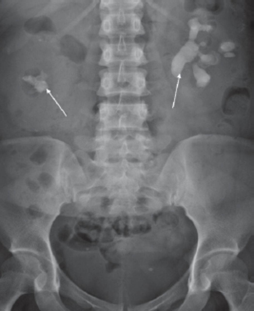
Plain film of the abdomen showing bilateral staghorn calculi (white arrows).

Figure 2
  • Download figure
  • Open in new tab
  • Download powerpoint
Figure 2

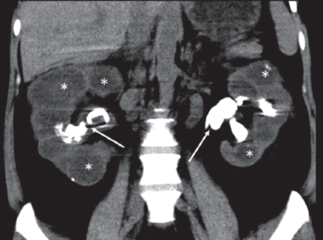
Coronal CT image showing bilaterally enlarged kidneys with bilateral staghorn calculi (white arrows), dilated calyces (asterisks) and paradoxical contracted renal pelves.

Questions

  1. What are the findings on plain film and CT of the abdomen.

  2. What radiological sign is seen on the CT?

  3. What is the diagnosis?

Answers

  1. The plain radiograph of the abdomen shows bilateral staghorn calculi with enlarged renal silhouette bilaterally. The CT shows dilatation of the renal calyces bilaterally with paradoxical contraction of the renal pelves.

  2. Dilatation of the renal calyces with paradoxical renal pelvis contraction gives the bilateral “bear paw” sign.

  3. Xanthogranulomatous pyelonephritis.

Discussion

Xanthogranulomatous pyelonephritis (XGP) is an uncommon form of bacterial pyelonephritis with an incidence of 1.4 per population of 100 000 per year.1 Almost all cases of XGP are unilateral. Bilateral involvement is extremely rare. Chronic obstruction of the urinary tract leads to destructive replacement of the renal parenchyma by granulomas, abscesses, and lipid laden macrophages, which is the hallmark of XGP. Diffuse XGP accounts for 90% of cases and focal XGP accounts for the remaining 10%. The typical CT findings in diffuse XGP are of dilated calyces with paradoxical contracted renal pelvis giving a ‘bear paw’ sign, with the dilated calyces resembling the toe pads of a bear’s paw.2 Focal XGP exhibits similar changes confined to an upper, or lower pole calyx. Although the CT findings highlighted in our case are typical, the CT findings in XGP have a wide range of findings such as diffuse renal enlargement, renal atrophy, hydronephrosis, focal masses and air within the urinary tract.3,4 Due to the wide range of CT findings, differentiating XGP from renal tumors and abscesses on imaging can be challenging preoperatively. The definitive treatment of diffuse XGP is complete nephrectomy and resection of any involved tissue. Partial nephrectomy has only been shown to be of benefit in focal XGP.5

In conclusion, XGP is an uncommon form of bacterial pyelonephritis with a wide range of CT findings. The bear paw sign describes the classic appearance of dilated calyces with relative sparing of the renal pelvis. Awareness of the imaging signs in XGP can lead to earlier diagnosis and appropriate treatment.

Case Reports

Case reports will only be considered for unusual topics that add something new to the literature. All Case Reports should include at least one figure. Written informed consent for publication must accompany any photograph in which the subject can be identified. Figures should be submitted with a 300 dpi resolution when submitting electronically. The abstract should be unstructured, and the introductory section should always include the objective and reason why the author is presenting this particular case. References should be up to date, preferably not exceeding 15.

Acknowledgement

The CT images were reviewed by Dr Raja Rizal Azman.

Footnotes

  • Notice: Authors are encouraged to submit quizzes for possible publication in the Journal. These may be in any specialty, and should approximately follow the format used here (maximum of 2 figures). Please address any submissions to: Editor, Saudi Medical Journal, Prince Sultan Military Medical City, PO Box 7897, Riyadh 11159, Kingdom of Saudi Arabia. Tel. +966 (11) 4777714 Ext. 42841.

  • Copyright: © Saudi Medical Journal

This is an open-access article distributed under the terms of the Creative Commons Attribution-Noncommercial-Share Alike 3.0 Unported, which permits unrestricted use, distribution, and reproduction in any medium, provided the original work is properly cited.

References

  1. ↵
    1. Parsons MA,
    2. Harris SC,
    3. Longstaff AJ,
    4. Grainger RG
    (1983) Xanthogranulomatous pyelonephritis: a pathological, clinical and aetiological analysis of 87 cases. Diagn Histopathol 6:203–219.
    OpenUrlPubMed
  2. ↵
    1. Dyer RB,
    2. Chen MY,
    3. Zagoria RJ
    (2004) Classic signs in uroradiology. Radiographics 24(Suppl 1):S247–S280.
    OpenUrlCrossRefPubMed
  3. ↵
    1. Loffroy R,
    2. Guiu B,
    3. Watfa J,
    4. Michel F,
    5. Cercueil JP,
    6. Krausé D
    (2007) Xanthogranulomatous pyelonephritis in adults: clinical and radiological findings in diffuse and focal forms. Clin Radiol 62:884–890.
    OpenUrlCrossRefPubMed
  4. ↵
    1. Kim JC
    (2001) US and CT findings of xanthogranulomatous pyelonephritis. Clin Imaging 25:118–121.
    OpenUrlCrossRefPubMed
  5. ↵
    1. Perez LM,
    2. Thrasher JB,
    3. Anderson EE
    (1993) Successful management of bilateral xanthogranulomatous pyelonephritis by bilateral partial nephrectomy. J Urol 149:100–102.
    OpenUrlPubMed
PreviousNext
Back to top

In this issue

Saudi Medical Journal: 37 (5)
Saudi Medical Journal
Vol. 37, Issue 5
1 May 2016
  • Table of Contents
  • Cover (PDF)
  • Index by author
Print
Download PDF
Email Article

Thank you for your interest in spreading the word on Saudi Medical Journal.

NOTE: We only request your email address so that the person you are recommending the page to knows that you wanted them to see it, and that it is not junk mail. We do not capture any email address.

Enter multiple addresses on separate lines or separate them with commas.
Adult female with acute renal failure and weight loss
(Your Name) has sent you a message from Saudi Medical Journal
(Your Name) thought you would like to see the Saudi Medical Journal web site.
Citation Tools
Adult female with acute renal failure and weight loss
Nor A. Abdul-Rahman, Raja R. Azman, Gnana Kumar
Saudi Medical Journal May 2016, 37 (5) 584-586; DOI: 10.15537/smj.2016.5.15042

Citation Manager Formats

  • BibTeX
  • Bookends
  • EasyBib
  • EndNote (tagged)
  • EndNote 8 (xml)
  • Medlars
  • Mendeley
  • Papers
  • RefWorks Tagged
  • Ref Manager
  • RIS
  • Zotero
Share
Adult female with acute renal failure and weight loss
Nor A. Abdul-Rahman, Raja R. Azman, Gnana Kumar
Saudi Medical Journal May 2016, 37 (5) 584-586; DOI: 10.15537/smj.2016.5.15042
Twitter logo Facebook logo Mendeley logo
  • Tweet Widget
  • Facebook Like
  • Google Plus One
Bookmark this article

Jump to section

  • Article
    • Clinical Presentation
    • Questions
    • Answers
    • Discussion
    • Acknowledgement
    • Footnotes
    • References
  • Figures & Data
  • eLetters
  • References
  • Info & Metrics
  • PDF

Related Articles

  • No related articles found.
  • PubMed
  • Google Scholar

Cited By...

  • No citing articles found.
  • Google Scholar

More in this TOC Section

  • The man who thought he was kicked from behind
  • Scalp roof tiles.
  • A child with congenital amputation of the foot
Show more Clinical Image

Similar Articles

CONTENT

  • home

JOURNAL

  • home

AUTHORS

  • home
Saudi Medical Journal

© 2025 Saudi Medical Journal Saudi Medical Journal is copyright under the Berne Convention and the International Copyright Convention.  Saudi Medical Journal is an Open Access journal and articles published are distributed under the terms of the Creative Commons Attribution-NonCommercial License (CC BY-NC). Readers may copy, distribute, and display the work for non-commercial purposes with the proper citation of the original work. Electronic ISSN 1658-3175. Print ISSN 0379-5284.

Powered by HighWire