


Abstract
Objectives: To determine the prevalence of inner ear anomalies and the frequency of different anomaly types among cochlear implant recipients.
Methods: This study included a retrospective chart review of all patients who received cochlear implants between January 2009 and January 2013 in King Abdulaziz University Hospital cochlear implant program in Riyadh, Saudi Arabia. All subjects underwent thin-cut CT of the temporal bone and MRI. The collected data included age, gender, and CT and MRI findings regarding temporal bone anomalies. Patients with any identified congenital inner ear anomalies were included in the study.
Results: In total, 316 patients’ cases were reviewed. Inner ear malformations were identified in 24 patients, which represented a prevalence of 7.5%. Among these 24 patients, 8 (33.3%) presented with a large vestibular aqueduct (LVA), 8 (33.3%) semicircular canal (SCC) dysplasia, 7 (29.1%) classical Mondini deformity, and one (4.1%) cochlear hypoplasia.
Conclusion: The prevalence of inner ear anomalies among cochlear implant recipients was 7.5%. This result is consistent with findings worldwide. The most common anomalies were LVA and SCC hypoplasia; by contrast, in other regions, the most common anomaly is either the Mondini deformity, or LVA.
Hearing loss management using cochlear implants in patients with inner ear anomalies has long been discussed in the otology community. In particular, hearing loss in children, which is a frequent clinical phenomenon in Saudi society,1,2 has a serious impact on patients and their families. However, no study has been performed to examine the prevalence of inner ear anomalies in our community, which features high rates of consanguinity and positive family histories of deafness.3,4 Magnetic resonances imaging and CT play important roles in the preoperative assessment of inner ear abnormalities such as cochlear nerve deficiency and variant anatomy; these abnormalities may affect not only the decision to perform the implantation procedure and the patient’s prognosis regarding auditory improvement, but also the risk of complications.5 The current study examined the prevalence of inner ear anomalies among cochlear implant recipients in the King Abdulaziz University Hospital (KAUH) cochlear implant program. Thus, the frequencies of different types of inner ear malformations are presented. This information can potentially help to predict malformations in hearing loss patients who will receive cochlear implants, leading to greater caution in the diagnosis and management of these patients.
Methods
A retrospective chart review of all patients who received cochlear implants in the Department of Otolaryngology and Head and Neck Surgery at KAUH in Riyadh between January 2009 and January 2013 was performed. All subjects underwent a high-resolution CT (HRCT) scan of their temporal bones and an MRI examination. The results of both types of imaging were reviewed by an expert otologist. The collected data included age, gender, and HRCT and MRI findings regarding temporal bone anomalies. Patients who were identified as having any type of congenital inner ear anomaly were included in the study. The cochleovestibular classification system proposed by Sennaroglu6 was used for this investigation. Additionally, large vestibular aqueduct (LVA) was assessed using the Cincinnati criteria.7
Results
In total, 316 patients received cochlear implants during the study period. Inner ear malformations were found in 24 patients, which represented a prevalence of 7.5%. The following anomalies were observed among these 24 patients: 8 (33.3%) patients exhibited semicircular canal (SCC) dysplasia (Figure 1), 8 (33.3%) patients exhibited LVA alone (Figure 2), 7 (29.1%) patients exhibited the classical Mondini (Figure 3) deformity (incomplete partition [IP] type 2, dilated vestibule and LVA), and 1 (4.1%) patient exhibited cochlear hypoplasia type 2 (Figure 4).

A CT showing one case of semicircular canal dysplasia (red asterisk) where the lateral semicircular canal fused with vestibule as one unite.

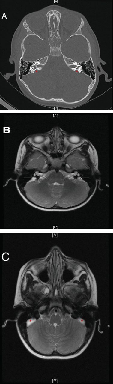
A) CT scan with bilateral large vestibular aqueduct (red asterisk). B) same patient with MRI showed large endolymphatic duct (white arrow). C) same MRI in other axial cut showed dilated endolymphatic sac (red asterisk).

Temporal bone CT scan with mondini A) shows 1: internal auditory canal. 2: dilated vestibule. 3: large vestibular aqueduct. B) apical cyst of cochlea (red asterisk)

Axial CT scan shows cochlear hypoplasia type II (black arrow).
Prevalence of various malformations
The vestibular aqueduct was normal in 9 (37.5%) cases and enlarged in 15 (62.5%) cases. Among the patients with LVA (including 8 cases of LVA alone and 7 cases of LVA associated with the Mondini deformity), bilateral enlargement was observed in 13 (54.1%) cases, and unilateral enlargement was observed (either isolated or coupled with the Mondini deformity) in 2 (8.3%) cases. The vestibule was normal in 15 (62.5%) cases and dilated in 9 (37.5%) cases (a condition associated with the Mondini deformity in 7 [29.1%] cases and involving fusion with the lateral SCC (LSCC) in 2 [8.3%] cases). The cochlea was normal in 16 (66.6%) cases, the Mondini deformity was observed in 7 (29.1%) cases, and cochlear hypoplasia was observed in one (4.1%) case. The SCC was normal in 16 (66.6%) patients and 8 (29.1%) patients exhibited SCC dysplasia. Moreover, there was one (4.1%) case each of an enlarged labyrinthine portion of the facial canal, a high jugular bulb, small malformed ossicles and rudimentary ossicles (Figure 5).

Prevalence of each inner ear anomaly among 316 patients received cochlear implants during the study period. LVA - large vestibular aqueduct, SCC - semicircular canal
Discussion
Mondini8 first described a cochlear anomaly involving one and a half turns, with loss of an interscalar septum in the apical turn. Subsequently, the term “Mondini deformity” has been used to refer to many different types of inner ear abnormalities. Jackler et al9 proposed an embryogenesis-based classification of congenital inner ear malformations using polytomography that remains widely accepted. Depending on the stage when development is arrested, inner ear malformations present along a spectrum that ranges from severe to mild abnormalities. Sennaroglu et al10 used HRCT to identify the radiological features of 2 completely different types of cochlear IP anomalies, which are referred to as IP-I and IP-II. Recently, the same authors identified X-linked deafness, which has been recognized as IP-III; a third type of IP.11
A major reason to measure the prevalence of inner ear anomalies is that our region has high rates of consanguinity and other risk factors for congenital anomalies.3,4 In addition, hearing impairment (HI) is common in this region. In one study, Al-Shaikh and Zakzouk12 measured the prevalence of HI in 4 major regions across Saudi Arabia and determined that the overall prevalence of sensorineural hearing loss (SNHL) was 1.5%. The prevalence of severe, or profound deafness was 0.72% among all screened children. According to the Central Saudi Department of Statistics and Information, 5,685,343 children (32.2% of the overall population) are of screening age,13 which is <15 years, and 0.72% of these children have severe to profound SNHL.12 A simple calculation reveals that there are 40,934 Saudi children with severe to profound SNHL.
In the current study, the prevalence of inner ear anomalies among cochlear implant recipients was 7.5%, which did not greatly differ from the corresponding prevalence in other regions of the world. Reports from the prior decade have indicated that the incidence of inner ear malformations ranges from 6.9-32%.14-18 A similar study conducted in the US (specifically, in North Carolina) by Buchman et al19 demonstrated that the prevalence of inner ear anomalies among patients who receiving cochlear implants was 8.8%. In our study, a male to female ratio of 3:2 was observed among hearing loss patients with inner ear anomalies; thus, males (60%) accounted for most patients. Al-Muhaimeed et al20 presented data from 117 Saudi patients with hearing loss who were undergoing cochlear implantation and demonstrated that most patients were males (n=70, 59.8%), rather than females (40.2%). In the current study, the individual frequencies of various inner ear anomalies were presented; in fact, these frequencies were one of the most important aspects of this report. The structures that were most commonly anomalous were the vestibular aqueduct, with SCC dysplasia and LVA accounting for 8 (33.3%) cases each, the Mondini deformity occurring in 7 (29.1%) cases, and cochlear hypoplasia observed in one (4.1%) case. There are differences between our study and other studies from different regions with respect to the frequencies of inner ear anomaly subtypes. In a 2010 review article on inner ear anomalies, Sennaroglu6 reported the following subtypes and frequencies: Michel deformity 6%, cochlear aplasia 5%, common cavity malformation 8%, cochlear hypoplasia 12%, IP 41% (IP-I [cystic cochleovestibular malformation], 20% IP-II [Mondini deformity] 19%, and IP-III [X-linked deafness] 2%), and LVA 15%. A literature review indicated that the most common anomalies and the frequencies of various anomalies differed among studies in different areas.
The present study reflects a significant percentage of patients with inner ear anomalies, although this percentage is within the range reported worldwide. This result has implications for cochlear implant surgery in patients with inner ear anomalies, which needs to be performed by qualified otology, or neuro-otology surgeons, as there is an increased chance of intraoperative difficulty.6 Both MRI and CT play important roles in the preoperative assessment of inner ear abnormalities. These tools can be extremely informative with respect to assessing a patient’s candidacy for cochlear implantation and anticipating risks and complications by emphasizing the potential risks for patients who will undergo this surgery. The findings presented here need to be emphasized more at different centers in different regions of Saudi Arabia and need to be applied accordingly via a national governmental plan to assess the cost-effectiveness based on the procedure and the outcome and to minimize patient risks by referring these cases to specialized, highly qualified centers.
In conclusion, severe to profound SNHL is a challenging medical problem, particularly if this condition is associated with an inner ear anomaly. Our study results demonstrated that the prevalence of inner ear anomalies among cochlear implant recipients was 7.5%. This result is consistent with findings worldwide. The most common types of anomalies were LVA and SCC hypoplasia; by contrast, in the literature, the most commonly observed anomalies have generally been either Mondini deformity, or LVA. Both CT and MRI images were used as diagnostic tools to identify different inner ear anomalies.
Footnotes
Disclosure. Authors have no conflict of interests, and the work was not supported or funded by any drug company.
- Received March 8, 2016.
- Accepted July 28, 2016.
- Copyright: © Saudi Medical Journal
This is an open-access article distributed under the terms of the Creative Commons Attribution-Noncommercial-Share Alike 3.0 Unported, which permits unrestricted use, distribution, and reproduction in any medium, provided the original work is properly cited.
References
In this issue


Jump to section
Related Articles
Cited By...
- No citing articles found.