
Main menu

  • Home
  • Content
    • Latest
    • Archive
    • home
  • Info for
    • Authors
    • Reviewers
    • Subscribers
    • Institutions
    • Advertisers
    • Join SMJ
  • About Us
    • About Us
    • Editorial Office
    • Editorial Board
  • More
    • Advertising
    • Alerts
    • Feedback
    • Folders
    • Help
  • Other Publications
    • NeuroSciences Journal

User menu

  • My alerts
  • Log in

Search

  • Advanced search
Saudi Medical Journal
  • Other Publications
    • NeuroSciences Journal
  • My alerts
  • Log in
Saudi Medical Journal

Advanced Search

  • Home
  • Content
    • Latest
    • Archive
    • home
  • Info for
    • Authors
    • Reviewers
    • Subscribers
    • Institutions
    • Advertisers
    • Join SMJ
  • About Us
    • About Us
    • Editorial Office
    • Editorial Board
  • More
    • Advertising
    • Alerts
    • Feedback
    • Folders
    • Help
  • Follow psmmc on Twitter
  • Visit psmmc on Facebook
  • RSS
OtherClinical Image
Open Access

Unusual skin rash in infant

Badi Alenazi, Fatima AlTamimi and Reem AlShammari
Saudi Medical Journal November 2017, 38 (11) 1153-1155; DOI: https://doi.org/10.15537/smj.2017.11.20357
Badi Alenazi
From the Department of Pediatrics, AlYamamah Hospital, Riyadh, Kingdom of Saudi Arabia
ABP, SBP
  • Find this author on Google Scholar
  • Find this author on PubMed
  • Search for this author on this site
  • For correspondence: [email protected]
Fatima AlTamimi
From the Department of Pediatrics, AlYamamah Hospital, Riyadh, Kingdom of Saudi Arabia
ABP, SBP
  • Find this author on Google Scholar
  • Find this author on PubMed
  • Search for this author on this site
Reem AlShammari
From the Department of Pediatrics, AlYamamah Hospital, Riyadh, Kingdom of Saudi Arabia
MBBS
  • Find this author on Google Scholar
  • Find this author on PubMed
  • Search for this author on this site
  • Article
  • Figures & Data
  • eLetters
  • Info & Metrics
  • References
  • PDF
Loading

Clinical Presentation

A 4-month old boy not known to have any medical illness, presented to the emergency with a history of skin rash for a day. The rash first appeared as a single red small spot on the chin, rapidly progressed to involve arms and legs within a 24 hour period, then enlarged and formed of vesicles with clear yellowish fluid, after that ruptured to leave thin brown crusts. Not involve scalp, neck, trunk, mucosal surface or genitalia. He had a recent upper respiratory tract infection and diarrhea for one week. No history of fever, poor feeding or decrease of activity. No history of contact with individuals who have similar complaints. No history of insect bites, trauma, skin disease or recent travel. Unremarkable past history. He was a product of full term at 38 weeks normal spontaneous vaginal delivery with a birth weight of 3 Kg. No neonatal complication. No maternal disease during pregnancy. Developmental history was appropriate for age. Baby was immunized up to 2 months. Unremarkable family and social history. He was not on any medication. The weight is 6 kg (on 50th percentile), height is 58 cm (on 25th percentile), and the head circumference is 40 cm (on 50th percentile). His vital signs were: temperature: 36.5, pulse rate 127/min, respiratory rate 26/min. He looks well, alert, active, not in respiratory distress, no pallor or jaundice, and no dysmorphic features. His skin examination revealed several erythematosus round well defined macules, distributed on the face, arms and legs (target lesions). Bullae on the face contains clear yellowish fluid and brown crust. The lesions were not warm, tender, blanching or pururitic. There is a moderate, non pitting symmetrical edema involves the dorsum of the feet. Other systemic examination were unremarkable. Investigations showed hemoglobin 11.3 g dl, white blood cel 15.9 x10³/µL, neutrophil 29%, and lymphocyte 61%. Serum urea and electrolytes were normal. Liver function tests were normal, renal profile and electrolyte were normal, urine and stool analysis were normal.

Figure 1
  • Download figure
  • Open in new tab
  • Download powerpoint
Figure 1

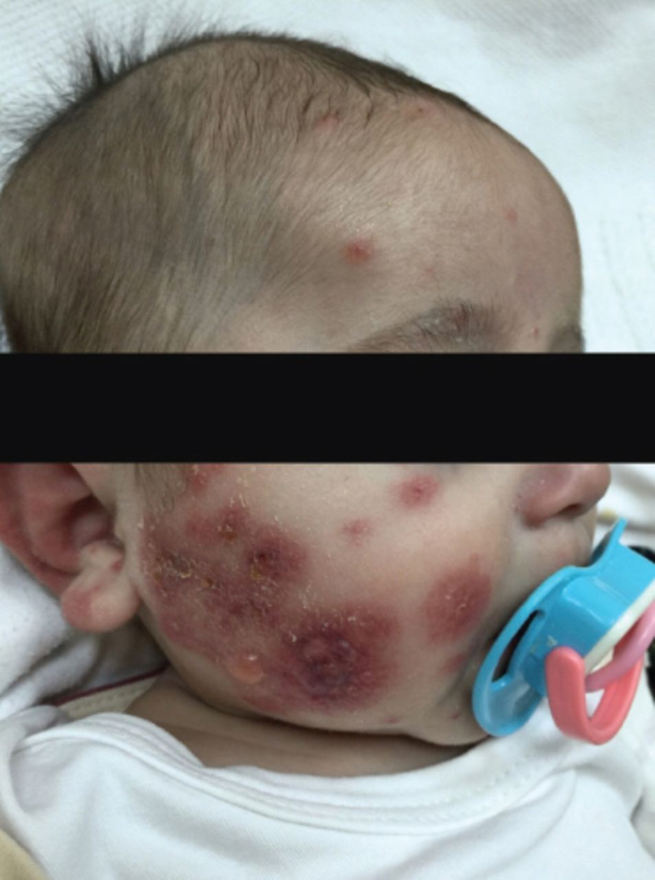
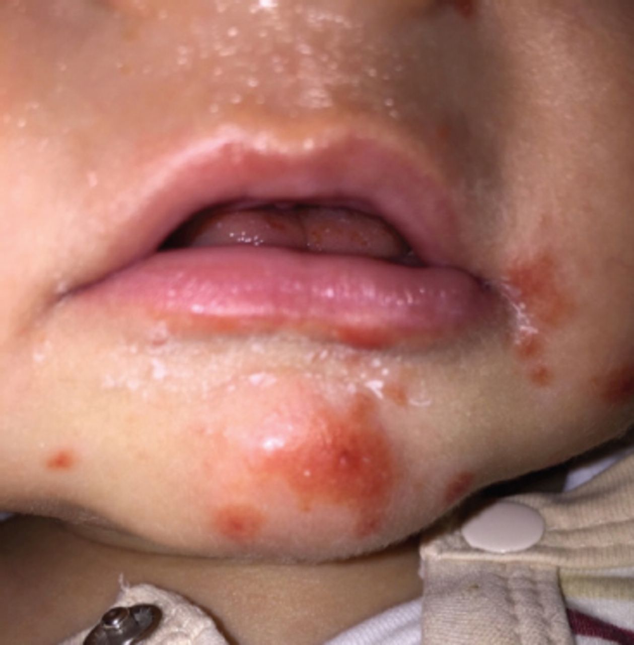
Several erythematous round well defined macules and bullae on the face.

Figure 2
  • Download figure
  • Open in new tab
  • Download powerpoint
Figure 2

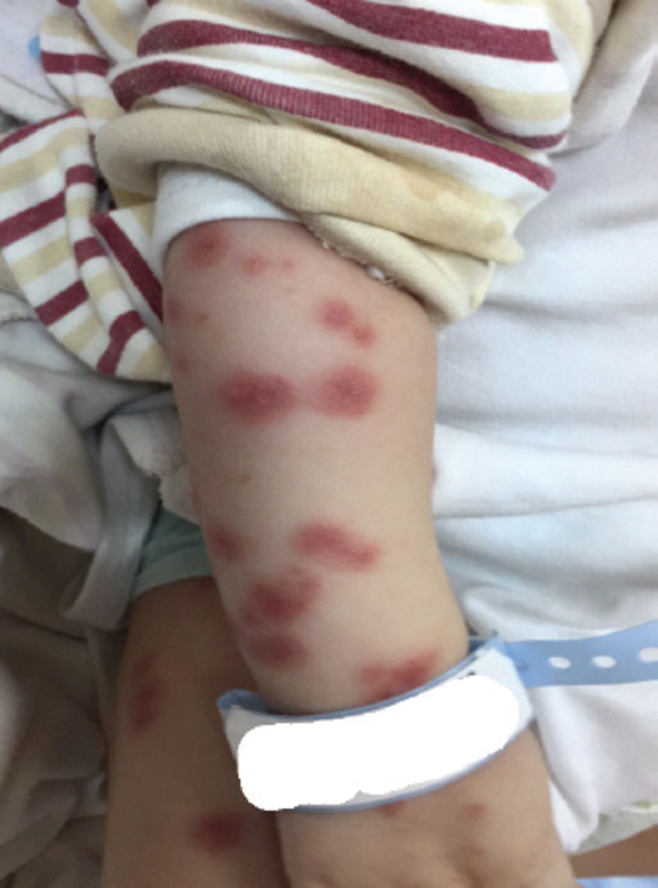
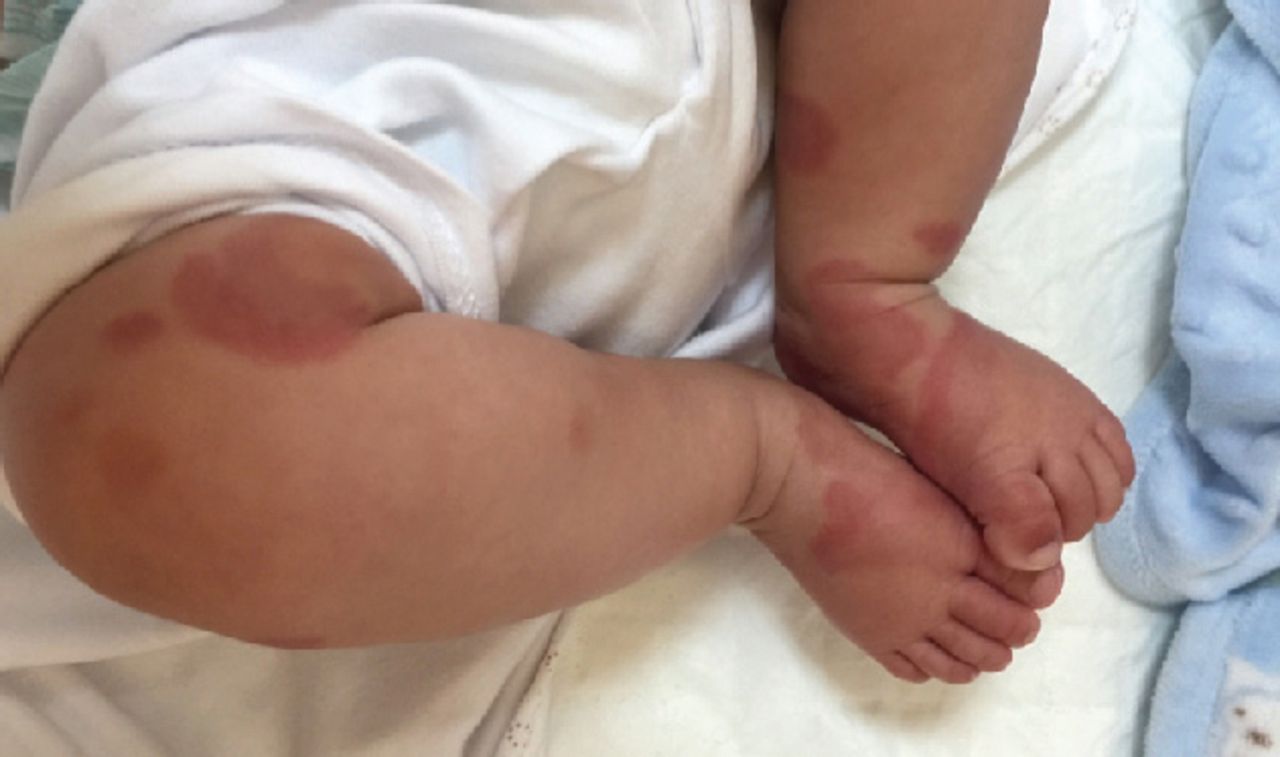
Several erythematous round well defined macules involving both arms.

Figure 3
  • Download figure
  • Open in new tab
  • Download powerpoint
Figure 3

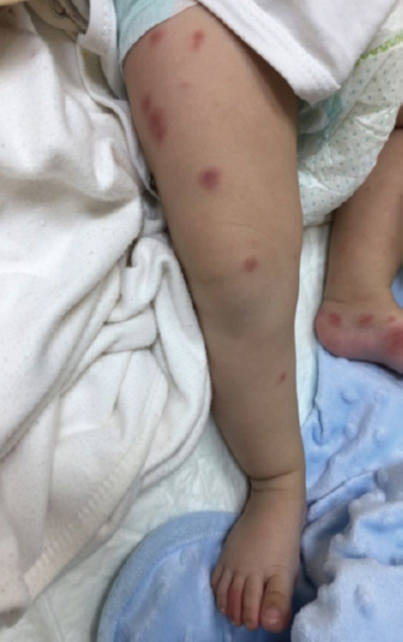
Several erythematous round well defined macules involving lower limbs.

Figure 4
  • Download figure
  • Open in new tab
  • Download powerpoint
Figure 4

Bullae on the face contains clear yellowish fluid with brown crust.

Figure 5
  • Download figure
  • Open in new tab
  • Download powerpoint
Figure 5

Several erythematous round well defined macules involving lower limbs and moderate, non pitting symmetrical edema involves the dorsum of feet.

Questions

  1. What are possible different diagnoses?

  2. What is the most likely diagnosis?

  3. What is the management of this case?

Answers

  1. The possible different diagnoses are:

    • Acute hemorrhagic edema of infancy

    • Henoch-Schonlein purpura (HSP)

    • Bullous impetigo

    • Drug Eruptions

    • Meningococcemia

    • Septicemia

  2. The most likely diagnosis is acute hemorrhagic edema of infancy

  3. Management of acute hemorrhagic edema of infancy is usually supportive

Discussion

Acute hemorrhagic edema of infancy (AHEI) is a rare entity of cutaneous small vessel vasculitis affecting infants between 4-24 months. It is immune complex-mediated vasculitis, which might be initiated from previous viral or bacterial infections (mostly upper respiratory tract infection, antibiotic and less likely after vaccination.1 It was initially considered as a variant of Henoch-Schönlein purpura (HSP); however, it is now considered a separate entity because of the lack of both visceral involvement and immunoglobulin A (IgA) skin depositions. The peak incidence in winters due to high rates of respiratory infections among infants.2 Acute hemorrhagic edema of infancy affects males are more than females.3 The differential diagnosis of AHEI includes HSP, erythema multiforme, meningococcemia and drug eruption.4 The classical presentation of AHEI including asymmetrical painless non-pitting edema of the face and sudden painful ecchymosis patches and plaques which appear on the face and extremities or large target shaped lesions.4 Acute hemorrhagic edema of infancy is usually diagnosed clinically after excluding other causes of purpura. Most AHEI infants are nontoxic in appearance. Histopathological findings in AHEI are very similar to HSP. However, IgA deposition is seen in only 10-35% cases of AHEI with less visceral complication. There is no effective treatment for AHEI and only symptomatic care is advised.5

Footnotes

  • Notice: Authors are encouraged to submit quizzes for possible publication in the Journal. These may be in any specialty, and should approximately follow the format used here. Please address any submissions to: Editor, Saudi Medical Journal, Prince Sultan Military Medical City, PO Box 7897, Riyadh 11159, Kingdom of Saudi Arabia. Tel. +966 (11) 4777714 Ext. 42840.

  • Copyright: © Saudi Medical Journal

This is an open-access article distributed under the terms of the Creative Commons Attribution-Noncommercial-Share Alike 3.0 Unported, which permits unrestricted use, distribution, and reproduction in any medium, provided the original work is properly cited.

References

  1. ↵
    1. Morrison RR,
    2. Saulsbury FT
    (1999) Acute hemorrhagic edema of infancy associated with pneumococcal bacteremia. Pediatr Infect Dis J 18:832–823.
    OpenUrlCrossRefPubMed
  2. ↵
    1. Moradinejad MH,
    2. Entezari P,
    3. Mahjoub F,
    4. Ziaee V
    (2011) Acute hemorrhagic edema of infancy;a report of five Iranian infants and review of the literature. Iran J Pediatr 21:107–112.
    OpenUrlPubMed
  3. ↵
    1. Legrain V,
    2. Lejean S,
    3. Taïeb A,
    4. Guillard JM,
    5. Battin J,
    6. Maleville J
    (1991) Infantile acute hemorrhagic edema of the skin: Study of ten cases. J Am Acad Dermatol 24:17–22.
    OpenUrlCrossRefPubMed
  4. ↵
    1. Millard T,
    2. Harris A,
    3. MacDonald D
    (1999) Acute infantile hemorrhagic oedema. J Am Acad Dermatol 41:837–839.
    OpenUrlCrossRefPubMed
  5. ↵
    1. Roy KP,
    2. Madke B,
    3. Kar S,
    4. Yadav N
    (2015) Acute hemorrhagic edema of infancy. Indian J Dermatol 60:624–625.
    OpenUrlPubMed
PreviousNext
Back to top

In this issue

Saudi Medical Journal: 38 (11)
Saudi Medical Journal
Vol. 38, Issue 11
1 Nov 2017
  • Table of Contents
  • Cover (PDF)
  • Index by author
Print
Download PDF
Email Article

Thank you for your interest in spreading the word on Saudi Medical Journal.

NOTE: We only request your email address so that the person you are recommending the page to knows that you wanted them to see it, and that it is not junk mail. We do not capture any email address.

Enter multiple addresses on separate lines or separate them with commas.
Unusual skin rash in infant
(Your Name) has sent you a message from Saudi Medical Journal
(Your Name) thought you would like to see the Saudi Medical Journal web site.
Citation Tools
Unusual skin rash in infant
Badi Alenazi, Fatima AlTamimi, Reem AlShammari
Saudi Medical Journal Nov 2017, 38 (11) 1153-1155; DOI: 10.15537/smj.2017.11.20357

Citation Manager Formats

  • BibTeX
  • Bookends
  • EasyBib
  • EndNote (tagged)
  • EndNote 8 (xml)
  • Medlars
  • Mendeley
  • Papers
  • RefWorks Tagged
  • Ref Manager
  • RIS
  • Zotero
Share
Unusual skin rash in infant
Badi Alenazi, Fatima AlTamimi, Reem AlShammari
Saudi Medical Journal Nov 2017, 38 (11) 1153-1155; DOI: 10.15537/smj.2017.11.20357
Twitter logo Facebook logo Mendeley logo
  • Tweet Widget
  • Facebook Like
  • Google Plus One
Bookmark this article

Jump to section

  • Article
    • Clinical Presentation
    • Questions
    • Answers
    • Discussion
    • Footnotes
    • References
  • Figures & Data
  • eLetters
  • References
  • Info & Metrics
  • PDF

Related Articles

  • No related articles found.
  • PubMed
  • Google Scholar

Cited By...

  • No citing articles found.
  • Google Scholar

More in this TOC Section

  • The man who thought he was kicked from behind
  • Scalp roof tiles.
  • A child with congenital amputation of the foot
Show more Clinical Image

Similar Articles

CONTENT

  • home

JOURNAL

  • home

AUTHORS

  • home
Saudi Medical Journal

© 2025 Saudi Medical Journal Saudi Medical Journal is copyright under the Berne Convention and the International Copyright Convention.  Saudi Medical Journal is an Open Access journal and articles published are distributed under the terms of the Creative Commons Attribution-NonCommercial License (CC BY-NC). Readers may copy, distribute, and display the work for non-commercial purposes with the proper citation of the original work. Electronic ISSN 1658-3175. Print ISSN 0379-5284.

Powered by HighWire