


Clinical Presentation
A rare complication of metastatic lung disease treatment
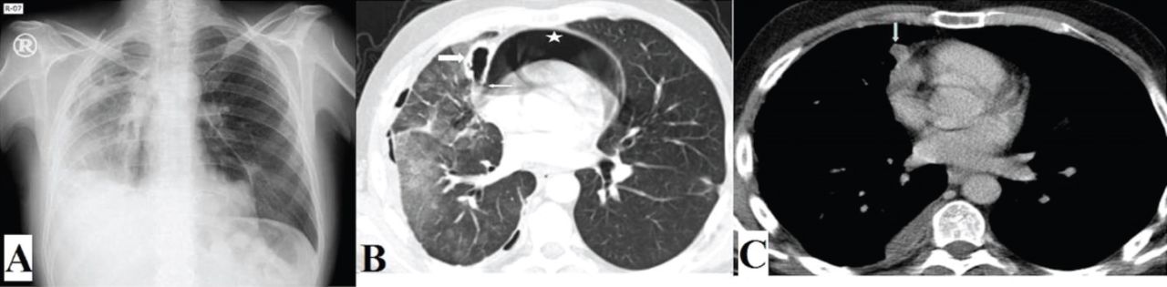
A 37-year-old male patient who had undergone partial glossectomy and neck dissection and who received chemotherapy and radiotherapy because of squamous cell carcinoma of the tongue base presented to our clinic with the complaint of hemoptysis. Chest radiography revealed pneumopericardium (Figure 1A). On thoracic CT, it was observed that the cavitary nodule was necrotic and had perforated the pericardium (Figure 1B). On a CT taken 2 months previously, there was a 20x12 mm solid nodule in the medial segment of the middle lobe of his right lung, neighboring the pericardium (Figure 1C). Since his echocardiograph revealed no pericardial tamponade, no surgical procedure was performed, and he was discharged with follow-up recommendations. It was his last admittance to our hospital. When the patient was questioned at the clinic where he was next presented, he received chemotherapy (etoposide and cisplatin). New lung metastases were diagnosed and erlotinib was added to his chemotherapy. His following controls were performed with repeated thorax CT. Approximately one month after his discharge from our hospital, we learned that he had died due to massive hemoptysis.

A) Chest x-ray: Pneumopericardium covered the heart, and volume loss due to right upper lobectomy is seen in the right hemithorax. B) Thoracic CT: There is a cavitary lesion (thick arrow) in the right paracardiac area and pneumopericardium (star). The image indicates a defect between the cavitary lesion and the pericardium (thin arrow). Additionally, in the right lung, widespread interstitial densities and minimal pneumothorax in the right hemithorax are seen. (After Erlotinib treatment.) C) Thoracic CT: A 20x12 mm diameter solid nodule neighboring the pericardium is seen in the right hemithorax (arrow). (Before Erlotinib treatment.)
Questions
Which is the first preferred radiological method to diagnose pneumopericardium?
Answers
Chest x-ray and/or CT are the first preferred radiological methods to diagnose pneumopericardium.
Discussion
Pneumopericardium is a rare condition that was first described by Bricketeau.1 Trauma and disease of organs neighboring the pericardium are the most common etiologies. Although pneumopericardium may develop due to high-pressure mechanical ventilation, mediastinal tumors, tuberculosis, or gastro pericardial fistula may also develop iatrogenically due to endomyocardial biopsy or after pacemaker implantation.2
Pneumopericardium develops due to fistulization between the pericardium and an air-containing organ or structure. These areas are mostly the pleural space, tracheobronchial tree, or the gastrointestinal tract.2,3 In the literature, there are only a few pneumopericardium cases caused by chemotherapy administered for a metastatic lung disease. Ours is one of them. The cases reported in the literature are mostly pneumothorax cases that developed due to necrosis of cystic subpleural metastases of the lung into the pleural space. In metastases in the lung parenchyma, tissue necrosis develops due to chemotherapy. The resultant bronchopleural or broncho pericardial fistula leads to pneumothorax or pneumopericardium.3,4 In our case, subpleural metastases located in the medial segment of the right middle lobe were adherent to the pericardium tissue due to the right upper lobectomy the patient previously underwent. After the administration of erlotinib, tissue necrosis developed in the metastases, and a fistulization occurred from lung parenchyma into the pericardium. Additionally, the occurrence and continuation of hemoptysis indicate that metastatic lesions underwent necrosis due to the erlotinib treatment.
Leprieur et al3 administered erlotinib to a patient with squamous cell lung carcinoma and detected right pneumothorax 12 days after treatment. Pneumothorax in such cases has been attributed to necrosis of the subpleural metastases. Similarly, Yang et al5 reported that a patient with metastatic colon adenocarcinoma developed right pneumothorax 7 days after bevacizumab treatment. In Yang et al’s article,5 3 mechanisms were proposed for the pneumothorax caused by lung metastases: 1) necrosis of subpleural metastases, 2) a check-valve mechanism caused by the obstruction of proximal and distal airways by tumor nodules, and 3) pulmonary infarct induced by tumor embolism.
In conclusion, pneumothorax and pneumopericardium can be observed during the treatment of metastatic disease of the lung. They usually occur through the necrosis of lesions in the parenchyma due to chemotherapy. Radiological imaging methods should be employed in patients who develop sudden chest pain and dyspnea during treatment and clinical follow-up.
The authors declare no conflict of interest with respect to the authorship and/or publication of this article. This manuscript or essence of its contents has not been previously published in part or in full on a website or printed in a journal in a language other than English.
Illustrations, Figures, Photographs
All figures or photographs should be submitted in a high resolution (minimum 300 DPI) electronic version saved in jpeg or tiff format. Original hard copies of all figures may be requested when necessary. Photographs will be accepted at the discretion of the Editorial Board. All lettering, arrows, or other artwork must be done by an artist or draftsman. If arrows are used please ensure they appear in a different color to the background color, preferably black with a white border, or white with a black border. If arrows distinguish different items on the figure then different arrow styles should be used ie. long, short, wide, narrow. Written informed consent for publication must accompany any photograph in which the subject can be identified. Written copyright permission, from the publishers, must accompany any illustration that has been previously published.
Footnotes
Notice: Authors are encouraged to submit quizzes for possible publication in the Journal. These may be in any specialty, and should approximately follow the format used here (maximum of 2 figures). Please address any submissions to: Editor, Saudi Medical Journal, Prince Sultan Military Medical City, PO Box 7897, Riyadh 11159, Kingdom of Saudi Arabia. Tel. +966 (11) 4777714 Ext. 42841.
- Copyright: © Saudi Medical Journal
This is an open-access article distributed under the terms of the Creative Commons Attribution-Noncommercial License (CC BY-NC), which permits unrestricted use, distribution, and reproduction in any medium, provided the original work is properly cited.
In this issue


Jump to section
Related Articles
Cited By...
- No citing articles found.