


Article Figures & Data
Figures
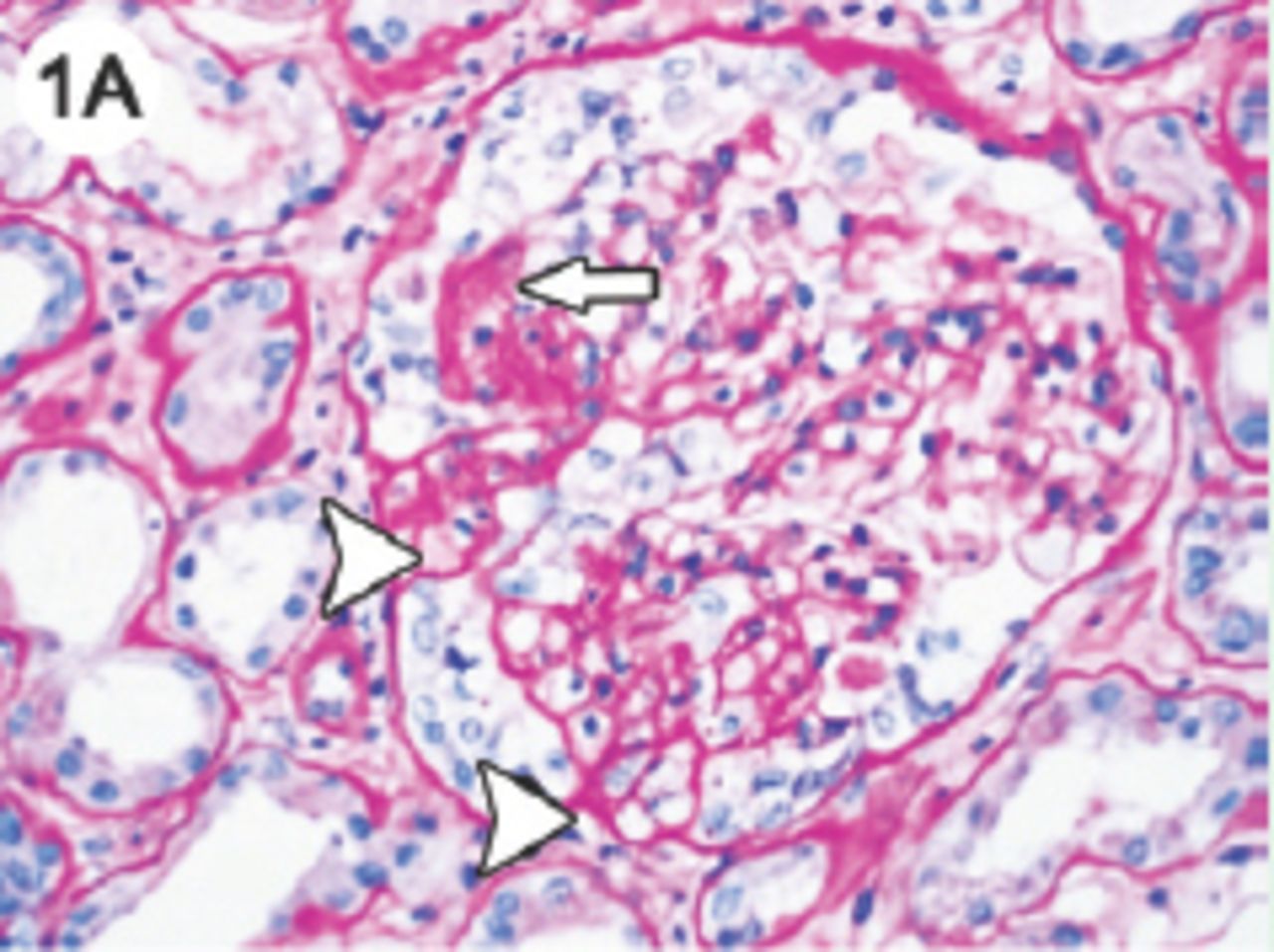
Light microscopy photomicrograph of a renal biopsy in a case of collapsing glomerulopathy shows segmental collapse of the glomerular tuft (arrow) with hyperplasia and hypertrophy of the overlying epithelial cells. Another tuft showing not otherwise specified type of segmental sclerosis with adhesion to the Bowman’s capsule is also noted (arrowhead). (Periodic Acid Schiff stain; original magnification x400.)
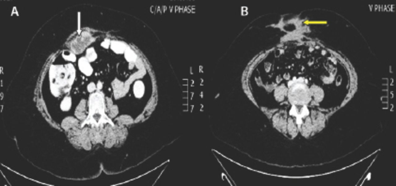
Computed tomography of the abdomen revealing A) right rectus abdominis muscle soft tissue mass suspicious of metastasis (white arrow) with peri-umbilical postoperative changes consistent with B) fibromatosis (yellow arrow)

In this issue


Jump to section
Related Articles
Cited By...
- No citing articles found.