


Abstract
Objectives: To emphasize different clinical features of tumor that can be misdiagnosed clinically.
Methods: A total of 8 cases operated between September 2009 and 2016 at the Celal Bayar University, Faculty of Medicine were included in the study. Patients’ clinicopathological features, type of surgery and follow up information were evaluated.
Results: Six patients were male. The average age was 75.50. The lesions were located on the head and neck, and chest wall. Six patients had a history of the rapid growth of lesion. There was no metastasis at the time of diagnosis. None of the patients needed adjuvant therapy. Mean follow up time was 19.37 months. None of the patients developed recurrence or metastasis.
Conclusion: This tumor resembles basal or squamous cell carcinoma. The histopathological evaluation may lead to misdiagnosis. Regional or distant metastasis is very rare. There is no consensus about adjuvant therapy. Screening for metastasis and close follow up are mandatory.
Trichilemmal carcinoma (TC) is one of the malignant tumors of hair follicles derived from outer root sheath epithelium. This tumor is the malignant form of “trichilemmoma” first described by Headington.1 Lesions occur mostly on the face and sometimes other sun-exposed areas of the body of older people. This tumor manifests as an exophytic or polypoid mass, with or without ulceration, scales, or rolled borders that resemble basal cell carcinoma (BCC), squamous cell carcinoma (SCC), keratoacanthoma, or proliferating pilar cyst.2 This malignant tumor is rare and can be aggressive. Recurrences, regional and distant metastasis have been reported.3-5 Limited studies are available investigating tumor clinical presentation, behavior, optimal treatment strategy, recurrence, metastatic potential and survival time. Most of the reports are case reports and a few have larger case numbers.6-12 In this study, we report a pigmented clinical variant and treatment outcomes of 8 cases diagnosed as TC. Time of occurrence and rapid growth phase of the tumor were interrogated. Since there are a few reports about this subject, we would like to spot light on the findings of our cases with additive information.
Methods
All authors state to the effect that the principles of the 1975 Declaration of Helsinki were followed during this research. The study was designed as a retrospective study. Patients operated at Faculty of Medicine, Celal Bayar University Hospital between September 2009 and September 2016, diagnosed as TC according to incisional or excisional biopsy results were included in the study. The age and gender of the patients, localization and size of the tumor, duration of tumor before the first examination and any previous surgery were recorded. All patients were asked about the time of occurrence of tumor and acceleration of growth at any time. Tumors were resected with 1 cm. margin and defect reconstructions were performed with a skin graft or local fasciocutaneous flaps. Histopathological examinations including immunohistochemical methods were performed. Patients were scanned with computed tomography (CT) for regional or distant metastasis and consulted to the Radiation Oncology department for adjuvant therapy. Patients were evaluated with ultrasonography (USG) monthly for the first three months, then at sixth month and at the end of the first year of the follow-up period. After the first year, yearly examinations were suggested. The reconstruction method, scan data for metastasis, the necessity of adjuvant therapy, follow up time and tumor recurrence data were recorded.
Results
Eight patients diagnosed with TC were included in the study. Six patients were male (M/F: 3/1). The mean age which was 75.50 (ranged between 56-93). One patient had previous surgery in another center but had no report. Duration of lesions until surgery was between 3 months to 20 years. Four patients had rapid growth of their lesions for the last couple of months. One of the lesions was located on the scalp, one of them was on the chest wall and the others were on the face. Dimensions of the tumors were between 3x2.5 cm and 10x10 cm. Ulceration was detected in the lesions of 4 patients (Table 1).
Gender, age, localization, size and duration of tumors, treatment methods and follow up information of the patients.
Histopathologically, tumors were connecting with the ulcerated epidermis and characterized by anastomosing trabecular, diffuse or lobular growth patterns. Tumors consisted of basaloid cells with peripheral palizading and large cells with clear cytoplasm. Pleomorphism and atypia were variable, but not high grade. Comedonecrosis was seen in the central part of tumor lobules. Trichilemmal keratinization was conspicuous. Only one patient had perineural invasion (Patient 3).
Defects of 6 patients were reconstructed with split-thickness skin graft (STSG) and defects of 2 patients were reconstructed with local flaps. Margins were tumor free in all patients. The follow up time ranged from 4-46 months (Mean=19.37 months) (Table 1). None of the patients had regional or distant metastasis at the time of diagnosis. None of the patients needed adjuvant therapy. One patient died as a result of myocardial infarction 8 months after surgery. There was no recurrence, regional or distant metastasis during follow up.
Discussion
Cutaneous adnexal carcinomas are rare and reported to represent 0.005% of all skin tumors. These carcinomas have a heterogenous origin and originate from undifferentiated stem cells.13 TCs are categorized in the follicle originated malign tumors according to their differentiation.14 They originate from outer root sheath epithelium. These tumors are seen on sun-exposed areas, mostly on the head and neck.2 Other possible risk factors are ionizing radiation, previous trauma or scar and genetic disorders. Clinical presentation may resemble BCC, SCC or keratoacanthoma and should be evaluated for differential diagnosis.
According to the literature, a slight majority of the cases were reported in men.12 In our patients, male dominance was seen in accordance with the literature. Macroscopically TCs were described as exophytic, polypoid, ulcerated or nodular lesions.11 In our study, most of the lesions were seen as described before. One of the lesions located on nasal dorsum was plain, irregular and pigmented resembling malignant melanoma (Figures 1-2).
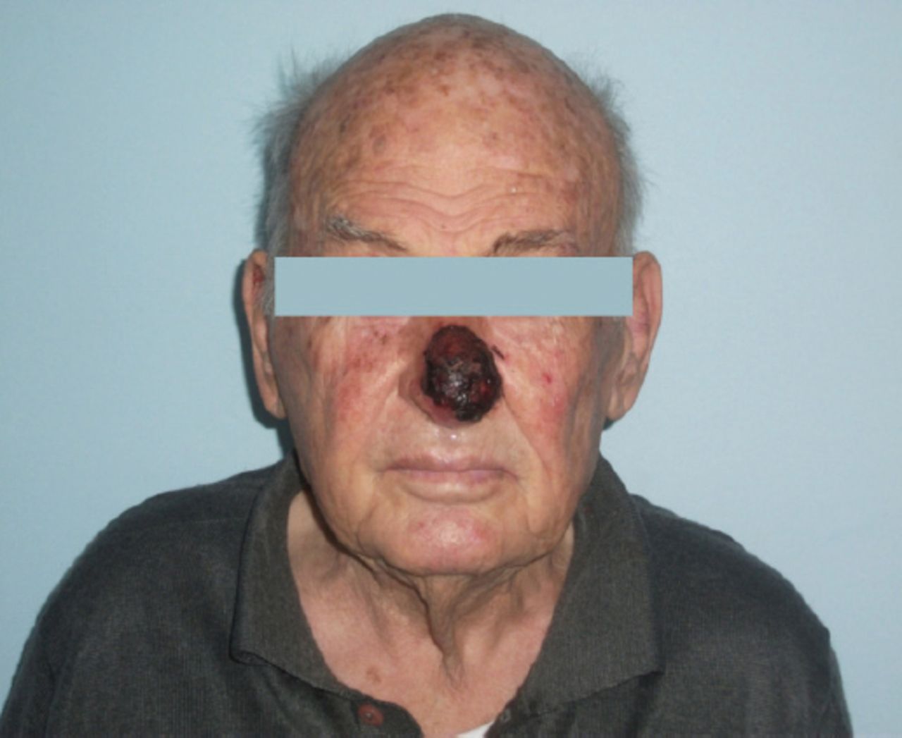
Preoperative view of pigmented TC located on nasal dorsum, resembling MM (Patient 2).

Postoperative view of the same patient 6 months after surgery.
This neoplasm is mostly located on head and neck. Extremities are other common localizations. Chest wall and abdomen are the other rare localizations reported before.12 In our study, one of the lesions was on the sternal notch with the noduloulcerative presentation. Incisional biopsy material of the patient taken and examined at another center had been reported as BCC. The localization was atypical for BCC. Formalin-fixed, paraffin-embedded blocks were obtained and reexamined at the Department of Pathology. After reexamination, the diagnosis was TC.
Four patients gave a history of rapid growth over the last couple of months. One patient had a surgery 6 years before the second surgery the representation of the mass for last 2 years. This should point out that malignant transformation of these tumors from benign precursors can occur.
Diagnosis of TC is based on histopathologic examination of hematoxylin-eosin stained slides. Trichilemmal keratinization and peripheral palizading are the signs which represent follicular root sheath origin of tumors. Nuclear atypia, marked cellular pleomorphism with atypical mitoses are associated with malignant behavior.14 The TC is an unusual malignant lesion with its histological characteristics suggesting an intermediate to high-grade malignancy but metastasis is uncommon and it is generally characterized by a benign neoplasm process that can be treated with complete excision.6 Surgery is considered as the treatment choice for TC, and periodic surveillance without adjuvant therapy is generally sufficient.7 Wide local excision has been suggested for TC but clear margins were mostly not described.9,11-12 Mohs micrographic surgery has been reported to be a successful technique for treating malign trichilemmal tumor.15 We treated all our patients with wide local excision with 1 cm. margin. Local cervical lymph node and distant metastases of TC have been rarely reported in the literature.9-11 There is no optimal treatment for this neoplasm with metastases and there is no established chemotherapy regimen in the literature. We scanned our patients with cranial, cervical and thoracoabdominal CT except one because he had refused further investigations. None of them had regional or distant metastasis and adjuvant RT was not adviced. Closed follow up was recommended to the patients by monthly visits for the first 3 months, at the 6th month, at the first year and once a year after then. Patients were scanned with cervical USG and no recurrence or metastasis was detected.
This study evaluates the clinical behavior of TC by investigating the rapid progression of the tumor before diagnosis. Four of the patients gave a history of the rapid growth of their tumors for a couple of months. This may be a sign of malignant transformation of benign tumors. This is one of the first reports like another one focusing on rapid progression before diagnosis.11,16 Also, we represent a different clinical form of TC with pigmentation. The main limitation of this study may be the shortness of follow-up time. Since our patients were elderly it was difficult to bring them to the hospital as they do not have any complaint.
In conclusions, TC is a rare malignant tumor of the hair follicles that can present with different clinical features. This tumor resembles SCC or BCC clinically. The histopathological evaluation may lead to misdiagnosis. Although this tumor is rarely reported, actual incidence is unknown. Regional or distant metastasis can be seen with this tumor. There is no consensus about adjuvant therapy but screening for metastasis and close follow up are mandatory.
Illustrations, Figures, Photographs
All figures or photographs should be submitted in a high resolution (minimum 300 DPI) electronic version saved in jpeg or tiff format. Original hard copies of all figures may be requested when necessary. Photographs will be accepted at the discretion of the Editorial Board. All lettering, arrows, or other artwork must be done by an artist or draftsman. If arrows are used please ensure they appear in a different color to the background color, preferably black with a white border, or white with a black border. If arrows distinguish different items on the figure then different arrow styles should be used ie. long, short, wide, narrow. Written informed consent for publication must accompany any photograph in which the subject can be identified. Written copyright permission, from the publishers, must accompany any illustration that has been previously published.
- Received October 4, 2017.
- Accepted December 20, 2017.
- Copyright: © Saudi Medical Journal
This is an open-access article distributed under the terms of the Creative Commons Attribution-Noncommercial-Share Alike 3.0 Unported, which permits unrestricted use, distribution, and reproduction in any medium, provided the original work is properly cited.
In this issue


Jump to section
Related Articles
Cited By...
- No citing articles found.