


Abstract
Objectives: To identify the occurrence rate of thyroid ultrasound abnormalities in asymptomatic subjects, and describe the features of detected nodules among university students.
Methods: The study is based on an observational research design that was conducted from April 2015 to May 2015. The study included 166 individuals, aged between 19 and 23 years. The subjects had their glands examined by ultrasound (US) scanning, using Philips ultrasound machine (5-12 MHz linear transducer).
Results: We recruited 90 (57.8%) females and 76 (42.2%) males without any indications of thyroid disease. Data estimated that 41 (24.7%) subjects had positive results on thyroid disease screening, 24 (70.6%) participants had solitary nodules, and 10 (29.4%) had multiple nodules. Thirty-four subjects revealed nodular presentation in the screening, among which 24 (70.6%) indicated solitary nodules and 10 (29.4%) had multiple nodules. Among 3 subjects, who indicated hypoechoic nodules, 2 (66.7%) underwent US-fine needle aspiration biopsy and received histological confirmation that they had papillary carcinomas.
Conclusion: Ultrasonography is a useful and effective technique for screening thyroid related diseases, and can be utilized as a routine practice for general population screening.
Thyroid nodules are identified as a frequent clinical problem, which usually leads to mild to severe complications. It is prevalent for the condition to be differentiated with thyroid carcinoma which is another rising concern. Expectedly, the incidence of papillary thyroid cancer will hit the third most commonly occurring cancer among females, consuming about 19 to 21 billion dollars of the healthcare budget in USA by the year 2019.1 Ultrasonography (US) is a common imaging technique that is significantly utilized for the diagnosis of thyroid diseases. Majority of thyroid nodules cases have been detected through US analysis; although, they can be identified by physical examination or palpations in 5% among females and 1% among males.2 A study conducted by Brander et al3 has indicated that thyroid nodules have a significant association with gender and age. The study presented that the incidence of the nodular presentation tends to be higher among females as compared to males.3 Incidence of asymptomatic thyroid conditions is another clinical issue that is increasingly reported. With the specification to thyroid nodules, the indications can be clinically insignificant and may provide no harm to the normal physiology of the human body. However, the chances of these factors preceding into a pathological state remain to be a possible risk. Therefore, screening of thyroid performance holds potential for health management; despite the inexpression related with signs and symptoms. Likewise, screening is also considered necessary in the developmental and transitional phases such as pregnancy and neonatal periods.4 Diagnosis of a thyroid nodule can be stressful for an individual. Although, a majority of the identifications turned out to be clinically insignificant and are presented as benign lesions.5 Past literature reflects that ultrasonography is a reliable and sensitive procedure as compared to the palpation. This study particularly aims to examine the university students for thyroid diseases at Applied Medical Sciences at King Abdulaziz University.
Methods
An observational study has been performed from April to May 2015. The protocol enrolled 166 undergraduate students from the age range 19-23 years. The participants were recruited on the basis of signs and symptoms of thyroid diseases and underwent sonographic scans at the Faculty of Applied Medical Sciences Department, King Abdulaziz University Hospital. The sample comprised of undergraduate students, as the students in this age are one of the important segments of Saudi society. Ethical approval was obtained and has been followed according to the Medical Research Department of the University.
Students were scanned by an advanced practitioner sonographer, followed by neck ultrasound. Conventional ultrasound imaging was done through Philips IU22 scheme, equipped with linear array transducer of 5 MHz to 12 MHz with subjects lying flat and their necks hyper extended. The ultrasound examination started with B-mode, and was followed by color Doppler imaging. The images of lobe were determined in longitudinal and transverse planes by placing the probe gently on the thyroid. The neck was cleaned after the completion of the thyroid scan.
Normal thyroid is characterized by homogenous soft tissue with medium echo-level texture. Moreover, enlarged cervical lymph nodes have been considered as the positive findings for the study. The number, size, site, vascularity, and echogenicity of nodules were reported during the examination. Descriptive statistics were performed using the software Statistical Package of Social Sciences version 16.0 (SPSS Inc., Chicago, IL, USA). Chi-square test has been applied for assessing the significance with regards to gender and ultrasound findings. Results were presented as frequencies and percentages. A p>0.05 was considered significant.
Results
Among 166 undergraduate students, 96 (57.8%) were females and 70 (42.2%) were males. Out of the total subjects, 41 (24.7%) participants presented positive results on thyroid disease screening (Table 1). Among them, 34 subjects had thyroid nodules and 6 had diffused abnormalities. Furthermore, 4 swelled structures were observed in the same location for parathyroid glands; among those, 3 had associated thyroid nodules.
The frequency distribution of the causes of burns in patients.
Incidental thyroid nodules were detected in 16 out of 70 (22.9%) of the males and 18 out of 96 (18.8%) of the females, with no significant difference between the genders (p=0.791). Among all patients, the total number of nodules was available in 34 patients, 24 (70.6%) of those patients had solitary nodule; while the remaining 10 (29.4%) patients had multiple nodules (Table 2).
Distribution of nodules among subjects according to the gender.
Nodules were seen either in only one lobe (right, left) or in both lobes. Among 41 patients (24.7%), 11 subjects presented one or more nodules in the right lobe (6.6%), 11 in the left lobe (6.6%), and 19 subjects presented the nodular indication in both lobes (11.4%) (Figure 1).
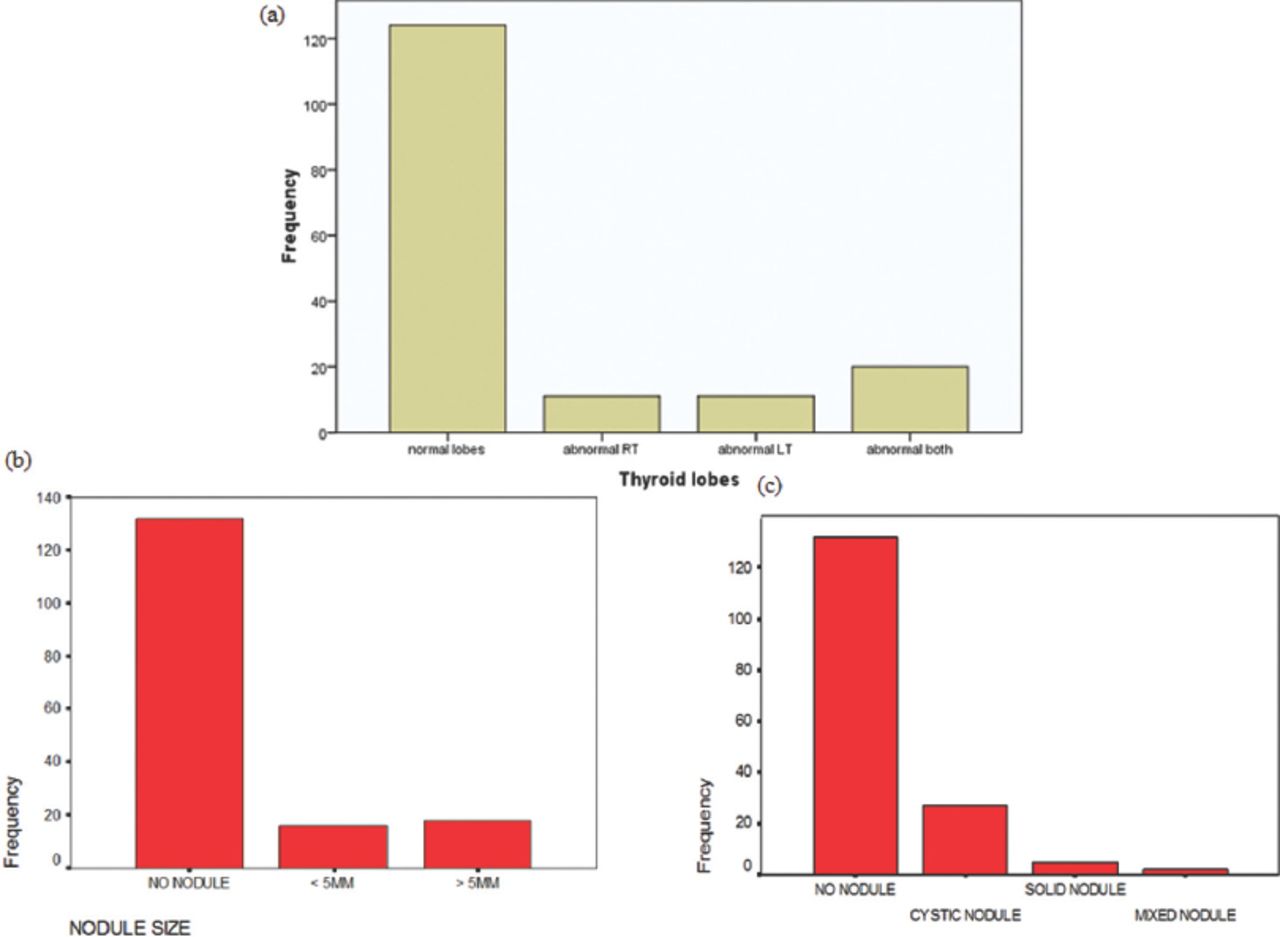
Thyroid diesease screening: a) location of nodule in the thyroid gland; b) sizes of the thyroid nodules detected in the study; c) sonographic features of the thyroid nodules found in the subject
The size of nodules was classified into 2 groups according to the diameters. The nodules with less than 5 mm diameter were considered as the smaller ones. While, the nodules with more than 5 mm diameter were considered as the larger nodules with the record observation of 16 mm at maximum. No statistical correlation had been established between the sizes of malignant nodules and benign nodules (Figure 1).
The data on the sonographic features of nodules was available for 34 patients, it revealed that 27 cases (79.4%) had anechoic, 3 (8.8%) had hypoechoic nodules, 2 (5.9%) had hyperechoic, and 2 (5.9%) had mixed echogenicity nodules (Figure 1). Among 3 subjects (2 females and 1 male), who indicated solid consistency, irregularity, ill-defined margins nodules, and hypoechoic texture, 2 (66.7%) showed micro-calcifications with the largest diameter more than 8 mm. The sonographic features for the 3 nodules have been observed to be linked with an amplified risk of thyroid malignancy. The 2 subjects with the presence of micro calcifications had fine needle aspiration (FNA) cytology, which was consistent with papillary carcinoma. This aspect was confirmed by surgical pathologic examination along with thyroidectomy for both groups. The FNA was not observed in the third nodule as it was very small; therefore, further follow-up was recommended.
Discussion
Neck palpitation provides the modality to screen and access the thyroid gland for the screening of thyroid nodules. However, the tactic presents scarce information about the severity of the condition.5 The significance of the approach can be determined by the results that have indicated a considerable number of asymptomatic individuals, who turned out to have positive results for the thyroid abnormalities. Shin et al6 has suggested that ultrasound screening has more predictive value in terms of treatment outcomes as compared to other alternatives. Kim et al7 has determined that ultrasonography is more useful in identifying diffused thyroidal diagnosis. The sonographic features also revealed the classification of nodules that suggested the occurrence of malignancy. The characteristics may include solid hypo-echogenicity or appearance, micro calcifications, increased vascularity, irregular margins, and the absence of a halo on the screening that have been consistently associated with the malignancy diagnosis.8
The estimated rate of occurrence is also noted by Olusola-Bello et al9, who determined the rate of 22.4% in the diagnosis incidental thyroid lesions among adult patients. Gnarini et al10 demonstrated the significant incidence of the thyroidal tumors, which tend to increase with the increase in age of the population. Thyroid issues are the second most prevalent endocrine condition that affect women of different ages. The concern may become strongly decisive during the phases like gestation that may highly elevate the risk of miscarriages, placental abruption, hypertension, and restriction of growth.11
In the present study, fine needle aspiration (FNA) cytology was not obtained for all small (<5 mm), solid, and mixed echogenicity suspicious nodules. Fine needle aspiration is a common technique, utilized in the approach despite of the ultrasound that is considered significant in the diagnostic domain. The diagnosis must be performed with respect to the standard procedure of the recommended guidelines, although the incidence of thyroid cancers remains low.12 Results displayed only 3 (8.8%) of the lesions that had sonographic features. It suggested a possibility of malignancy; therefore, FNA was not performed in all the 3 cases. Fine needle aspiration has been applied in 2 of the cases; whereas, FNA was not proposed for the third case due to the small diameter of the nodule. However, follow-up ultrasonography has been recommended in every 6 to 12 months for preventing any recurrence of the nodular growth.
There is 3-fold increase in the occurrence of thyroid cancer from 1975 to 2009. The modern medical practices have helped in the diagnosis of small and asymptomatic thyroid cancers. Treatment related discussions for the thyroid cancers needs to be based on mortality, rather than the survival data.13 A study conducted by Smith-Bindman et al14 stated that thyroid ultrasound imaging can be used for diagnosis of thyroid cancer among patients with low risk of cancer. These patients can be deferred for biopsy later. The trends observed in thyroid cancer are reflected through the increase in the thyroid nodules. Majority of the thyroid nodules do not present any symptoms as a result of physical examination/palpitations.15
One of the limitations of the study was the recruitment of dense population into the evaluation due to the selected age range of the group. Another limitation was the short time of the study period that was proposed on the basis data availability on pathology for all nodules having sonographic features and possible presence of malignancy.
In conclusion, the study has shown that ultrasonography is a useful and effective technique for the screening of thyroidal diseases. It has successfully determined the tumor for asymptomatic thyroid gland condition in the past, and the procedure has now become more effectual with the modern up-gradation. Therefore, it is suggested that the thyroid ultrasound scanning can be utilized as a routine practice for general population screening, particularly for the women in their reproductive phase. For future prospects, it is recommended to conduct similar studies and apply more approaches with a bigger sample size that may reveal more information about the predictors and prognosis of the disease
Acknowledgmet
This project was funded by the Deanship of Scientific Research (DSR), King Abdulaziz University, Jeddah, Kingdom of Saudi Arabia under grant No. G-84/142/1434. The authors, therefore, acknowledge DSR for the technical and financial support.
- Received August 24, 2017.
- Accepted January 10, 2018.
- Copyright: © Saudi Medical Journal
This is an open-access article distributed under the terms of the Creative Commons Attribution-Noncommercial-Share Alike 3.0 Unported, which permits unrestricted use, distribution, and reproduction in any medium, provided the original work is properly cited.
In this issue


Jump to section
Related Articles
Cited By...
- No citing articles found.