


Article Figures & Data
Figures
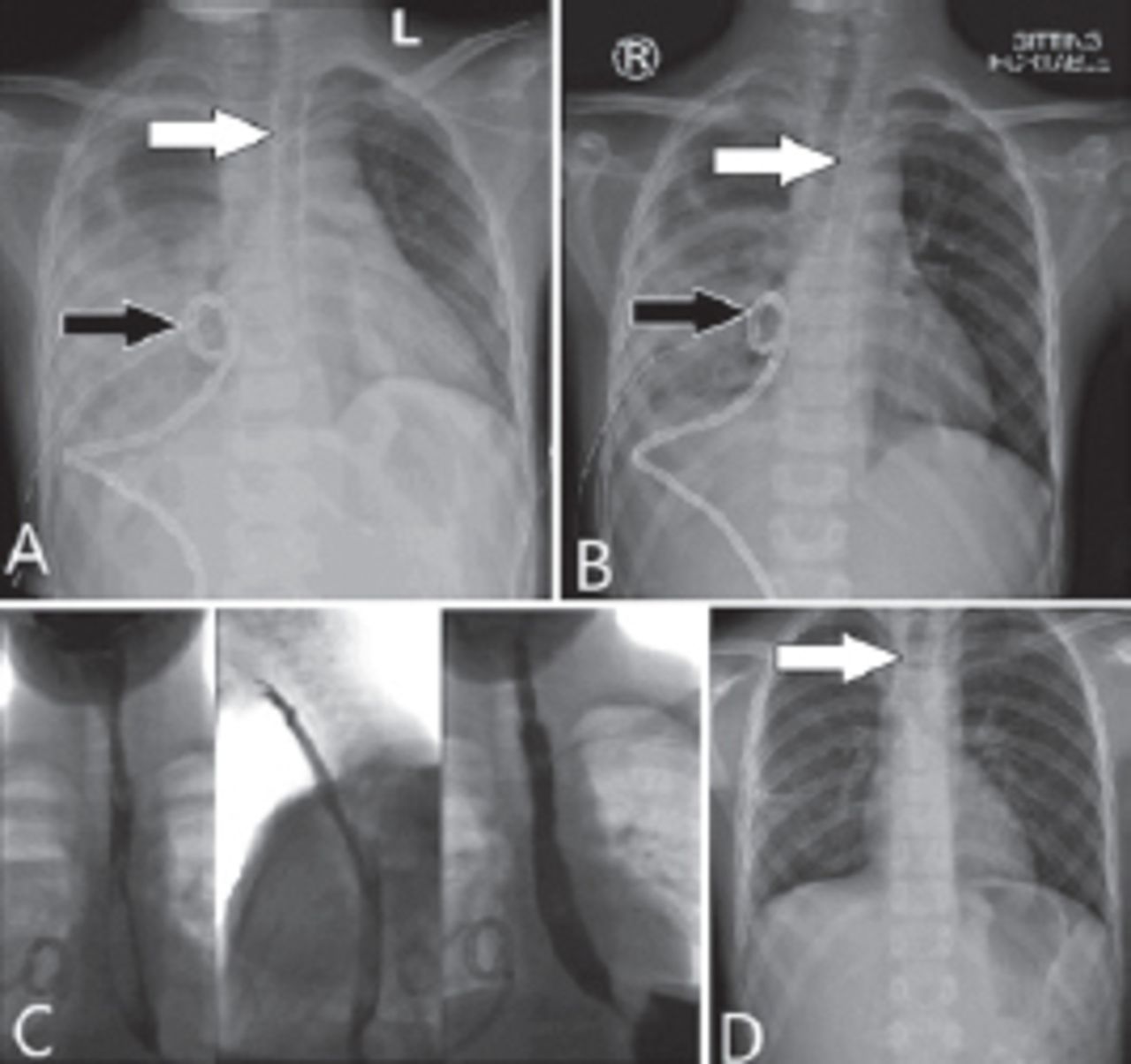
A) X-ray of a patient with esophageal perforation with right pleural effusion, 48-hours post perforation, B) after chest tube insertion. C) CT scan shows right pleural effusion arrow indicate site of perforation and D) esphagogram white arrow shows the site of perforation and leak
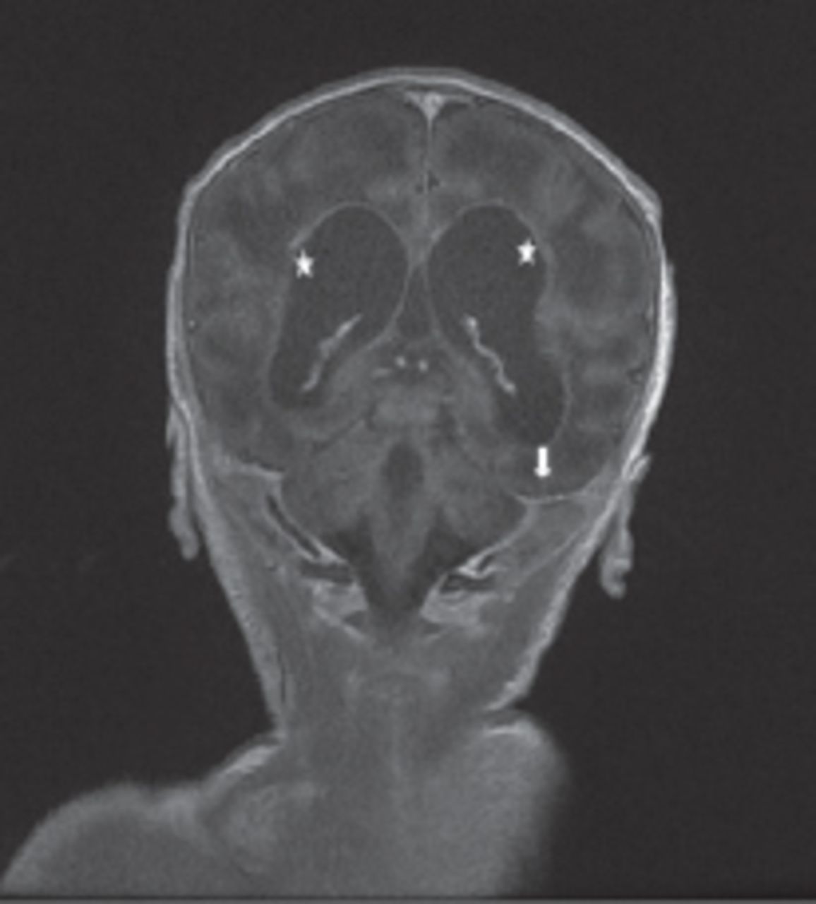
Magnetic resonance imaging coronal T1 post contrast image showing basal meningeal enhancement (arrow) and ependymal enhancement of the lateral ventricular wall (star)

Tables
In this issue


Jump to section
Related Articles
Cited By...
- No citing articles found.