


Article Figures & Data
Figures
- Figure 1
- Flow diagram of studies’ screening and inclusion. VHL: Virtual Health Library
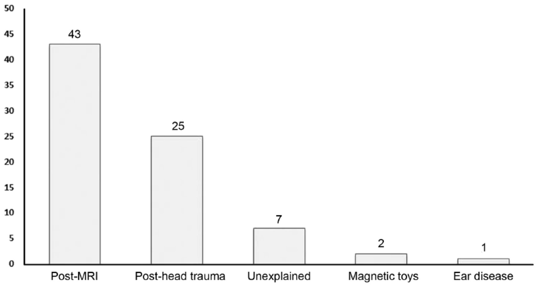
- Figure 2
- Reasons for magnet migration in accordance with the authors’ reporting.

Tables
Author, year Country Study design Sample size Number of MM/RSM cases Device type Etiology of hearing loss QA score Bawazeer et al 201918 Saudi Arabia and France Case series 6 1 CI422 Congenital deafness 10 Broomfield et al 201823 Australia Case report 1 1 Nucleus CI512 Advanced otosclerosis from the age of 15 years. 11 Chan & Wu 201113 Taiwan Case report 1 1 Nucleus freedom, Cochlear limited, Australia - 7 Cuda et al 201326 Italy Case report 1 1 Nucleus 5, CI512;Cochlear LTD Bilateral SNHL 10 Demir et al 201932 Turkey Case report 1 1 Nucleus, CI24RE, Bilateral profound SNHL 10 Deneuve et al 20086 France Case report 1 1 CI24RCS Bilateral SNHL 9 Di Nardo et al 201231 Italy Case report 1 1 Advanced Bionics Hi-Res 90K array with Harmony speech processor Bilateral SNHL 11 Epperson et al 201916 USA Case report 1 1 Cochlear 532 Bilateral SNHL 11 Keereweer et al 201433 Netherlands Case report 1 1 - - 11 Leong & Yeon 20189 Singapore Case report 1 1 HiRes 90 K SNHL 10 Mickelson & Kozak 200834 Canada Case report 1 1 Nucleus contour 24R device SNHL 10 Nichani et al 200635 UK Case series 4 4 Nucleus 24 Contour Softip implant SNHL 10 Özgür et al 201936 Turkey Case report 1 1 Nucleus freedom straight CI24RE SNHL 10 Raghunandhan et al 201037 India Case report 1 1 - Congenital bilateral hearing loss 10 Stokroos & Dijk 200712 Netherlands Case report 2 2 Nucleus CI24R Bilateral SNHL 9 Wild et al 201038 Switzerland Case report 3 3 Nucleus Freedom,CI24RECA, Cochlear Corporation Bilateral profound hearing loss (n=1), Profound hearing loss due to a mutation in gene 26 (n=2) 10 Wilkinson et al 200439 USA Case report 1 1 Nucleus CI24RCS device (Cochlear Corporation, Englewood, CO, USA). Bilateral profound SNHL 10 Yun et al 200511 USA Case report 3 3 Nucleus CI24R - 9 Bhadania et al 201840 India RCR 250 2 (MedEl, Cochlear, Advanced Bionics) and underwent surgery via - 7 Brown et al 200941* USA RCR 44 3 - - 8 Cullen et al 200842 USA RCR 93 2 - - 7 Hashemi & Bahrani 201219 Iran RCR 11 2 - - 8 Hassepass et al 201428 Germany RCR 2027 22 (6) CI512, (5) Cochlear Nucleus Freedom, (1) CI422 - 7 Jiang et al 201643 China RCR 1,065 1 - - 7 Sefein 201844 Egypt RCR 112 1 - 8 Kim et al 200845 South Korea RCR 720 2 CI24R - 8 Loundon et al 201046 France RCR 434 (43 complication) 3 - - 7 Migirov et al 201047* Israel RCR 320 3/1* - - 7 Orhan et al 201248 Turkey RCR 344 2 Nucleus (Cochlear Limited, Lane Cove, Australia) - 8 Qiu et al 201149 China RCR 416 1 - 8 Leinung et al 202017 Germany RCR 9 9 (5) CI 512, (2) CI 24RE, (1) CI 532, (1) HiRes 90 k - 8 Tam et al 202050 Australia PCR 76 2 - - 9 Tarkan et al 201351 Turkey RCR 475 1 - - 8 Kim et al 20157 South Korea RCR 18 1 - - 8 Young et al 201652 USA RCR 12 1 - - 8 Brian et al 201320 USA RCR 12 1 - - 10 Receiver-stimulator migration cases. MM/RSM: magnet migration/receiver-stimulator migration; SNHL: sensorineural hearing loss; RCR: retrospective cohort review; PCR: prospective cohort review; CI: cochlear implant; MRI: magnetic resonance imaging; (-): data were not available.
- Table 2
- Clinical characteristics of magnet displacement according to the reason of migration.
Author, year Cause of migration Cases (n) Gender Mean age (SD) (years) MRI dose/indication Head bandage/pain during the MRI Clinical presentation Clinical examination Bawazeer et al 201918 MRI 1 - 28 1.5T -/Y Progressive neurologicalillness - Brian et al 201320 MRI 1 Male 4 1.5 T/Spine and brain tumor Y/Y - - Broomfield et al 201323 MRI 1 Female 64 1.5 T/spinal cord compression suspection -/Y Gait disturbance upper limb weakness Bilateral skin reactions Cuda et al 201326 MRI 1 Male 72 1.5 T/biliary duct pathology Y/- Pain and hotness Focal skin alteration over the left inner coil Demir et al 201932 MRI 1 Female 7 1.5 T/congenital scoliosis follow up N/Y Inability to use the implant due to a wound Redness, wound scarring and edema on the implant body and magnet site Deneuve et al 20086 MRI 1 Male 8 1.5 T/neurologic disorder progression Y/Y local erythema with edema and tenderness local erythema, edema, tenderness, the magnet was outside the SR Di Nardo et al 201231 MRI 1 Female 31 1.5 T/64-MHz brain MRI/mitochondrial myopathy sudden detorioation N/Y Pain and a burning sensation Bulge in the receiver-stimulator Epperson et al 201916 MRI 1 Female 10 1.5 T/central hypothyroidism suspection Y/Y Intermittent fever and tenderness over the processor/magnet site. - Leinung et al 202017 MRI 9 Male (22%) 37.2 (21.7) Y (n=6)/- Pain (4), swelling (5), redness (2), palpable displacement (3), inability to wear the CI processor (7) Leong et al 20189 MRI 1 Male 67 1.5 T/suspected cervical and lumbar radiculopathy Y/Y Discomfort and a bulge - Kim et al 20157 MRI 1 Female 25 1.5 T/malignant ependymoma Y/Y - - Özgür et al 201936 MRI 1 Male 4 3 T/suspected diabetes insipidus Y/Y Swelling over the magnet site The magnet had turned upside down, the external part was reversed (inside facing out) and still attracting the internal part. Tam et al 202050 MRI 2 Female 36 and 74 - - - - Young et al 201652 MRI 1 Female 11.6 1.5 T Y/- Discomfort and swelling of the soft tissue overlying the portion of the device. - Hassepass et al 201428 3 cases post-head trauma,19 cases post-MRI 22 - - - - - - Loundon et al 201046 1 case post-MRI, 2 cases post-head trauma 3 - - - -/Y - - Bhadania et al 201840 Head trauma 2 - - - - - - Jiang et al 201643 Head trauma 1 - - - - - - Keereweer et al 201433 Head trauma 1 Male 1.5 - - The sound processor could no longer connect to the CI Diffuse swelling without erythema of the skin overlying the CI. Kim et al 200845 Head trauma 2 Female 4 and 6 - - - - Mickelson et al 200834 Head trauma 1 Male 1.8 - - - The magnet was palpable anteroinferiorly. Migirov et al 201047 Head trauma 3 - - - - - - Nichani et al 2006 35 2 cases post head trauma2 unexplanied reasons, without history of apparent trauma 4 Male (100%) 3 (82) - - (2) erythema and swelling of the scalp over the RS site (1) swelling over the magnet site(3) erythema and swelling of the scalp over the RS site. Orhan et al 201248 Head trauma 2 - - - - - - Stokroos et al 200712 Head trauma 1 Female 44 y - - Known to have seizures A slight bulge over the processor part of the implant and some local tenderness with a small, firm, palpable but less-well-defined mass. Head trauma 1 Male 3 y - - Loss of the function of the implant A slight bulge of the skin was visible over the implant site, and a small, firm swelling was felt over the processor part. Tarkan et al201351 Head trauma 1 - - - - - - Wilkinson et al 200439 Head trauma 1 Male 13 m - - No response in the external coil - Chan et al 201113 No apparent trauma 1 Male 4 y - - Poor response to sound Small and firm bulge over the processor part of the implant. Qiu et al 201149 Unexplained reasons 1 - - - - - - Raghunandhan et al 201037 Unexplained reasons 1 Female 13 y - - Rapid deterioration in auditory verbal skills A small boggy swelling in the mastoid region over the internal RS coil site. Sefein et al 201844 Associated with chronic suppurative otitis media 1 Male - - - - - Yun et al 200511 Head trauma 1 Male 70 m - - - Magnet was external to the SR by palpation. No apparentcause 1 Male 31 m - - Swelling precludng use of the external device. Ridge was palpable over the anterior body of the SR. Erythema without fluctuance Head trauma 1 Male 28 m - - Tender knot over the RS Magnet was found to be freely mobile under the flap Wild et al 201038 No apparentcause 1 Male 34 m - - - Skin irritation over the implant site Playing with magnetic toys 1 Female 67 m - - - Dislocated magnet lateral to the receiver aerial Playing with magnetic toys 1 Female 56 m - - Dislocated magnet - Y, yes; N, no; y, years; m, months; CI, cochlear implant; RS, receiver-stimulator MRI; magnetic resonance imaging.
Author, year Cases (n) Investigation Management Tool Finding Post MRI Bawazeer et al 201918 1 CT Magnet rotation without total implant displacement within the cochlea Emergency surgery Walker et al 201320 1 - - Spontaneous reduction Broomfield et al 201323 1 Radiography Left magnet displaced Magnet replacement with titanium spacers. Cuda et al 201326 1 Radiography Partial magnet migration on the left side Surgical exploration and magnet repositioning Demir et al 201932 1 Examination There was a hard spot consistent with the contour of the magnet under the scar. The magnet was excised from the s ubcutaneous tissue without compromising the integrity of the skin. Deneuve et al 20086 1 Examination Magnet was palpable Removal under local anesthesia followed by repositioning after 5 days Di Nardo et al 201231 1 Radiography Magnet displacement Manual maneuver for repositioning Epperson et al 201916 1 Radiography and CT Normal findings after the initial examination, but magnet angulation was noted on re-evaluation Repositioning of the magnet with a CI 500 series replacement Leinung et al 202017 9 4 Radiography 5 CT Magnet displacement Surgical repositioning Leong et al 20189 1 Radiography Dislocated from its slot in the receiver stimulator Endoscopic repositioning Kim et al 20157 1 Radiography The internal magnet was displaced outside the receiver container Reinsertion of the magnet into the retainer using a microelevator and repositioning Özgür et al 201936 1 Radiography Magnet displacement Surgical repositioning Tam et al 202050 2 - - Surgical revision and magnet repositioning; subsequent infection led to device loss in one case Young et al 201652 1 Radiography 90-degree rotation of the magnet Surgical replacement Post head trauma Bhadania et al 201840 2 Radiography Magnet displacement Surgical replacement Jiang et al 201643 1 - - Surgical replacement Keereweer et al 201433 1 Radiography Magnet displacement (on top of the titanium housing of the receiver-stimulator) Surgical replacement Kim et al 200845 2 Radiography Floating magnet from the device well Revision surgery and surgical repositioning Mickelson et al 200834 1 Radiography Magnet displacement Surgical magnet repositioning Recurrence after 3 yr and treated by surgical lasso technique Orhan et al 201248 2 Radiography Magnet displacement Reinsertion of the magnet by (1) sub-periosteal temporal pocket technique, (2) standard technique Stokroos et al 200712 2 Radiography Luxation and anterior displacement of the magnet Surgical exploration and magnet repositioning Tarkan et al 201351 1 - - Revision surgery Wilkinson et al 200439 1 Radiography Magnet migration outside the antenna coil to a position compromising normal function of the device. Surgical replacement Nichani et al 2006*35 4 Radiography Magnet displacement Surgical repositioning Yun et al 200511 2 Examination Magnet was palpable Surgical replacement Unexplained reasons and other conditions Chan et al 201113 1 Radiography Magnet migration from the silicon pocket toward the antenna Surgical exploration Cullen et al 200842 2 - - Revision surgery Hashemi et al 201219 2 Neuroresponse telemetry Poor response - Qiu et al 201149 1 Radiography Magnet displacement Revision surgery without re-implantation Raghunandhan et al 201037 1 Radiography Magnet migration from its socket in the receiver-stimulator coil Surgical exploration and repositioning Sefein et al 201844 1 - - Surgical repositioning Wild et al 201038 3 - - Revision surgery and surgical replacement Yun et al 200511 1 Examination Magnet was palpable Magnet reduction ↵* Two cases were due to unexplained reasons, without a history of apparent trauma
In this issue


Jump to section
Related Articles
Cited By...
- No citing articles found.