


Article Figures & Data
Figures
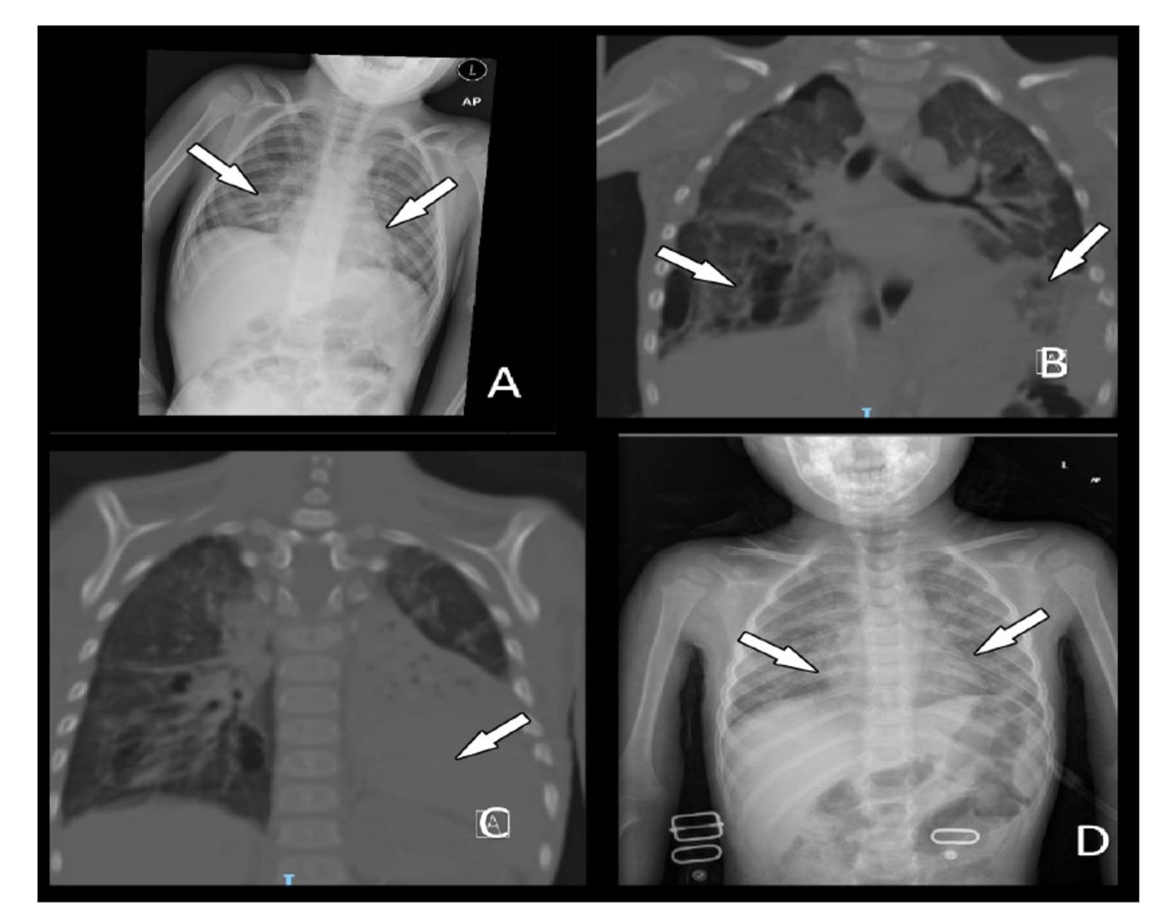
- Figure 1
- Progression of chest radiography (CXR) and computed tomography (CT) findings in a pediatric COVID-19 patient. A) CXR of the patient on admission showed bilateral mid-lung zone opacifications. B&C) During admission to the pediatric intensive care unit, coronal CT images of the same patient showed multiple right sided pneumatoceles, bilateral ground-glass opacification, and left lower lobe consolidation. D) CXR of the patient at the 3-month post-discharge follow-up showed recovering bilateral ground-glass opacifications.
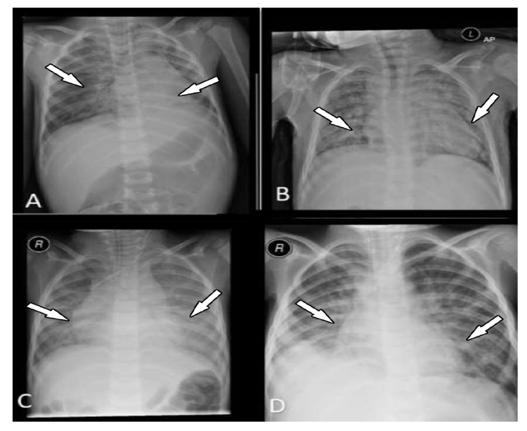
- Figure 2
- Chest radiographs of 4 pediatric patients with positive COVID-19 results. A) A 3-year-old male patient with COVID-19; initial chest radiography after intubation shows bilateral central ground-glass opacities with left lower lobe consolidation. B) An 8-year-old male patient with underlying severe bronchopulmonary dysplasia was admitted with COVID-19; chest radiography showed bilateral ground-glass opacities (more on the left). C) Chest radiography of a 4-year-old patient showing bilateral ground-glass opacity. D) Chest radiography of a 5-year-old showing bilateral consolidation with right lower lobe localization. This patient eventually died.


Tables
Variables n (%) Age in months, median (IQR) 24 (12-72) Age categories Infants 11 (32.4) Preschool age 15 (44.0) School age 8 (23.5) Gender (male) 16 (47.0) Symptoms and signs GI symptoms (vomiting/diarrhea) 20 (59.0) SpO2 on room air, median (IQR) 75 (70-82) Respiratory failure requiring mechanical ventilation 12 (35.0) Signs of shock or multi-organ failure 2 (5.9) Comorbidities 31 (91.2) Vital signs at admission, median (IQR) Heart rate 147.5 (120-181) FiO2 100 (80-100) Values are presented as a number and percentage (%) or as median and interquartile range (IQR). GI: gastrointestinal, SpO2: peripheral oxygen saturation, FiO2: the fraction of inspired oxygen
Blood and biochemical tests n Children’s reference range Values White blood cells (×109/L) 34 4500-11000 9.21 (6-12.9) Absolute lymphocyte count (per mm3) 31 1000-4800 2340 (1540-4610) Absolute neutrophil count (per mm3) 31 1800-7700 4740 (2470-10300) Hemoglobin (g/dL) 34 12.0-16.0 12 (10.5-13) Platelets (×109/L) 34 150,000-450,000 281 (186-334) CRP (mg/dL) 22 <0.30 5 (0.5-14.5) ESR (mm/hr) 29 0-13 32 (15-55) Ferritin, µg/l 8 30-300 351 (144-1293) Procalcitonin, µg/l 13 0.00-0.08 0.34 (0.04-5.5) Troponin, ng/ml 12 <2.0 3.8 (1.5-46) D-dimer (µg/L) 14 <500 970 (725-3003) BUN (mg/dL) 31 8.0-25 13 (8-19) Creatinine (mg/dL) 31 0.30-1.00 0.4 (0.3-0.5) Sodium (mEq/L) 31 135-145 137 (134-139) ALT (IU/L) 29 10-55 29 (18-43.5) AST (IU/L) 29 9.0-32 29 (22-53.5) Albumin (g/dL) 26 3.4-5.4 3.1 (2.8-3.7) Total bilirubin (mg/dL) 19 0.0-1.0 0.5 (0.3-1) LDH (IU/L) 17 13-60 472 (310.5-716) Values are presented as median and interquartile range (IQR). CRP: C-reactive protein, ESR: erythrocyte sedimentation rate, BUN: blood urea nitrogen, ALT: alanine transaminase, AST: aspartate transaminase, LDH: lactate dehydrogenase
Variables Values Medical treatment Inotropes 7 (20.6) Systemic steroid 20 (58.8) IVIG 12 (35.3) Aspirin 3 (8.8) Azithromycin 34 (100) ≥2 antibiotics 34 (100) Favipiravin 32 (94.1) Hydroxychloroquine 9 (26.5) Respiratory support HFNC 31 (91.2) MV 11 (32.4) Days on HFNC 3 (1-20) Days on MV 10 (1-60) Days on PICU 7 (1-91) Total oxygen days during the hospitalization 7 (3-80) The total days of hospital stay 10 (3-105) Values are presented as a number and precentage (%) or as median and interquartile range (IQR). IVIG: intravenous immunoglobulin, HFNC: high-flow nasal cannula therapy, MV: mechanical ventilation, PICU: pediatric intensive care unit
Variables Values Complications Multiorgan failure 4 (11.8) Seizures 5 (14.8) ARDS 13 (38.2) Air leak 4 (11.8) Outcomes at the time of discharge Tracheostomized 3 (8.8) Oxygen dependent 6 (17.6) Discharged alive 32 (94.0) A 3-months follow-up 26 (76.5) Values are presented as a number and precentage (%).
ARDS: acute respiratory distress syndrome
In this issue


Jump to section
Related Articles
Cited By...
- No citing articles found.