


Abstract
Objectives: To analyze the data of patients with otorhinolaryngological foreign bodies and to evaluate the management and outcomes of foreign bodies to prevent complications.
Methods: A retrospective study was conducted over 8 years at Aseer Central Hospital to examine all admitted cases with foreign bodies in the ear, nose, throat, esophagus and bronchus during the period from January 2011 to January 2019. Patient demographic data, type of foreign body, and most common site were analyzed.
Results: A total of 184 patients were admitted, including 72 (39.1%) males and 112 (60.9%) females. The age range was from one year old to 70 years old; the mean±standard deviation of age was 10.6±12.55 years. Foreign bodies were most commonly located in the esophagus (n=97, 52.7%), followed by the bronchus (n=55, 29.9%). A statistically significant difference was found, with a p-value of 0.00001. The most common site in children was the bronchus (n=39, 21%); the most common site in adults was the esophagus (n=18, 72%).
Conclusion: Otorhinolaryngological foreign bodies are found most frequently in preschool-aged children. The most common site in children was the bronchus, and the most common site in adults was the esophagus. Prevention measures are essential to reduce the risk of ingestion and admission, which can be challenging.
Otorhinolaryngological foreign bodies are relatively common problems that are frequently encountered in emergency situations.1 Foreign bodies may affect any part of the head and neck, such as the nose, ears, throat, hypopharynx, bronchus and oesophagus.2-4 Common foreign bodies include food, plastics, beads, papers and toys.5,6 The presence and removal of foreign bodies, especially in the airway, can be challenging to operators and may lead to death.7,8
The aim of this study was to collect and review the data of patients with otorhinolaryngological foreign bodies with respect to the site of the foreign body, age of the patient, type of foreign body and method of removal and to evaluate the management and outcome of foreign bodies with the aim of minimizing the harmful consequences.
Methods
Approval from the Institutional Research and Ethics Committee was obtained from the College of Medicine, King Khalid University, Abha, Kingdom of Saudi Arabia. We reviewed the medical records of all patients who were admitted at Aseer Central Hospital with foreign bodies in the ear, nose, throat, esophagus or bronchus at our institute between January 2011 and January 2019. Patient age, gender, type of foreign body and most common site were analyzed. Patients were evaluated by complete history, physical examination and radiological investigation such as soft tissue neck lateral view and chest x-ray.
Inclusion criteria for our patients of foreign body in ear, nose, throat, bronchus, trachea and esophagus over the last 8 years at Aseer Central Hospital. The exclusion criteria include patients less than one year of age or more than 70 years old and patient with hematological disorder.
All procedures were carried out under general anesthesia using rigid endoscopy for removal of foreign body for both esophagus and respiratory tract. Patients were followed immediately after the surgery to assess any complications and at the first week postoperatively. The management and outcome of procedure was assisted by group discussion of all authors.
Data analyses were performed by using Statistical Package for Social Sciences for Windows, version 22.0 (SPSS Inc., Chicago, IL, USA). Fisher’s 95% confidence intervals were calculated for the proportions. Pearson’s Chi-square test was used as test of significance at the 5%.
Results
There were 184 patients admitted to Aseer Central Hospital; most patients were admitted through the emergency department. The patients included 72 males (39.1%) and 112 females (60.9%). Their ages ranged from 1-70 years old with an average age of 10.45 ± 12.65 years and a median age of 6 years. The most common site in children was the bronchus (n=39, 21%), and the most common site in adults was the esophagus (n=18, 72%) (Table 1).
Distribution of the study sample by age and site of the foreign bodies.
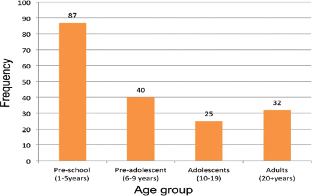
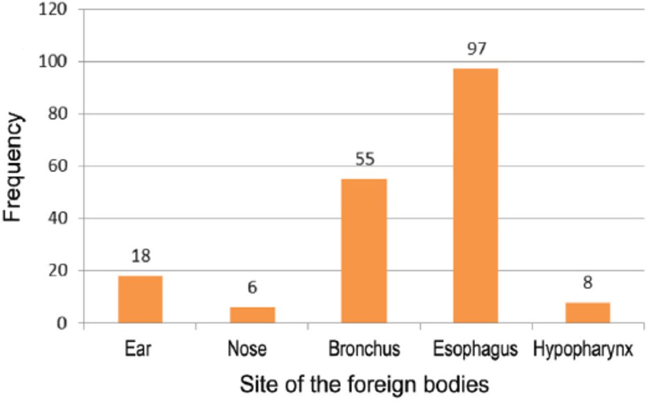
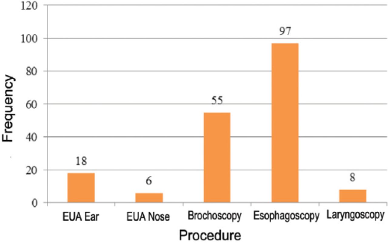
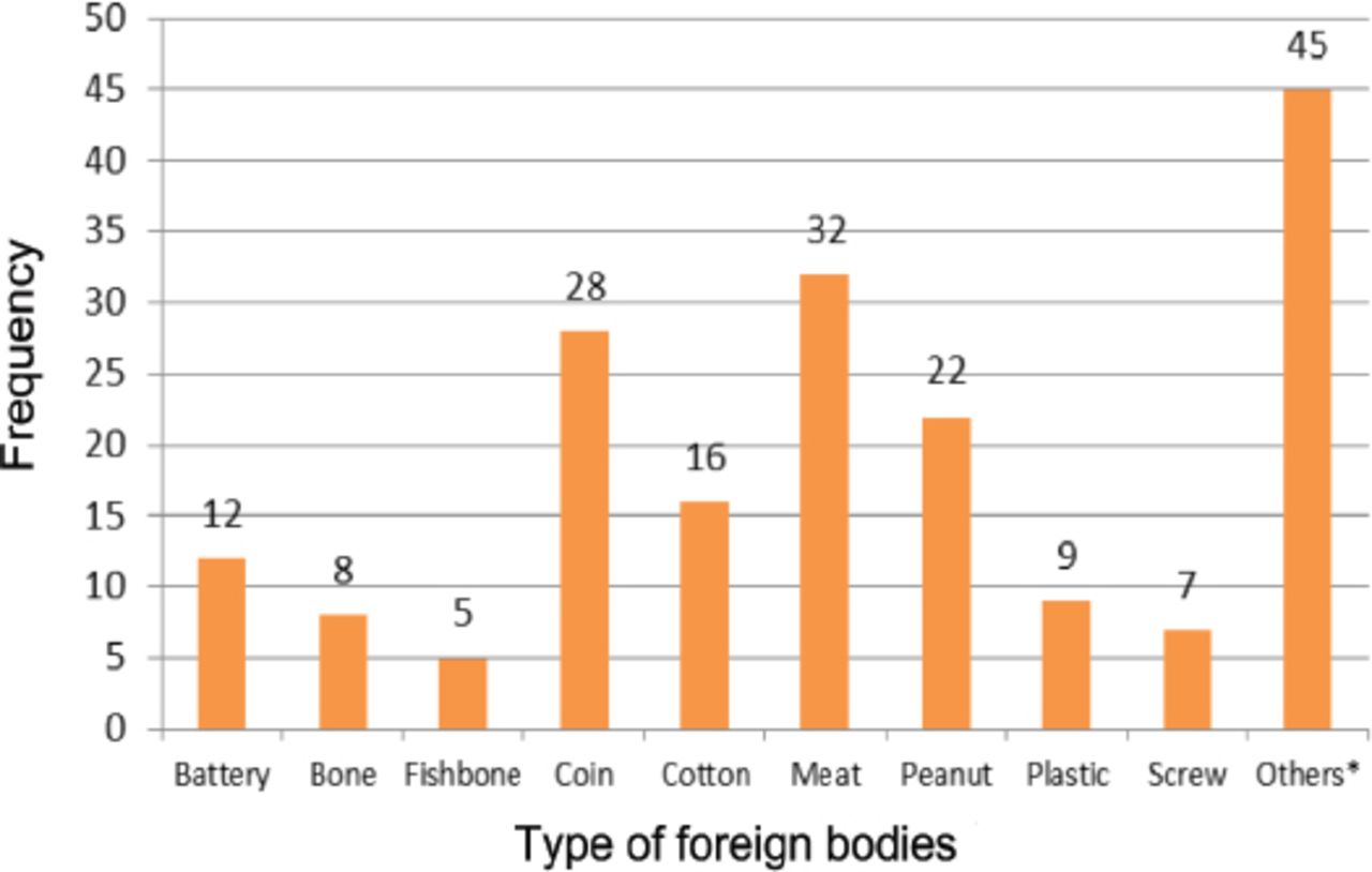
Regarding the distribution of the study sample by age group, preschool-aged children (1-5 years old) were most prevalent (n=87, 47.3%), followed by preadolescents aged 6-9 years old (n=40, 21.7%) and adolescents aged 10-19 years old (n=25, 13.6%). There were 32 adults (>20 years old), accounting for 17.4% of the sample. The distribution of age groups did not significantly differ between genders (p=0.431) (Figure 1). The most common site was the esophagus (n=97, 53%), followed by the bronchus (n=55, 30%). The highest prevalences of esophageal foreign bodies were observed among adolescents (n=18, 72%) and adults (n=23, 71%). The lowest prevalences of esophageal foreign bodies were observed among preschool children (n=29, 33.3%) and preadolescents (n=27, 67.5%). The difference in the prevalences of esophageal foreign bodies between ages groups was statistically significant (p=0.001). On the other hand, the highest prevalences of bronchial foreign bodies were observed among preschool-aged children (n=39, 44.8%) and preadolescents (n=9, 22.5%). The lowest prevalences of bronchial foreign bodies were observed among adolescents (n=5, 20%) and adults (n=2, 6.3%). The difference in the prevalences of bronchial foreign bodies between ages groups was statistically significant (p=0.001) (Figure 2). The most frequent procedure was esophagoscopy (n=97), followed by bronchoscopy (n=55) (Figure 3). The most common foreign body was meat (n=32), followed by coins (n=28) and peanuts (n=22) . Other foreign bodies found in the study (n=45) included teeth, toothpicks, buttons, wood, pearls, batteries, nails, pencils, rings, screws, seeds and earrings. Regarding the presence of meat as a foreign body, the highest prevalences were observed among adults (n=21, 65.5%) and adolescents (n=5, 12.5%). The lowest prevalence was observed among preschool-aged children (n=2, 2.2%).

Distribution of the study sample by age group.

Distribution of the study sample by site of the foreign bodies.

Distribution of the study sample by procedure.
The difference in the prevalences of meat as a foreign body between ages groups was statistically significant (p=0.001) (Figure 4).

Distribution of the study sample by type of foreign body.
Regarding the presence of coins as foreign bodies, the highest prevalences were observed among preadolescents (n=13, 32.5%) and adolescents (n=5, 20%). The lowest prevalence was observed among preschool-aged children (n=10, 11.4%). No coins were found among adults. The difference in the prevalences of coins as foreign bodies between ages groups was statistically significant (p=0.001).
Discussion
Foreign bodies in the ears, nose or throat are commonly observed in otorhinolaryngology emergency services.9 They vary widely in shape, size, and composition. The symptoms may range from asymptomatic to acute life-threatening symptoms.10 In our study, the most common age group affected was preschool-aged children; the bronchus was found to be the most common site of foreign bodies in children, while the esophagus was the most common site in adults. In children, it could be due to the late arrival of patients to our tertiary hospital which may take 6-12 hours from insertion of foreign body to the hospital visit, this also could affect the site of foreign bodies in children. Impacted meat was the most common foreign body seen in adults specifically in handicap patients or those with esophageal abnormality associated with anatomic or motor abnormality like strictures, web achalasia, esophageal spasm and diverticula, this is consistent with other study,11 while coins were the most common foreign body in preschool-aged children. Children are mostly affected due to their tendency to put things in their mouth, their inability to masticate well, their inadequate control of deglutition, as well as their tendency to cry, shout and play during eating.12,13 In some studies, fish bone was the most common type discovered in oropharynx and hypopharynx, especially in both tonsils and piriform fossa.14 However, in our study, it was found that the most common type of foreign bodies were in the esophagus in adults and coins in the bronchus in children. This could be due to several factors, including the geographic location, nutritional habits of the patient and if the service is provided in a secondary or tertiary hospital. Most of fish bone foreign bodies were removed in secondary hospitals before the patient was referred to a tertiary care hospital. In general, ear foreign bodies are the most common foreign body encountered in children followed by nose and pharynx.15 This different from our study because our hospital is tertiary hospital which received difficult cases of foreign bodies of esophagus and trachea from secondary hospital of Aseer region. In elderly patients, edentate and poor masticating habits are predisposing factors.16-18 Foreign bodies are usually ingested accidentally, but they may occasionally be ingested with homicidal or suicidal intent. Most nasal and aural foreign bodies can be easily removed in the emergency room or out patient department, but in some cases, the operating room may be required for simple foreign body dislodgement, especially in children.19-21 On the other hand, foreign bodies in the bronchus or esophagus must be treated in the operating room. In our study, the most frequent procedure was rigid esophagoscopy (n=97, 53%), followed by rigid bronchoscopy (n=55, 30%). The management plan of foreign bodies’ removal of upper airway depends on many factors like availability of wide range of instruments, endoscopic baskets, to achieve better outcome,22 general condition of the patients, severity of symptoms, size and type of foreign bodies and experience of surgeons. In our study, all procedures were carried out under general anesthesia using rigid endoscopy. Some foreign bodies are frustrating and challenging especially large items and those with sharp edges are most likely to become impacted in the larynx. In the other hand, inhaled foreign bodies that are round and non-compressible with smooth, slippery surfaces pose most risk of complete airway obstruction and death. Another important issue of controversy in management of foreign bodies in trachea is the type of anesthesia, there is a wide consensus on using sevoflurane or halothane as inhalational agents for induction of anesthesia and spontaneous breathing as a maintenance technique of choice. Urgent bronchoscopy is recommended for diagnostic or therapeutic purposes. These procedures may lead to serious complications, including upper airway obstruction, bradycardia, bronchospasm, laryngeal edema, pneumothorax, bleeding, esophageal perforation, mediastinitis, tooth breakdown, great vessel injury and increased risk of fatality.23-26 In our study, 4 patients out of 184 had complications with an incidence of 2%, one child developed laryngeal spasm and emergency tracheostomy was carried out, foreign body was removed from trachea and he made uneventful recovery and discharge home after one week, other 3 patients developed laryngeal edema and hoarsens responded well to dexamethasone and racemic epinephrine.
Study limitations
Pediatric cases were transferred to a new hospital in Abha called Abha Children Hospital.
In conclusion, one of the success factor of management is making plan with an anesthetist to secure air way and to remove foreign body without complications like laryngeal spasm that may need emergency tracheostomy.
Acknowledgment
The authors would like to thank American Journal Experts (www.aje.com) for English language editing and author services for research publication.
Footnotes
Disclosure. Authors have no conflict of interests, and the work was not supported or funded by any drug company.
- Received May 8, 2020.
- Accepted May 23, 2020.
- Copyright: © Saudi Medical Journal
This is an open-access article distributed under the terms of the Creative Commons Attribution-Noncommercial-Share Alike 3.0 Unported, which permits unrestricted use, distribution, and reproduction in any medium, provided the original work is properly cited.
References
In this issue


Jump to section
Related Articles
Cited By...
- No citing articles found.