
Main menu

  • Home
  • Content
    • Latest
    • Archive
    • home
  • Info for
    • Authors
    • Reviewers
    • Subscribers
    • Institutions
    • Advertisers
    • Join SMJ
  • About Us
    • About Us
    • Editorial Office
    • Editorial Board
  • More
    • Advertising
    • Alerts
    • Feedback
    • Folders
    • Help
  • Other Publications
    • NeuroSciences Journal

User menu

  • My alerts
  • Log in

Search

  • Advanced search
Saudi Medical Journal
  • Other Publications
    • NeuroSciences Journal
  • My alerts
  • Log in
Saudi Medical Journal

Advanced Search

  • Home
  • Content
    • Latest
    • Archive
    • home
  • Info for
    • Authors
    • Reviewers
    • Subscribers
    • Institutions
    • Advertisers
    • Join SMJ
  • About Us
    • About Us
    • Editorial Office
    • Editorial Board
  • More
    • Advertising
    • Alerts
    • Feedback
    • Folders
    • Help
  • Follow psmmc on Twitter
  • Visit psmmc on Facebook
  • RSS
Review ArticleReview Article
Open Access

Pediatric brucellosis

An update review for the new millennium

Elham E. Bukhari
Saudi Medical Journal April 2018, 39 (4) 336-341; DOI: https://doi.org/10.15537/smj.2018.4.21896
Elham E. Bukhari
From the Infectious Disease Section, Department of Pediatrics, College of Medicine, King Saud University Medical City, King Saud University, Riyadh, Kingdom of Saudi Arabia
MD
  • Find this author on Google Scholar
  • Find this author on PubMed
  • Search for this author on this site
  • For correspondence: [email protected]
  • Article
  • Figures & Data
  • eLetters
  • Info & Metrics
  • References
  • PDF
Loading

Abstract

Brucellosis remains the most common bacterial zoonotic infection in many countries worldwide. Despite being long recognized and controllable, the disease still causes substantial morbidity, affecting especially the young population. The aim of this review is to provide insight to the epidemiology, etiology, clinical features, diagnosis, and management of childhood brucellosis.

Brucellosis is one of the most common zoonotic bacterial infections and causes disease worldwide.1 This disease is a major threat to global public health and is one of the greatest socioeconomic problems in many developing countries.2 Brucellosis is caused by the bacterial genus Brucella. It affects people from all age groups, including the pediatric population. In the reported literature discussing brucellosis in children there are many discrepancies concerning the epidemiological and clinical features as well as the outcome rates.3 The purpose of this paper is to review and determine general trends with regards to the epidemiology, clinical features, diagnosis, and treatment of childhood brucellosis.

Epidemiology

Human brucellosis is amongst the most common zoonotic diseases with an average yearly global incidence over 500,000 and a prevalence of more than 10/100,000 population in some endemic countries.4 Human brucellosis remains a major human health problem in many developing regions, especially in the Mediterranean basin, North and East Africa, the Middle East, the Arabian Peninsula, the Indian subcontinent and parts of South America and central Asia.5-6 In contrast, brucellosis has been reduced or eliminated in many developed countries, including many Northern European countries. For example, only 22-47 annual cases were reported between 2010 and 2015 in Germany. Most of these cases were associated with travel to brucellosis-endemic countries surrounding the Mediterranean Sea (namely, Italy, Spain, Turkey).7-9

All age groups and both men and women are susceptible to human brucellosis.10-11 However, about 11-56% of patients affected by brucellosis are younger than 14 years in endemic regions.12 Brucellosis may be more common in children in developing countries due to lack of pasteurization of milk and exposure to animals in an agrarian society.12

Brucella species are encapsulated Gram-negative coccobacilli known to affect wild and domestic animals (namely, bovines, camels, sheep, and goats), causing abortion and infertility. B.abortus, B.melitensis, B.canis and B.suis represent the primary Brucella spp. capable of causing disease in humans. Brucella melitensis mainly infects sheep and goats and is the most virulent and primary causative agent for human brucellosis. Infection can be transmitted to humans through direct contact with infected animals or their secretions, consumption of raw milk and dairy products, and inhalation of aerosols.13 Less common routes of transmission include breast feeding (mother to child), consumption of uncooked meat, and sexual contact.14-16 In addition, brucellosis is considered one of the most common laboratory-transmitted infectious diseases, accounting for 2% of all infections.17-18 In Saudi Arabia, brucellosis is an endemic zoonotic disease. Ministry of Health reported incidence of 18/100,000 population/year in 2011. A number of reports from endemic areas exhibited a high percentage of pediatric patients (20-30% of affected patients).19

A recent study has determined that that brucellosis is a major health problem in the Kingdom of Saudi Arabia (KSA).20 Although the incidence rate of brucellosis has fallen between year 2004-2012, however it was still higher than most other developed countries and developing countries. The prevalence of brucellosis among those aged <14 years was lower than other age groups. Male Saudi citizens aged 15-44 years had the highest risk of acquiring brucellosis and those aged <one year had the lowest prevalence. Al-Qassim had the greatest number of cases and was followed by Aseer in the South, Hail, and the Northern borders. The Western part had the fewest number of cases compared to other areas.20

Etiology

Brucella spp. are small Gram-negative, intracellular, nonmotile, nonsporulating, nontoxigenic, nonfermenting, facultative coccobacilli (Figure 1). Currently, based on host preferences and phenotypic differences, Brucella encompasses over 10 species, B.melitensis, B.abortus, B.suis, B.canis, B.ovis, B.neotomae, B.pinnipedialis, B.microti, B.ceti, and B.inopinata.21-22 Brucella melitensis is maintained in nature by sheep and goats, B.abortus by bovines, B.suis by swine, and B.canis by dogs.

Figure 1
  • Download figure
  • Open in new tab
  • Download powerpoint
Figure 1

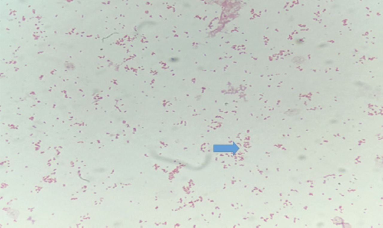
Gram Stained, gram negative coccobacilli (Brucella).

Although B.pinnipediae and B.cetaceae typically affect marine animals, they are now known to be capable of causing disease in humans, mainly neurobrucellosis.21

Recently, a novel species, B.inopinata (strain BO1), which is associated with a breast implant infection in a patient in Oregon, was isolated from a wild rodent in Australia.22

Clinical presentation of brucellosis in children

Brucellosis exhibits protean clinical and laboratory characteristics that can mimic other infectious and non-infectious conditions. Patients commonly have a wide range of symptoms including undulant fever, headache, chills, myalgia, and arthralgia. The most common clinical manifestations of brucellosis are fever (87.5-90%) and fatigue (70-75%) followed by sweating, myalgia, and weakness.23 Brucellosis often results in systemic infections with an acute (<2 months), subacute (2-12 months), or chronic relapsing course (>one year) with severe complications.24-25 Brucellosis is also associated with arthritis, spondylitis, epididymo-orchitis, acute renal failure, endocarditis, splenic abscess, abortion, and neurobrucellosis.3,26-27 Childhood brucellosis produces mild to moderate disease and rarely progresses to chronicity.28 Most pediatric reviews have reported a wide range frequencies of clinical manifestations in children with brucellosis. Fever and constitutional symptoms, consisting of chills, sweating, fatigue, malaise, anorexia, weight loss, abdominal pain, headaches, myalgias, and arthralgias, are amongst the most common symptoms in children.29 In a Macedonian study, children comprised 317 (18.7%) of the 1,691 patients with brucellosis. The patients had a median age of 9 years, age range of 7 months to 14 years, and 201 (63.4%) patients were males. Family history of similar disease was present in 197 (62.1%) patients. The predominant clinical manifestations were fever (248, 78.2%), joint pain (228, 71.9%), and hepatomegaly (216, 68.1%).30

Arthritis was reported in 24 out of 96 (25%) child patients diagnosed with brucellosis from a children medical center in Tehran. Monoarthritis was recorded in 15 patients (62.5%) with involvement of the knee (8, 45%), hip (5, 29%), and ankle (2, 8%), while 9 (37.5%) patients suffered from polyarthritis.31 In addition to osteoarticular involvement, children with brucellosis often suffer from hematologic abnormalities, such as anemia, thrombocytopenia, and pancytopenia. Disseminated intravascular coagulation and leucopenia have been reported. Brucellosis should be considered during diagnosis of patients with pancytopenia and immune thrombocytopenic purpura in endemic regions.32 Liver involvement of hepatomegaly and splenomegaly with mild to moderate elevation in liver transaminase elevations are also common clinical symptoms of children with brucellosis. Recently, one study reported rates of 55% hepatomegaly and 60% splenomegaly within a US population.33 There were many rare complications of brucellosis that have been described in children such as cardiac complications such as endocarditis and myocarditis.34 Neurobrucellosis is also a rare complication which can be detected in 2–7% of child cases. The most common clinical forms of neurobrucellosis are meningitis, encephalitis, and myelitis while brain abscesses are extremely rare.35-36 Brucellosis may also manifest symptoms in the eyes such as uveitis, keratitis, conjunctivitis, and neuro-ophthalmic defects. However, ocular brucellosis is rare among children.37

Diagnosis

Laboratory diagnosis of brucellosis relies on 3 approaches: 1) culture of Brucella bacteria from blood, bone marrow, tissue samples, or cerebrospinal fluid and other body fluids; 2) a compatible clinical picture, such as arthralgia, fever, sweating, chills, headache, and malaise, which is supported by the detection of specific antibodies at significant titers; 3) nucleic acid amplification detection methods. An adequate response to anti-brucellosis therapy was also accepted for diagnosis in those who were seronegative and did not yield samples with culture positive for Brucella.3,38-39 Culturing Brucella is one of the most effective diagnostic methods for human brucellosis. Blood, tissue samples, pus and cerebrospinal, joint, or pleural fluid can be used to isolate Brucella. Automated culture systems (for example, BACTEC 9240, BacT/Alert, and Vital systems) are safe and fast methods for diagnosis and are instituted in most clinical microbiology laboratories. They enable detection of Brucella in more than 95% of positive cultures within a routine 1-week incubation period.40-42 The current gold standard for brucellosis diagnosis depends on isolation of Brucella spp. from samples. However, it requires level 3 biocontainment facilities and highly skilled technical personnel to handle samples and live bacteria for eventual identification and biotyping of Brucella species.43 Alternative brucellosis diagnostic methods include serological tests such as the Rose Bengal test, the serum agglutination test (SAT), and the antiglobulin or Coombs’ test, which are based on antibody reactivity against smooth lipopolysaccharide (LPS). The veterinarian Rose Bengal slide agglutination test is used to screen sera for IgG, IgM, and IgA antibodies against Brucella and shows remarkable sensitivity and specificity. A reciprocal titer >160, in the presence of a compatible clinical picture, is considered diagnostic. Because IgM antibodies tend to persist for prolonged periods, even in successfully treated patients, IgG antibodies are then titered after degrading IgM antibodies with 2-mercaptoethanol or dithiothreitol. Declining IgG titers indicate successful eradication of the organism, while persisting or increasing titers may indicate recrudescence of the disease. A newly developed Enzyme-linked immune-sorbent assay (ELISA), Brucellacapt (Vircell SL, Granada, Spain) has demonstrated improved sensitivity compared to traditional agglutination methods.40,42 The most prominent laboratory abnormalities seen in acute and subacute cases were lymphomonocytosis, anemia, leukopenia, thrombocytopenia, elevated C reactive protein and ESR.44 Classical biochemical methods to identify Brucella are time-consuming and only provide species-level information. The development of high resolution molecular methods (for instance, singleplex and multiplex PCR) have become important for Brucella spp. identification.45-47 Moreover, rapid genus-level and species-level identification of Brucella is possible via 16S rRNA (ribosomal RNA) gene sequencing and real-time PCR-based high resolution melt (HRM) analysis.48-50 Several genus-specific multiplex PCR systems have been developed based on primer pairs that target the IS711, IS650, 16SRNA, BCPS31, and omp2a sequences. Polymerase chain reaction can also be used for assessing treatment efficacy, species differentiation, and biotyping of isolates.43,51 A study comparing the blood culture Bactec system and whole blood and serum PCR method determined that both methods (whole blood and serum PCR) were similarly sensitive and specific for diagnosing human brucellosis.52 In another study, the specificity and sensitivity of SAT, Coombs Wright test, 2-mercaptoethanol test (2ME), ELISA (IgG and IgM), and PCR were compared using serum samples. Among the applied methods of diagnosis, the SAT displayed the lowest positivity rate and ELISA test had the highest efficiency. Also, the sensitivity of the PCR method was lower in comparison to ELISA.53

Treatment

Over the past few years, several meta-analyses of randomized controlled trials and systematic reviews on the treatment of human brucellosis (that included mostly adult patients) have been published.54-55 The optimal antimicrobial treatment for brucellosis is frequently hampered by the requirement for prolonged antibiotic administration and the need to use combination therapy. Systematic review of the literature demonstrated that antibiotic treatment should be administered for 6 weeks or longer to reduce the risk of relapse, and the authors concluded that a dual or triple antimicrobial regimen with an aminoglycoside (either streptomycin or gentamicin) for the first 2-3 weeks is preferable. The choice and duration of therapy are related to patient characteristics and the presence of a focal disease. A 3-drug regimen including aminoglycosides is advised for patients with endocarditis or meningitis.55 Other regimens include a combination of doxycycline plus trimethoprim-sulfamethoxazole or a fluoroquinolone plus rifampicin. The presence of spondylitis or endocarditis usually indicates that the treatment will need to be a longer duration.56 For the treatment of brucellosis in children, combination treatment regimens that include TMP-SMX, doxycycline, and rifampicin are recommended. Doxycycline is recommended only for children over 8 years old, as children younger than 8 years may be more sensitive to the side effects of doxycycline, especially tooth discoloration.

There are 2 effective treatment regimens for different age groups. For children over 8 years old, oral doxycycline (4 mg/kg/day) and rifampicin (20 mg/kg/day) are typically prescribed, and for children under 8 years old, oral trimethoprim TMP (6-8 mg/kg/day), sulphamethoxazole SMX (30-40 mg/kg/day), and rifampicin (20 mg/kg/day) are typically prescribed. Both are prescribed for 6-8 weeks. Complications and relapse can be successfully treated with triple-drug regimens. Pediatricians involved in the management of children with brucellosis should encourage compliance with the prescribed antibiotic regimen through education of patients and their families and assess treatment results through rigorous long-term follow-ups. Even when patients are adequately treated, relapses of the disease, usually milder than the initial episode, may occur at some time during the following year. Several studies reported that combined treatment of childhood brucellosis lasting at least 4 weeks results with a wide range frequency of relapses (0-32%).57-58 Data on treatment of brucellosis among Saudi children is limited. A study investigating the clinical and therapeutic features of brucellosis in 163 Saudi brucellosis patients were treated successfully with antimicrobial therapy consisting of doxycycline, rifampicin, streptomycin, tetracycline and trimethoprim/sulfamethoxazole (TMP/SMX) in varying combinations. Relapse rates were 3.6% and treatment failure rates were 2.1%. Doxycycline-rifampin and doxycycline-streptomycin were the most commonly prescribed drug regimens for adults and children older than 8 years, and rifampin-sulfamethoxazole-trimethoprim for children younger than 8 years old. All treatment failures and relapses occurred among children <10 years of age or adults >45 years old.59

Congenital brucellosis is a rare condition associated with significant morbidity and mortality. Clinical manifestations of neonatal brucellosis can vary; in areas where brucellosis is endemic, brucellosis should be suspected after excluding other microbial infections. A variety of drugs have been recommended for treatment of neonatal brucellosis. Treatment with rifampicin and trimethoprim/sulfamethoxazole for both mother and the neonate effectively relieves brucellosis without any complications. Favorable outcomes from combined use of rifampicin and trimethoprim/sulfamethoxazole for the treatment of brucellosis with has also been previously reported.60

In conclusion, in this updated review, we described the main epidemiological, clinical, and laboratory features, treatment options of brucellosis in children.

Statistics

Excerpts from the Uniform Requirements for Manuscripts Submitted to Biomedical Journals updated November 2003.

Available from www.icmje.org

Describe statistical methods with enough detail to enable a knowledgeable reader with access to the original data to verify the reported results. When possible, quantify findings and present them with appropriate indicators of measurement error or uncertainty (such as confidence intervals). Avoid relying solely on statistical hypothesis testing, such as the use of P values, which fails to convey important information about effect size. References for the design of the study and statistical methods should be to standard works when possible (with pages stated). Define statistical terms, abbreviations, and most symbols. Specify the computer software used.

Acknowledgment

This work was supported by the College of Medicine Research Center, Deanship of Scientific Research, King Saud University, Riyadh, Saudi Arabia.

Footnotes

  • Disclosure. Authors have no conflict of interests, and the work was not supported or funded by any drug company.

  • Copyright: © Saudi Medical Journal

This is an open-access article distributed under the terms of the Creative Commons Attribution-Noncommercial-Share Alike 3.0 Unported, which permits unrestricted use, distribution, and reproduction in any medium, provided the original work is properly cited.

References

  1. ↵
    1. Yagupsky P
    (2011) Pediatric brucellosis: an (almost) forgotten disease. Adv Exp Med Biol 719:123–132.
    OpenUrlPubMed
  2. ↵
    1. Musallam II,
    2. Abo-Shehada MN,
    3. Hegazy YM,
    4. Holt HR,
    5. Guitian FJ
    (2016) Systematic review of brucellosis in the Middle East: disease frequency in ruminants and humans and risk factors for human infection. Epidemiol Infect 144:671–685.
    OpenUrlCrossRef
  3. ↵
    1. Buzgan T,
    2. Karahocagil MK,
    3. Irmak H,
    4. Baran AI,
    5. Karsen H,
    6. Evirgen O,
    7. et al.
    (2010) Clinical manifestations and complications in 1028 cases of brucellosis: a retrospective evaluation and review of the literature. Int J Infect Dis 14:e469–478.
    OpenUrlCrossRefPubMed
  4. ↵
    1. Mirnejad R,
    2. Jazi FM,
    3. Mostafaei S,
    4. Sedighi M
    (2017) Molecular investigation of virulence factors of Brucella melitensis and Brucella abortus strains isolated from clinical and non-clinical samples. Microb Pathog 109:8–14.
    OpenUrl
  5. ↵
    1. Rahman MS,
    2. Faruk MO,
    3. Her M,
    4. Kim JY,
    5. Kang SI,
    6. Jung SC
    (2011) Prevalence of brucellosis in ruminants in Bangladesh. Vet Med 56:379–385.
    OpenUrl
  6. ↵
    1. Zapata ZM,
    2. Santos JS
    (2014) Brucelosis. Medicine-Programa de formación medica continuada 11:3045–3053.
    OpenUrl
  7. ↵
    1. Grunow R,
    2. Jacob D,
    3. Klee S,
    4. Schlembach D,
    5. Jackowski-Dohrmann S,
    6. Loenning-Baucke V,
    7. et al.
    (2016) Brucellosis in a refugee who migrated from Syria to Germany and lessons learnt, 2016. Euro Surveill, 21.
    1. Garofolo G,
    2. Fasanella A,
    3. Di Giannatale E,
    4. Platone I,
    5. Sacchini L,
    6. Persiani T,
    7. et al.
    (2016) Cases of human brucellosis in Sweden linked to Middle East and Africa. BMC Res Notes 9:277.
    OpenUrlCrossRef
  8. ↵
    1. Norman FF,
    2. Monge-Maillo B,
    3. Chamorro-Tojeiro S,
    4. Pérez-Molina JA,
    5. López-Vélez R
    (2016) Imported brucellosis: A case series and literature review. Travel Med Infect Dis 14:182–199.
    OpenUrlCrossRef
  9. ↵
    1. Mirnejad R,
    2. Mohammadi M,
    3. Majdi A,
    4. Taghizoghi N,
    5. Piranfar V
    (2013) Molecular typing of brucella melitensis and b. abortus from human blood samples using PCR-RFLP method. Jundishapur J Microbiol 6:1–5.
    OpenUrl
  10. ↵
    1. Bosilkovski M,
    2. Krteva L,
    3. Dimzova M,
    4. Vidinic I,
    5. Sopova Z,
    6. Spasovska K
    (2010) Human brucellosis in macedonia–10 years of clinical experience in endemic region. Croat Med J 51:327–336.
    OpenUrlPubMed
  11. ↵
    1. Akbayram S,
    2. Dogan M,
    3. Akgun C,
    4. Peker E,
    5. Parlak M,
    6. Caksen H,
    7. et al.
    (2011) An analysis of children with brucellosis associated with pancytopenia. Pediatr Hematol Oncol 28:203–208.
    OpenUrlPubMed
  12. ↵
    1. Aypak C,
    2. Altunsoy A,
    3. Kutta Çelik A
    (2012) Epidemiological and clinical aspects of human brucellosis in eastern anatolia. J Nippon Med Sch 79:343–348.
    OpenUrlPubMed
  13. ↵
    1. Dogan DG,
    2. Aslan M,
    3. Menekse E,
    4. Yakinci C
    (2010) Congenital brucellosis: case report. Ann Trop Paediatr 30:229–231.
    OpenUrlCrossRefPubMed
    1. Cacace ML,
    2. Claros EA,
    3. Erazu KA,
    4. Escobar GI,
    5. Lucero NE
    (2013) Congenital brucellosis in an infant. Vector Borne Zoonotic Dis 13:513–515.
    OpenUrl
  14. ↵
    1. Mangalgi SS,
    2. Sajjan AG,
    3. Mohite ST,
    4. Gajul S
    (2016) Brucellosis in occupationally exposed groups. J Clin Diagn Res 10:24–27.
    OpenUrl
  15. ↵
    1. Lowe CF,
    2. Showler AJ,
    3. Perera S,
    4. McIntyre S,
    5. Qureshi R,
    6. Patel SN,
    7. et al.
    (2015) Hospital-associated transmission of Brucella melitensis outside the laboratory. Emerg Infect Dis 21:150–152.
    OpenUrl
  16. ↵
    1. Centers for Disease Control and Prevention
    Brucellosis USA: Centers for disease control and prevention, Available at http://www.cdc.gov/brucellosis/. [Accessed 31 March 2014].
  17. ↵
    1. Alshaalan MA,
    2. Alalola SA,
    3. Almuneef MA,
    4. Albanyan EA,
    5. Balkhy HH,
    6. AlShahrani DA,
    7. et al.
    (2014) Brucellosis in children: prevention, diagnosis and management guidelines for general pediatricians endorsed by the Saudi Pediatric Infectious Diseases Society (SPIDS). Int J Pediatr Adolesc Med 1:40–46.
    OpenUrl
  18. ↵
    1. Aloufi AD,
    2. Memish ZA,
    3. Assiri AM,
    4. McNabb SJ
    (2016) Trends of reported human cases of brucellosis, Kingdom of Saudi Arabia, 2004-2012. J Epidemiol Glob Health 6:11–18.
    OpenUrlCrossRef
  19. ↵
    1. Pappas G
    (2010) The changing Brucella ecology: novel reservoirs, new threats. Int J Antimicrob Agents 36:8–11.
    OpenUrlCrossRef
  20. ↵
    1. Tiller RV,
    2. Gee JE,
    3. Frace MA,
    4. Taylor TK,
    5. Setubal JC,
    6. Hoffmaster AR,
    7. et al.
    (2010) Characterization of novel Brucella strains originating from wild native rodent species in North Queensland, Australia. Appl Environ Microbiol 76:5837–5845.
    OpenUrlAbstract/FREE Full Text
  21. ↵
    1. Beheshti S,
    2. Rezaian GR,
    3. Azad F,
    4. Faghiri Z,
    5. Taheri F
    (2010) Seroprevalence of brucellosis and risk factors related tohigh risk occupational groups in Kazeroon, South of Iran. Int J Occup Environ Med 1:62–68.
    OpenUrl
  22. ↵
    1. Xavier MN,
    2. Paixão1 TA,
    3. den Hartigh AB,
    4. Tsolis RM,
    5. Santos RL
    (2010) Pathogenesis of Brucella spp. Open Vet Sci J 9:109–118.
    OpenUrl
  23. ↵
    1. Galińska EM,
    2. Zagórski J
    (2013) Brucellosis in humans etiology, diagnostics, clinical forms. Ann Agric Environ Med 20:233–238.
    OpenUrl
  24. ↵
    1. Dagli O,
    2. Dokur M,
    3. Guzeldag G,
    4. Ozmen Y
    (2011) Acute renal failure due to Brucella melitensis. J Infect Dev Ctries 5:893–895.
    OpenUrlPubMed
  25. ↵
    1. Zhong Z,
    2. Yu S,
    3. Wang X,
    4. Dong S,
    5. Xu J,
    6. Wang Y,
    7. et al.
    (2013) Human brucellosis in the People's Republic of China during 2005-2010. Int J Infect Dis 17:289–292.
    OpenUrl
  26. ↵
    1. Sasan MS,
    2. Nateghi M,
    3. Bonyadi B,
    4. Aelami MH
    (2012) Clinical features and long term prognosis of childhood brucellosis in northeast Iran. Iran J Pediatr 22:319–325.
    OpenUrl
  27. ↵
    1. Okur M,
    2. Erbey F,
    3. Bektas MS,
    4. Kaya A,
    5. Dogan M,
    6. Acar MN,
    7. et al.
    (2012) Retrospective clinical and laboratory evaluation of children with brucellosis. Pediatr Int 54:215–218.
    OpenUrlPubMed
  28. ↵
    1. Bosilkovski M,
    2. Krteva L,
    3. Caparoska S,
    4. Labacevski N,
    5. Petrovski M
    (2015) Childhood brucellosis: Review of 317 cases. Asian Pac J Trop Med 8:1027–1032.
    OpenUrl
  29. ↵
    1. Zamani A,
    2. Kooraki S,
    3. Adabi Mohazab R,
    4. Zamani N,
    5. Matloob R,
    6. Hayatbakhsh MR,
    7. et al.
    (2011) Epidemiological and clinical features of Brucella arthritis in 24 children. Ann Saudi Med 31:270–273.
    OpenUrlPubMed
  30. ↵
    1. Citak EC,
    2. Citak FE,
    3. Tanyeri B,
    4. Arman D
    (2010) Hematologic manifestations of brucellosis in children:5 years experience of an anatolian center. J Pediatr Hematol Oncol 32:137–140.
    OpenUrlCrossRefPubMed
  31. ↵
    1. Logan LK,
    2. Jacobs NM,
    3. McAuley JB,
    4. Weinstein RA,
    5. Anderson EJ
    (2011) A multicenter retrospective study of childhood brucellosis in Chicago, Illinois from 1986 to 2008. Int J Infect Dis 15:812–817.
    OpenUrl
  32. ↵
    1. Tasdemir K,
    2. Kaya MG,
    3. Mavili E,
    4. Gunebakmaz O,
    5. Ozbek A,
    6. Sarli B,
    7. et al.
    (2010) Surgical approach to the management of Brucella endocarditis. Eur J Cardiothorac Surg 37:1021–1024.
    OpenUrlCrossRefPubMed
  33. ↵
    1. Guven T,
    2. Ugurlu K,
    3. Ergonul O,
    4. Celikbas AK,
    5. Gok SE,
    6. Comoglu S,
    7. et al.
    (2013) Neurobrucellosis: clinical and diagnostic features. Clin Infect Dis 56:1407–1412.
    OpenUrlCrossRefPubMed
  34. ↵
    1. Yilmaz S,
    2. Avcu G,
    3. Beyazal M,
    4. Arslan M
    (2016) A rare cause of seizures: brucellar brain abscess. Braz J Infect Dis 20:310–311.
    OpenUrl
  35. ↵
    1. Mohammadi Z,
    2. Dehghani A,
    3. Ghanbari HO,
    4. Akhlaghi MR,
    5. Nasrollahi K,
    6. Salam H
    (2014) Ocular manifestations in a child with systemic brucellosis. J Res Med Sci 19:677–679.
    OpenUrl
  36. ↵
    1. Bosilkovski M,
    2. Katerina S,
    3. Zaklina S,
    4. Ivan V
    (2010) The role of Brucellacapttest for follow-up patients with brucellosis. Comp Immunol Microbiol Infect Dis 33:435–442.
    OpenUrlCrossRefPubMed
  37. ↵
    1. Mile B,
    2. Valerija K,
    3. Krsto G,
    4. Ivan V,
    5. Ilir D,
    6. Nikola L
    (2012) Doxycycline rifampin versus doxycycline-rifampin-gentamicin in treatment of human brucellosis. Trop Doct 42:13–17.
    OpenUrlCrossRefPubMed
  38. ↵
    1. Gwida M,
    2. Dahouk SA,
    3. Melzer F,
    4. Rösler U,
    5. Neubauer H,
    6. Tomaso H
    (2010) Brucellosis–Regionally emerging zoonotic disease? Croat Med J 51:289–295.
    OpenUrlCrossRefPubMed
    1. Baddour MM
    (2012) Diagnosis of brucellosis in humans: a review. J Vet Adv 2:149–156.
    OpenUrl
  39. ↵
    1. Christopher S,
    2. Umapathy BL,
    3. Ravikumar KL
    (2010) Brucellosis: review on the recent trends in pathogenicity and laboratory diagnosis. J Lab Physicians 2:55–60.
    OpenUrlCrossRefPubMed
  40. ↵
    1. Dahouk S,
    2. Nöckler K
    (2011) Implications of laboratory diagnosis on brucellosis therapy. Expert Rev Anti Infect Ther 9:833–845.
    OpenUrlCrossRefPubMed
  41. ↵
    1. Okur M,
    2. Erbey F,
    3. Bektas MS,
    4. Kaya A,
    5. Dogan M,
    6. Acar MN,
    7. et al.
    (2012) Retrospective clinical and laboratory evaluation of children with brucellosis. Pediatr Int 54:215–218.
    OpenUrlPubMed
  42. ↵
    1. Mustafa AS,
    2. Habibi N,
    3. Osman A,
    4. Shaheed F,
    5. Khan MW
    (2017) Species identification and molecular typing of human Brucella isolates from Kuwait. PLoS One 12:0182111.
    OpenUrl
    1. LoÂpez-Goñi I,
    2. GarcõÂa-Yoldi D,
    3. MarõÂn CM,
    4. de Miguel MJ,
    5. Barquero-Calvo E,
    6. GuzmaÂn-Verri C,
    7. et al.
    (2011) New Bruce-ladder multiplex PCR assay for the biovar typing of Brucella suis and the discrimination of Brucella suis and Brucella canis. Vet Microbiol 154:152–155.
    OpenUrlCrossRefPubMed
  43. ↵
    1. Yu WL,
    2. Nielsen K
    (2010) Review of detection of Brucella spp. by polymerase chain reaction. Croat Med J 51:306–313.
    OpenUrlCrossRefPubMed
  44. ↵
    1. Singh A,
    2. Gupta VK,
    3. Kumar A,
    4. Singh VK,
    5. Nayakwadi S
    (2013) 16S rRNA and omp31 gene based molecular characterization of field strains of B melitensis from aborted foetus of goats in India. ScientificWorldJournal 2013:160376.
    OpenUrl
    1. Winchell JM,
    2. Wolff BJ,
    3. Tiller R,
    4. Bowen MD,
    5. Hoffmaster AR
    (2010) Rapid identification and discrimination of Brucella isolates by use of real-time PCR and high resolution melt analysis. J Clin Microbiol 48:697–702.
    OpenUrlAbstract/FREE Full Text
  45. ↵
    1. Mohamed Zahidi J,
    2. Bee Yong T,
    3. Hashim R,
    4. Mohd Noor A,
    5. Hamzah SH,
    6. Ahmad N
    (2015) Identification of Brucella spp. isolated from human brucellosis in Malaysia using high- resolution melt (HRM) analysis. Diagn Microbiol Infect Dis 81:227–233.
    OpenUrl
  46. ↵
    1. Coelho AC,
    2. Garcia Diez J
    (2015) Biological risks and laboratory-acquired infections: A reality that cannot be ignored in health biotechnology. Front Bioeng Biotechnol 3:56.
    OpenUrl
  47. ↵
    1. Alikhani MY,
    2. Hashemi SH,
    3. Naseri Z,
    4. Farajnia S,
    5. Peeri-Dogaheh H
    (2012) Diagnosis of human brucellosis by blood culture (BACTEC) and PCR method via whole blood and serum. Jundishapur J Microbiol 6:248–251.
    OpenUrl
  48. ↵
    1. Hajia M,
    2. Fallah F,
    3. Angoti G,
    4. Karimi A,
    5. Rahbar M,
    6. Gachkar L,
    7. et al.
    (2013) Comparison of methods for diagnosing brucellosis. Laboratory Medicine 44:29–33.
    OpenUrlCrossRef
  49. ↵
    1. Yousefi-Nooraie R,
    2. Mortaz-Hejri S,
    3. Mehrani M,
    4. Sadeghipour P
    (2012) Antibiotics for treating human brucellosis. Cochrane Database Syst Rev 10:CD007179.
    OpenUrlPubMed
  50. ↵
    1. Solís García del Pozo J,
    2. Solera J
    (2012) Systematic review and meta-analysis of randomized clinical trials in the treatment of human brucellosis. PLoS One 7:e32090.
    OpenUrlCrossRefPubMed
  51. ↵
    1. Al-Tawfiq JA,
    2. Memish ZA
    (2013) Antibiotic susceptibility and treatment of brucellosis. Recent Pat Antiinfect Drug Discov 8:51–54.
    OpenUrl
  52. ↵
    1. Ulug M,
    2. Yaman Y,
    3. Yapici F,
    4. Can-Ulug N
    (2011) Clinical and laboratory features complications and treatment outcome of brucellosis in childhood and review of the literature. Turk J Pediatr 53:413–424.
    OpenUrlPubMed
  53. ↵
    1. Khalili M,
    2. Sharifi-Mood B,
    3. Metanat M,
    4. Salehi M
    (2014) Epidemiology, clinical presentation, and treatment outcome of brucellosis among children. Int J Infect 1:e21570.
    OpenUrl
  54. ↵
    1. Somily AM,
    2. Alahaideb A,
    3. Ridha A,
    4. Syyed S,
    5. Shakoor Z,
    6. Fatani A,
    7. et al.
    (2017) Clinical and therapeutic features of brucellosis: An 11-year study at a tertiary care hospital in Riyadh, Saudi Arabia. Trop Biomed 34:1–6.
    OpenUrl
  55. ↵
    1. Alnemri AR,
    2. Hadid A,
    3. Hussain SA,
    4. Somily AM,
    5. Sobaih BH,
    6. Alrabiaah A,
    7. et al.
    (2017) Neonatal brucellosis: A case report. J Infect Dev Ctries 11:199–202.
    OpenUrl
PreviousNext
Back to top

In this issue

Saudi Medical Journal: 39 (4)
Saudi Medical Journal
Vol. 39, Issue 4
1 Apr 2018
  • Table of Contents
  • Cover (PDF)
  • Index by author
Print
Download PDF
Email Article

Thank you for your interest in spreading the word on Saudi Medical Journal.

NOTE: We only request your email address so that the person you are recommending the page to knows that you wanted them to see it, and that it is not junk mail. We do not capture any email address.

Enter multiple addresses on separate lines or separate them with commas.
Pediatric brucellosis
(Your Name) has sent you a message from Saudi Medical Journal
(Your Name) thought you would like to see the Saudi Medical Journal web site.
Citation Tools
Pediatric brucellosis
Elham E. Bukhari
Saudi Medical Journal Apr 2018, 39 (4) 336-341; DOI: 10.15537/smj.2018.4.21896

Citation Manager Formats

  • BibTeX
  • Bookends
  • EasyBib
  • EndNote (tagged)
  • EndNote 8 (xml)
  • Medlars
  • Mendeley
  • Papers
  • RefWorks Tagged
  • Ref Manager
  • RIS
  • Zotero
Share
Pediatric brucellosis
Elham E. Bukhari
Saudi Medical Journal Apr 2018, 39 (4) 336-341; DOI: 10.15537/smj.2018.4.21896
Twitter logo Facebook logo Mendeley logo
  • Tweet Widget
  • Facebook Like
  • Google Plus One
Bookmark this article

Jump to section

  • Article
    • Abstract
    • Acknowledgment
    • Footnotes
    • References
  • Figures & Data
  • eLetters
  • References
  • Info & Metrics
  • PDF

Related Articles

  • No related articles found.
  • PubMed
  • Google Scholar

Cited By...

  • Brucellosis of the Spine in Children--An Analysis of 5 Children From an Endemic Setting
  • Host F-box protein 22 enhances the uptake of Brucella by macrophages and drives a sustained release of pro-inflammatory cytokines through degradation of the anti-inflammatory effector proteins of Brucella
  • Google Scholar

More in this TOC Section

  • Harnessing artificial intelligence for infection control and prevention in hospitals
  • Effects of antidiabetic drugs on the level of serum uric acid in patients who have type 2 diabetes
  • The future of personalized medicine in Saudi Arabia
Show more Review Article

Similar Articles

CONTENT

  • home

JOURNAL

  • home

AUTHORS

  • home
Saudi Medical Journal

© 2025 Saudi Medical Journal Saudi Medical Journal is copyright under the Berne Convention and the International Copyright Convention.  Saudi Medical Journal is an Open Access journal and articles published are distributed under the terms of the Creative Commons Attribution-NonCommercial License (CC BY-NC). Readers may copy, distribute, and display the work for non-commercial purposes with the proper citation of the original work. Electronic ISSN 1658-3175. Print ISSN 0379-5284.

Powered by HighWire