
Main menu

  • Home
  • Content
    • Latest
    • Archive
    • home
  • Info for
    • Authors
    • Reviewers
    • Subscribers
    • Institutions
    • Advertisers
    • Join SMJ
  • About Us
    • About Us
    • Editorial Office
    • Editorial Board
  • More
    • Advertising
    • Alerts
    • Feedback
    • Folders
    • Help
  • Other Publications
    • NeuroSciences Journal

User menu

  • My alerts
  • Log in

Search

  • Advanced search
Saudi Medical Journal
  • Other Publications
    • NeuroSciences Journal
  • My alerts
  • Log in
Saudi Medical Journal

Advanced Search

  • Home
  • Content
    • Latest
    • Archive
    • home
  • Info for
    • Authors
    • Reviewers
    • Subscribers
    • Institutions
    • Advertisers
    • Join SMJ
  • About Us
    • About Us
    • Editorial Office
    • Editorial Board
  • More
    • Advertising
    • Alerts
    • Feedback
    • Folders
    • Help
  • Follow psmmc on Twitter
  • Visit psmmc on Facebook
  • RSS
Research ArticleOriginal Article
Open Access

Screening for Q fever

A tertiary care hospital-based experience in central Saudi Arabia

Abdulkarim F. Alhetheel, Khalifa Binkhamis, Ali Somily, Mazin Barry and Zahid Shakoor
Saudi Medical Journal December 2018, 39 (12) 1195-1199; DOI: https://doi.org/10.15537/smj.2018.12.23695
Abdulkarim F. Alhetheel
From the Department of Pathology (Alhetheel, Binkhamis, Somily, Shakoor) and Department of Infectious Diseases (Barry), College of Medicine, King Khalid University Hospital, King Saud University, Riyadh, Kingdom of Saudi Arabia
MSc, PhD
  • Find this author on Google Scholar
  • Find this author on PubMed
  • Search for this author on this site
  • For correspondence: [email protected]
Khalifa Binkhamis
From the Department of Pathology (Alhetheel, Binkhamis, Somily, Shakoor) and Department of Infectious Diseases (Barry), College of Medicine, King Khalid University Hospital, King Saud University, Riyadh, Kingdom of Saudi Arabia
MD, FRCPC
  • Find this author on Google Scholar
  • Find this author on PubMed
  • Search for this author on this site
Ali Somily
From the Department of Pathology (Alhetheel, Binkhamis, Somily, Shakoor) and Department of Infectious Diseases (Barry), College of Medicine, King Khalid University Hospital, King Saud University, Riyadh, Kingdom of Saudi Arabia
MD, FRCPC
  • Find this author on Google Scholar
  • Find this author on PubMed
  • Search for this author on this site
Mazin Barry
From the Department of Pathology (Alhetheel, Binkhamis, Somily, Shakoor) and Department of Infectious Diseases (Barry), College of Medicine, King Khalid University Hospital, King Saud University, Riyadh, Kingdom of Saudi Arabia
MD, FRCPC
  • Find this author on Google Scholar
  • Find this author on PubMed
  • Search for this author on this site
Zahid Shakoor
From the Department of Pathology (Alhetheel, Binkhamis, Somily, Shakoor) and Department of Infectious Diseases (Barry), College of Medicine, King Khalid University Hospital, King Saud University, Riyadh, Kingdom of Saudi Arabia
MD, PhD
  • Find this author on Google Scholar
  • Find this author on PubMed
  • Search for this author on this site
  • Article
  • Figures & Data
  • eLetters
  • Info & Metrics
  • References
  • PDF
Loading

Abstract

Objectives: To evaluate the presence of Coxiella burnetii (C. brunetii) infection among patients presenting with fever of unknown origin (FUO).

Methods: A cross-sectional study of 100 patients (54 men and 46 women; mean age: 34.3 ± 19.2 years) with FUO was conducted at King Khalid University Hospital, Riyadh, Saudi Arabia between March 2015 and June 2016. Phase 1 and phase 2 C. burnetii-specific antibodies in serum samples were detected by enzyme-linked immunosorbent assay.

Results: Coxiella burnetii phase 1 and phase 2 antibodies were detected in 16% of the patients. Phase 2 IgM was present in 2% of the patients, whereas phase 2 IgG antibodies were detected in 11% of the patients. Coxiella burnetii-specific phase 1 IgG was found in 2% of the patients, and 8% of the patients harbored phase 1 IgA antibodies in their serum.

Conclusion: The presence of C. burnetii-specific antibodies in many patients suffering from FUO highlights the importance of Q fever screening among patients presenting with febrile illness.

Q fever is a zoonotic disease caused by Coxiella burnetii and is found worldwide. This disease may present as an acute or chronic febrile illness and has the potential to cause outbreaks.1,2 Coxiella burnetii is a pleomorphic, obligate intracellular coccobacilli with a gram-negative cell wall. This spore-forming organism is able to survive for long periods in food products and under hostile environmental conditions. Notably, C. burnetii undergoes well-defined phase variation.3

Lipopolysaccharide (LPS) is a major virulence determinant of C. burnetii.4 Following isolation from animals or humans, C. burnetii expresses phase I antigens and is highly infectious (a single bacterium may infect a human). Phase I LPS, with its extended carbohydrate structure, sterically blocks access of an antibody to surface proteins thus allowing bacterial persistence after acute infection.4 Sub-culturing of C. burnetii has been shown to induce antigenic shift in LPS to phase II which is a relatively less infectious form and is accessible to antibodies.4,5 Lipopolysaccharide seems to be the only antigenic difference between phase I and II6 and is extremely valuable for the serological differentiation between acute and chronic Q fever.

Coxiella burnetii is found in arthropods, rodents, and other animals.7 This disease has been documented in Saudi Arabia and other Arabian Gulf and Middle Eastern countries (UAE and Oman) in humans and animals;8-14 however, data are scarce. Infected domestic animals such as goats, cattle, sheep, cats, and dogs3 shed desiccation-resistant organisms in urine, feces, milk, and other products for human consumption. Recent reports have indicated that the prevalence of C. burnetii among domestic livestock in Saudi Arabia is approximately 30%, indicating that Q fever is a potential threat to the local population.15

Coxiella burnetii can be transmitted to humans by direct contact or by airborne, vector-borne, and common vehicle routes. Q fever is mostly asymptomatic; however, patients occasionally become ill with severe retrobulbar headache, fever, chills, fatigue, and myalgia. Patient with Q fever can present acutely with pneumonia, hepatitis, or chronically with endocarditis. Acute Q fever is traditionally treated with either tetracycline or doxycycline for over a period of 2 weeks.16

Q fever is usually diagnosed by serological tests, including microagglutination, complement fixation, radioimmunoassay, indirect immunofluorescence assay (IFA), indirect hemolysis test, enzyme-linked immunosorbent assay (ELISA), enzyme-linked immunosorbent fluorescence assay, dot immunoblotting, and western immunoblotting.17 However, additional, more accurate methods are needed to identify C. burnetii in different phases.

In this study, we aimed to screen serum samples from patients presenting with fever to determine the presence of phase 1 and phase 2 C. burnetii-specific antibodies.

Methods

This cross-sectional study was conducted at King Khalid University Hospital (KKUH), Riyadh, Saudi Arabia between March 2015 and June 2016. In total, 100 patients were enrolled in this study, including 54 men and 46 women (mean age: 34.3 ± 19.2 years). The inclusion criteria were as follows: patients presenting with fever of unknown origin (FUO) with negative test results for blood culture, antibody detection for brucellosis, typhoid fever, and serological tests for other bacterial and viral infections. A single blood specimen was collected from patients for screening because the symptom duration among all patients was over one month. Clinical details of the patients were not available due to lack of access to patient records. All patients were tested for phase 1 IgA and IgG and for phase 2 IgM and IgG C. burnetii antibodies. The study was performed according to the ethical standards of the Declaration of Helsinki. The study was approved by the Institutional Review Board of the College of Medicine, King Saud University, Saudi Arabia.

Detection of Coxiella burnetii antibodies

Coxiella burnetii-specific antibodies were detected by ELISA using commercial kits (SERION, Germany) in accordance with the manufacturers’ instructions. For detection of phase 1 IgA and IgG, 10 µL serum was diluted with 1000 µL sample diluent. For detection of phase 2 IgG C. burnetii-specific IgG antibodies, 10 µL patient serum was diluted in 1000 µL sample diluent, and an aliquot of 50 µL was further diluted in 200 µL of the same buffer for detection of phase 2 IgG. For detection of phase 2 IgM, 10 µL patient serum was treated with 1000 µL of 1:4 diluted rheumatoid factor absorption solution. Following sample preparation, all tests were performed under similar conditions. Briefly, 100 µL of diluted samples or controls was dispensed in a 96-well plate precoated with C. burnetii antigens. The plate was then incubated for 30 min at 37°C to allow formation of antigen-antibody complexes. The plate was washed 4 times, and alkaline phosphatase conjugate was added and incubated for an additional 30 min at 37°C. Thereafter, a washing step was performed, and 100 µL substrate solution (para-nitrophenylphosphate) was added. Finally, the reaction was stopped using 100 µL stop solution (<0.1 N sodium hydroxide, 40 mM ethylenediaminetetraacetic acid), and the colorimetric signal was determined by measuring the absorbance at 405 nm using a spectrophotometer. Since repeat sampling was not possible, any sample yielding a borderline result was tested for a second time and was reported as negative if the result remained the same.

Results

Among the 100 patients suffering from FUO screened for C. burnetii-specific antibodies, 16 (16%) patients (8 men and 8 women) were found to harbor phase 1 and phase 2 antibodies (Table 1). Figure 1 shows data for C. burnetii-specific phase 2 IgG and IgM antibodies of the 100 patients tested, only 2 tested positively for phase 2 IgM, and 11 patients had phase 2 IgG C. burnetii antibodies in serum samples. Phase 1 testing for C. burnetii-specific antibodies revealed that phase 1 IgG antibodies were present only in 2 patients, whereas 8 patients had phase 1 IgA C. burnetii-specific antibodies in serum samples (Figure 2). Phase 2 IgG and phase 1 IgA antibodies were detected together in 5 patients, whereas phase 2 IgG, phase 1 IgA, and phase 1 IgG were detected together in only 2 patients (Table 1).

View this table:
  • View inline
  • View popup
  • Download powerpoint
Table 1

Fever of unknown origin cases.

Figure 1
  • Download figure
  • Open in new tab
  • Download powerpoint
Figure 1

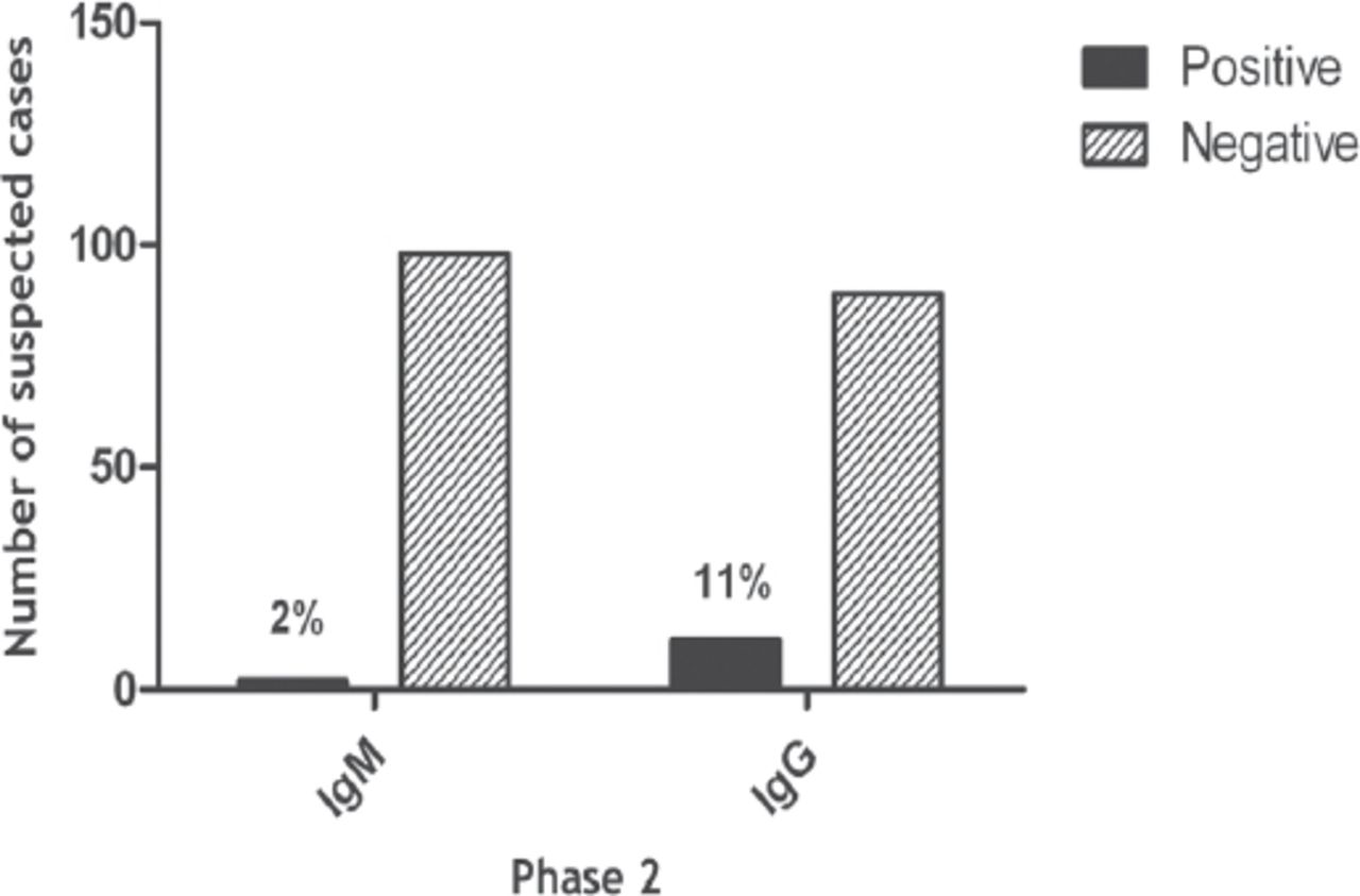
Coxiella burnetii-specific phase 2 IgG and IgM antibodies.

Figure 2
  • Download figure
  • Open in new tab
  • Download powerpoint
Figure 2

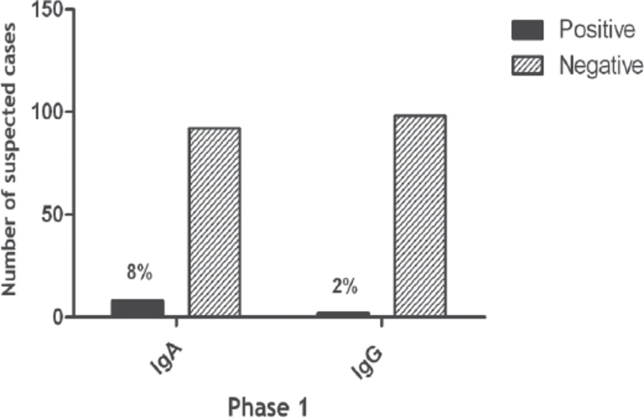
Coxiella burnetii-specific phase 1 IgG and IgM antibodies.

Discussion

Based on the detection of phase 1 and phase 2 C. burnetii-specific antibodies, 16% of patients harbored evidence of C. burnetii infection in the present study. In the northeastern region of Spain, the seroprevalence of C. burnetii in humans was reported to be 15.3%.18 In contrast, in this study, the study population comprised patients with FUO, and the actual seroprevalence in the local population appears to be lower than 16%. A seroprevalence of 4.8% was reported among 459 unexposed women from Denmark.19 Moreover, in Western Kenya, the prevalence rate has been reported to be as low as 2.5%.20 Seven years after an outbreak of Q fever in the Netherlands, the seroprevalence of Q fever remains high, at 33.8%.21 Collectively, these observations indicated regional variations in the prevalence of Q fever that may be due to differences in the prevalence rates among animals, as well as differences in human exposure, previous outbreaks of Q fever in the community, selection of the study population, and the methods used for detection of C. burnetii infection.

Coxiella burnetii-specific antibodies in the serum of patients suffering from FUO were detected by ELISA in the present study. Using IFAs, a prevalence of 35.2% has previously been reported from the Kingdom of Saudi Arabia in a similar study population.22 The prevalence of Q fever observed in the present study (16%) was almost half that reported previously in the Kingdom of Saudi Arabia. Although IFAs have been shown to detect higher percentage of C. burnetii-specific phase 1 and phase 2 antibodies compared with ELISA,23 there is evidence refuting these claims.24 Two consecutive studies, including the present study, have clearly demonstrated the presence of serological evidence of C. burnetii in the local human population. These observations have emphasized the need for large-scale epidemiological studies using more than one testing modality among the local human and animal populations to assess the prevalence of C. burnetii in Saudi Arabia.

Both phase 2 and phase 1 C. burnetii-specific antibodies were detected in the present study. At least 13% of patients with Q fever are believed to evade diagnosis either due to no attempt to screen the patients for the presence of C. burnetii specific antibodies or screening for C. burnetii in the early stages of infection before the appearance of detectable titers of C. burnetii specific antibodies. Phase 2 IgM appear within first 2 weeks after the onset of symptoms, reaching a maximum concentration by 4-8 weeks and remain detectable for a relatively short period.25 Phase 2 IgG antibodies appear later as the disease progresses and tend to persist for years or even for life.26 These observations suggest that among the local population, all patients suffering from FUO should be offered C. burnetii screening to avoid Q fever-associated morbidity and mortality.

Phase 1 C. burnetii IgA antibodies were detected in 8% of the study population in the present study. These antibodies have been shown to exhibit a strong correlation with Q fever in general, particularly with endocarditis.27,28 For the same reason, the presence of IgA phase 1 antibodies has been considered as a diagnostic marker of chronic Q fever.27 However, there is evidence demonstrating that phase 1 C. burnetii antibodies may contribute little to the diagnosis of Q fever28 but can serve as a useful marker for the follow-up of treated patients.29 The absence of a 4-fold decrease in phase 1 IgG and IgA antibodies and the presence of phase 2 IgM antibodies at the end of 1 year are among the determinants associated with mortality in Q fever.30

Despite the important findings of our study, the results should be interpreted based on the limitation that few cases were repeatedly within the gray zone for a single antibody marker, hindering interpretation of the findings; these patients could not be reached to collect additional samples. Thus, further studies with more patients are needed to confirm these findings.

In conclusion, phase 1 and phase 2 C. burnetii antibodies were detected in a relatively large proportion of patients with FUO. Evidence continues to emerge documenting the presence of Q fever in livestock and humans in the Kingdom of Saudi Arabia. These observations indicate the necessity for national investigations to determine the exact prevalence of Q fever among humans and animals in Saudi Arabia and to introduce Q fever screening for suspected cases on a regular basis.

Acknowledgment

The authors would like to thank the Faculty of Medicine, King Khalid University Hospital; and the Deanship of Scientific Research and Researchers Support & Services Unit, King Saud University, Riyadh, KSA for their technical support.

Footnotes

  • Disclosure. This study was approved by the Institutional Review Board of the College of Medicine, King Saud University, Riyadh, Saudi Arabia (Project no. E14-1135).

  • Received August 21, 2018.
  • Accepted October 21, 2018.
  • Copyright: © Saudi Medical Journal

This is an open-access article distributed under the terms of the Creative Commons Attribution-Noncommercial-Share Alike 3.0 Unported, which permits unrestricted use, distribution, and reproduction in any medium, provided the original work is properly cited.

References

  1. ↵
    1. Faix DJ,
    2. Harrison DJ,
    3. Riddle MS,
    4. Vaughn AF,
    5. Yingst SL,
    6. Earhart K,
    7. et al.
    (2008) Outbreak of Q fever among US military in western Iraq, June-July 2005. Clin Infect Dis 46:e65–e68.
    OpenUrlCrossRefPubMed
  2. ↵
    1. Karagiannis I,
    2. Schimmer B,
    3. Van LA,
    4. Timen A,
    5. Schneeberger P,
    6. Van RB,
    7. et al.
    (2009) Investigation of a Q fever outbreak in a rural area of The Netherlands. Epidemiol Infect 137:1283–1294.
    OpenUrlCrossRefPubMed
  3. ↵
    1. Baca OG,
    2. Paretsky D
    (1983) Q fever and Coxiella burnetii:a model for host-parasite interactions. Microbiol Rev 47:127–149.
    OpenUrlFREE Full Text
  4. ↵
    1. Hackstadt T
    (1990) The role of lipopolysaccharides in the virulence of Coxiella burnetii. Ann N Y Acad Sci 590:27–32.
    OpenUrlCrossRefPubMed
  5. ↵
    1. Toman R,
    2. Skultety L
    (1996) Structural study on a lipopolysaccharide from Coxiella burnetii strain nine mile in avirulent phase II. Carbohydr Res 283:175–185.
    OpenUrlCrossRefPubMed
  6. ↵
    1. Amano K,
    2. Williams JC
    (1984) Chemical and immunological characterization of lipopolysaccharides from phase I and phase II Coxiella burnetii. J Bacteriol 160:9940–1002.
    OpenUrl
  7. ↵
    1. Babudieri B
    (1959) Q fever:a zoonosis. Adv Vet Sci Cop Med 5:82–182.
    OpenUrl
  8. ↵
    1. Afzal M,
    2. Sakkir M
    (1994) Survey of antibodies against various infectious disease agents in racing camels in Abu\Dhabi, United Arab Emirates. Rev Sci Tech 13:787–792.
    OpenUrlPubMed
    1. Al-Hajjar S,
    2. Hussain Qadri SM,
    3. al-Sabban E,
    4. Jager C
    (1997) Coxiella burnetii endocarditis in a child. Pediatr Infect Dis J 16:911–913.
    OpenUrlCrossRefPubMed
    1. Bannatyne RM,
    2. Qasem L
    (2000) Aetiology of community acquired pneumonia:fashionable or familiar. Saudi Med J 21:104.
    OpenUrlPubMed
    1. Gelpi AP
    (1966) Q fever in Saudi Arabia. Am J Trop Med Hyg 15:784–798.
    OpenUrlPubMed
    1. Greth A,
    2. Calvez D,
    3. Vassart M,
    4. Lefevre PC
    (1992) Serological survey for bovine bacterial and viral pathogens in captive Arabian oryx (Oryx leucoryx Pallas, 1776). Rev Sci Tech 11:1163–1168.
    OpenUrlPubMed
    1. Lippi M,
    2. Sebastiani A,
    3. el-Mutabakani H
    (1968) Detection of serum antibodies against Reoviruses, Adenoviruses and Coxiella burnetii in a group of inhabitants of Riyad, Saudi Arabia. Arch Ital Sci Med Trop Parassitol 49:129–136.
    OpenUrlPubMed
  9. ↵
    1. Scrimgeour EM,
    2. Johnston WJ,
    3. Al Dhahry SH,
    4. El-Khatim HS,
    5. John V,
    6. Musa M
    (2000) First report of Q fever in Oman. Emerg Infect Dis 6:74–76.
    OpenUrlPubMed
  10. ↵
    1. Jarelnabi AA,
    2. Alshaikh MA,
    3. Bakhiet AO,
    4. Omer SA,
    5. Aljumaah RS,
    6. Harkiss GD,
    7. et al.
    (2018) Seroprevalence of Q fever in farm animals in Saudi Arabia. Biomed Res 29:895–900.
    OpenUrl
  11. ↵
    1. Marrie TJ
    1. Yeamen MR,
    2. Baca OG
    (1990) in Q Fever. Volume 1:The Disease, Antibiotic susceptibility of Coxiella burnetii, ed Marrie TJ (CRC Press, Florida (USA)), pp 213–224.
  12. ↵
    1. Murray PR,
    2. Baron EJ,
    3. Jorgensen JH,
    4. Landry ML,
    5. Pfaller MA,
    6. et al.
    1. Brouqui P,
    2. Marrie T,
    3. Raoult D
    (2007) in Manual of Clinical Microbiology, Coxiella, eds Murray PR, Baron EJ, Jorgensen JH, Landry ML, Pfaller MA, et al. (ASM Press, Washington (DC)), pp 1062–1069.
  13. ↵
    1. Cardenosa N,
    2. Sanfeliu I,
    3. Font B,
    4. Munoz T,
    5. Nogueras MM,
    6. Segura F
    (2006) Short report:seroprevalence of human infection by Coxiella burnetii in Barcelona (northeast of Spain). Am J Trop Med Hyg 75:33–35.
    OpenUrlAbstract/FREE Full Text
  14. ↵
    1. Nielsen SY,
    2. Molbak K,
    3. Nybo Andersen AM,
    4. Brink HT,
    5. Kantso B,
    6. Krogfelt KA,
    7. et al.
    (2013) Prevalence of Coxiella burnetii in women exposed to livestock animals, Denmark, 1996 to 2002. Euro Surveill 18:20528.
    OpenUrl
  15. ↵
    1. Wardrop NA,
    2. Thomas LF,
    3. Cook EA,
    4. de Glanville WA,
    5. Atkinson PM,
    6. Wamae CN,
    7. et al.
    (2016) The sero-epidemiology of Coxiella burnetii in humans and cattle, Western Kenya:evidence from a cross-sectional study. PLoS Negl Trop Dis 10:e0005032.
    OpenUrl
  16. ↵
    1. Morroy G,
    2. van der HW,
    3. Albers J,
    4. Coutinho RA,
    5. Bleeker-Rovers CP,
    6. Schneeberger PM
    (2015) Population screening for chronic Q-fever seven years after a major outbreak. PLoS One 10:e0131777.
    OpenUrlCrossRef
  17. ↵
    1. Almogren A,
    2. Shakoor Z,
    3. Hasanato R,
    4. Adam MH
    (2013) Q fever:a neglected zoonosis in Saudi Arabia. Ann Saudi Med 33:464–468.
    OpenUrlPubMed
  18. ↵
    1. Wegdam-Blans MC,
    2. Wielders CC,
    3. Meekelenkamp J,
    4. Korbeeck JM,
    5. Herremans T,
    6. Tjhie HT,
    7. et al.
    (2012) Evaluation of commonly used serological tests for detection of Coxiella burnetii antibodies in well-defined acute and follow-up sera. Clin Vaccine Immunol 19:1110–1115.
    OpenUrlAbstract/FREE Full Text
  19. ↵
    1. Szymanska-Czerwinska M,
    2. Galinska EM,
    3. Niemczuk K,
    4. Knap JP
    (2015) Prevalence of Coxiella burnetii infection in humans occupationally exposed to animals in Poland. Vector Borne Zoonotic Dis 15:261–267.
    OpenUrl
  20. ↵
    1. Dupuis G,
    2. Peter O,
    3. Peacock M,
    4. Burgdorfer W,
    5. Haller E
    (1985) Immunoglobulin responses in acute Q fever. J Clin Microbiol 22:484–487.
    OpenUrlAbstract/FREE Full Text
  21. ↵
    1. Dupont HT,
    2. Thirion X,
    3. Raoult D
    (1994) Q fever serology:cutoff determination for microimmunofluorescence. Clin Diagn Lab Immunol 1:189–196.
    OpenUrlPubMed
  22. ↵
    1. Peacock MG,
    2. Philip RN,
    3. Williams JC,
    4. Faulkner RS
    (1983) Serological evaluation of Q fever in humans:enhanced phase I titers of immunoglobulins G and A are diagnostic for Q fever endocarditis. Infect Immun 41:1089–1098.
    OpenUrlAbstract/FREE Full Text
  23. ↵
    1. Raoult D,
    2. Etienne J,
    3. Massip P,
    4. Iaocono S,
    5. Prince MA,
    6. Beaurain P,
    7. et al.
    (1987) Q fever endocarditis in the south of France. J Infect Dis 155:570–573.
    OpenUrlCrossRefPubMed
  24. ↵
    1. Raoult D,
    2. Houpikian P,
    3. Tissot DH,
    4. Riss JM,
    5. Arditi-Djiane J,
    6. Brouqui P
    (1999) Treatment of Q fever endocarditis:comparison of 2 regimens containing doxycycline and ofloxacin or hydroxychloroquine. Arch Intern Med 159:167–173.
    OpenUrlCrossRefPubMed
  25. ↵
    1. Million M,
    2. Thuny F,
    3. Richet H,
    4. Raoult D
    (2010) Long-term outcome of Q fever endocarditis:a 26-year personal survey. Lancet Infect Dis 10:527–535.
    OpenUrlCrossRefPubMed
PreviousNext
Back to top

In this issue

Saudi Medical Journal: 39 (12)
Saudi Medical Journal
Vol. 39, Issue 12
1 Dec 2018
  • Table of Contents
  • Cover (PDF)
  • Index by author
Print
Download PDF
Email Article

Thank you for your interest in spreading the word on Saudi Medical Journal.

NOTE: We only request your email address so that the person you are recommending the page to knows that you wanted them to see it, and that it is not junk mail. We do not capture any email address.

Enter multiple addresses on separate lines or separate them with commas.
Screening for Q fever
(Your Name) has sent you a message from Saudi Medical Journal
(Your Name) thought you would like to see the Saudi Medical Journal web site.
Citation Tools
Screening for Q fever
Abdulkarim F. Alhetheel, Khalifa Binkhamis, Ali Somily, Mazin Barry, Zahid Shakoor
Saudi Medical Journal Dec 2018, 39 (12) 1195-1199; DOI: 10.15537/smj.2018.12.23695

Citation Manager Formats

  • BibTeX
  • Bookends
  • EasyBib
  • EndNote (tagged)
  • EndNote 8 (xml)
  • Medlars
  • Mendeley
  • Papers
  • RefWorks Tagged
  • Ref Manager
  • RIS
  • Zotero
Share
Screening for Q fever
Abdulkarim F. Alhetheel, Khalifa Binkhamis, Ali Somily, Mazin Barry, Zahid Shakoor
Saudi Medical Journal Dec 2018, 39 (12) 1195-1199; DOI: 10.15537/smj.2018.12.23695
Twitter logo Facebook logo Mendeley logo
  • Tweet Widget
  • Facebook Like
  • Google Plus One
Bookmark this article

Jump to section

  • Article
    • Abstract
    • Methods
    • Results
    • Discussion
    • Acknowledgment
    • Footnotes
    • References
  • Figures & Data
  • eLetters
  • References
  • Info & Metrics
  • PDF

Related Articles

  • No related articles found.
  • PubMed
  • Google Scholar

Cited By...

  • No citing articles found.
  • Google Scholar

More in this TOC Section

  • Psychological stress and its association with bronchial asthma in Saudi Arabia
  • The factors affecting comfort and the comfort levels of patients hospitalized in the coronary intensive care unit
  • Exploring communication challenges with children and parents among pharmacists in Saudi Arabia
Show more Original Article

Similar Articles

CONTENT

  • home

JOURNAL

  • home

AUTHORS

  • home
Saudi Medical Journal

© 2025 Saudi Medical Journal Saudi Medical Journal is copyright under the Berne Convention and the International Copyright Convention.  Saudi Medical Journal is an Open Access journal and articles published are distributed under the terms of the Creative Commons Attribution-NonCommercial License (CC BY-NC). Readers may copy, distribute, and display the work for non-commercial purposes with the proper citation of the original work. Electronic ISSN 1658-3175. Print ISSN 0379-5284.

Powered by HighWire