


Article Figures & Data
Figures
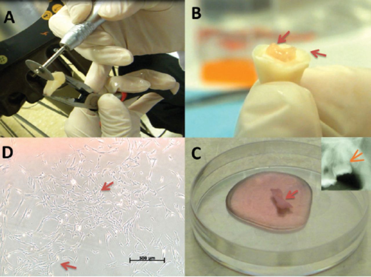
- Figure 1
Collecting pulp tissue from extracted teeth. A) Stable finger support while using a diamond disc to create a 360° grove at 2 mm depth under the cemento-enamel junction. B) The crown was separated from the root (arrows) with minimum debris by wedging the chisel in the groove and applying gentle force with a hammer. C) The exposed pulp tissue (arrow) was collected with a hemostat and Endodontic K-files, and placed in 4°C Dulbecco’s Modified Eagle’s Medium (DMEM) supplemented with 45 mg/L D-glucose, 4 mM L-glutamine, and 110 mg/L sodium pyruvate (Gibco, Loughborough, UK). The culture medium also contained a 10% penicillin-streptomycin solution (Pen-Strep; 10 units penicillin and 10 µg streptomycin per µL, Gibco), Selecting teeth with a large pulp chamber (arrow) ensured the removal of pulp tissue in one piece with minimal debris. D) Dental pulp cells formed visible colonies at day 14 as viewed under an inverted light microscope (arrows).
- Figure 2
Isolated dental pulp stem cells characterization (DPSCs) showing A) Positive mesenchymal stem markers (CD105, CD90, CD73, CD13, CD29 and CD44) and negative for hematopoietic and endothelial markers (CD34, CD45, CD14, and CD31) as well as for the MHC class II marker human leukocyte antigen - antigen d related. B) Positive immunostaining for 4’, 6-diamidino-2-phenylindole (DAPI) and localization of Vimentin in DPSCs. C) Positive immunostaining for DAPI and negative for localization of Vimentin in control.
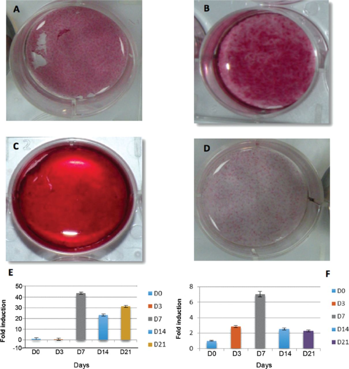
- Figure 3
Dental pulp stem cells osteogenic differentiation showing A) day 7, B) day 14 positive ALP staining. C) day 14 positive alizarin red staining, D) control, E) representative graph of RT-polymerase chain reaction of alkaline phosphatase gene expression, and F) RUNX-2 gene expression
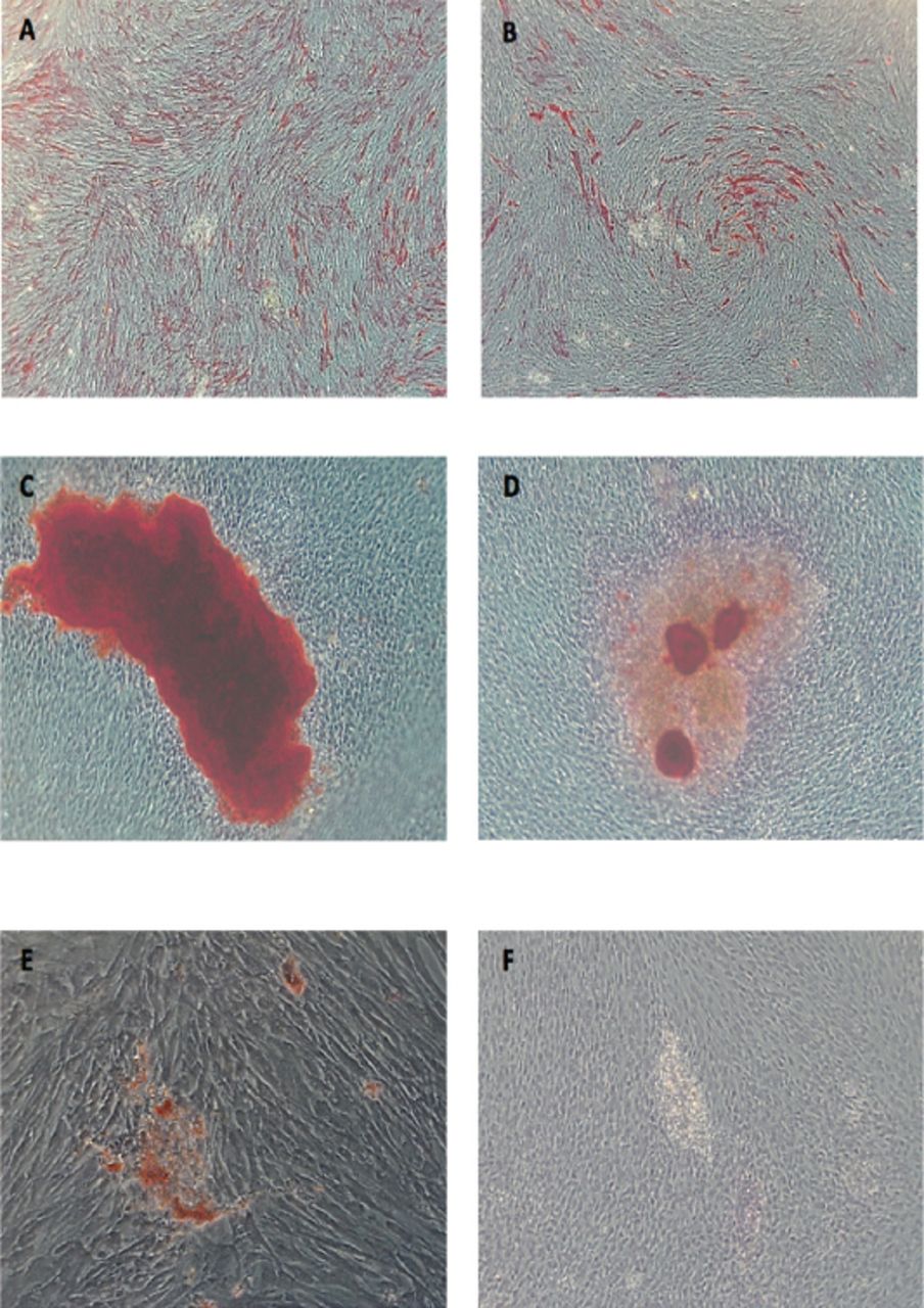
- Figure 4
Post-thaw dental pulp stem cells osteogenic, odontogenic, adipogenic differentiation showing A) day 7 ALP activity after osteogenic induction (X5), B) odontogenic induction, C) Day 14 alizarin red S stain (X10) in osteogenic, D) odontogenic induction media, E) day 21 oil red O stained droplet (X20) after adipogenic induction, (F) Control
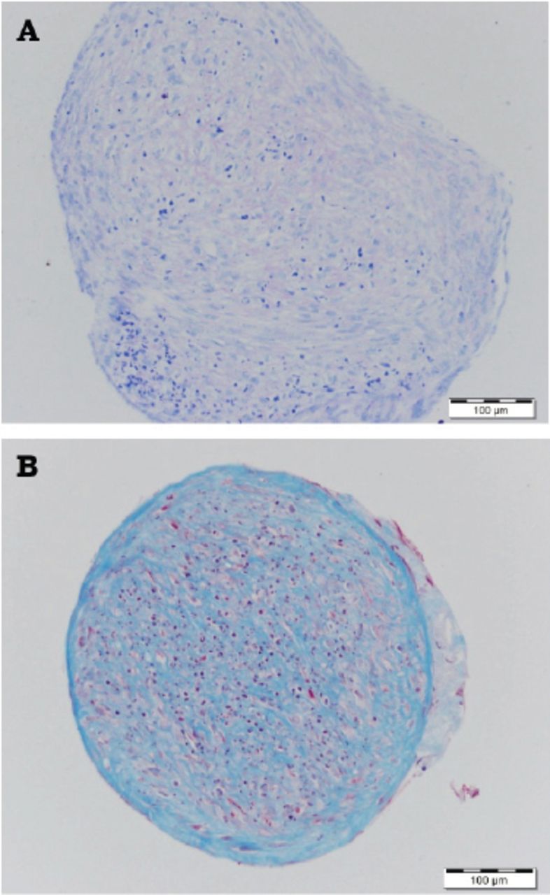
- Figure 5
Post-thaw dental pulp stem cells chondrogenic differentiation showing A) Day 35 proteoglycans were positive to Toluidine blue stain, B) Day 50 collagen fibers were positive to Masson’s trichrome stain.
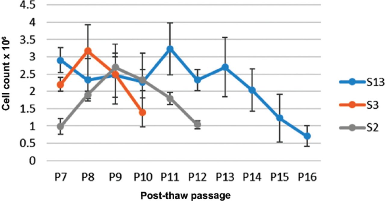
- Figure 6
Cell growth curves for 3 dental pulp stem cells line cultured until senescence


Tables
In this issue


Jump to section
Related Articles
Cited By...
- No citing articles found.