


Abstract
Objectives: To define the dimensions of the frontal sinus in groups standardized for age and gender and to discuss the reasons and the effects of the variations.
Methods: Frontal sinus measurements were obtained from paranasal CT scans of 180 males and 180 females in the Radiology Department of Dursun Odabas Medical Center of Yuzuncu Yil University, Van, which is located in Eastern Turkey, between February and March 2016. The width and height of sinuses were measured on a coronal plane, and the anteroposterior length was measured on an axial plane. Volumes were calculated using the Hospital Information Management Systems and Image Archiving and Management System program. The Statistical Package of the Social Science version 13 was used for statistical analyses.
Results: We determined differences in the frontal sinus measurements of different age groups in a Turkish adult population. Frontal sinus dimensions were usually higher in females and lower in males after 40-49 years of age than their younger counterparts, but the measurements were lower in females and higher in males in 70≤ years of age group than 60-69 years of age. Left frontal sinus was dominant in young age groups but right frontal sinus was dominant in groups 40-49 years of age or older.
Conclusion: We observed crossing of the measurements between the different age groups, which we could not find clear explanations. The results of such studies may affect forensic identification from frontal sinus measurements.
Frontal sinuses are paired lobulated cavities located behind the superciliary arches in the frontal bone. Each frontal sinus opens into the middle meatus on the same side via the infundibulum.1 Frontal sinuses are not radiographically visible at birth. They begin to develop during the second year of life, and they can be detected radiographically after the age of 5.2-4 The radiographic pattern of frontal sinuses are unique to every individual, even among monozygotic twins.3 There is consensus among forensic scientists concerning the usability of antemortem and postmortem radiographs and CT scans of frontal sinuses for forensic personal identification today. Superimposing the antemortem and postmortem radiographs of a person for positive identification is an accepted procedure in forensic medicine.2,3,5 Authors have typically accepted that the development of the frontal sinus is completed by approximately 20 years of age and remains stable until further enlargement may occur from bone resorption during advanced ages, except in rare events such as fractures, severe infections, or tumors.2,3 Since the frontal sinuses are unique to every individual, some authors have considered using them as a substitute for fingerprints. Yoshino et al2 proposed a classifying system for frontal sinuses utilizing area, asymmetry, scalloping, septa, and cells as criteria. Riberio Fde6 suggested a method of obtaining standard measurements from the radiographs of frontal sinuses and storing the acquired information in a computer databank. In 2007, Tatlisumak et al7 reported on the presence or absence of frontal sinus, septum, scalloping (FSS) system as a simple and useful method to use in frontal sinus CT scans. Recently, some authors published papers supporting the usability of the FSS system for forensic identification.8-10 Other systems were also proposed.11 The use of frontal sinus radiographs for personal identification relies on the stability of frontal sinuses throughout the lifespan or at least for a long period of time. McLaughlin et al12 proposed that frontal sinuses continue to expand until the age of 40 years due to the mechanical stresses of mastication and growth hormone levels. Tatlisumak et al13 reported that most of the frontal sinus measurements reached their highest values between 31 and 40 years of age or between 41 and 50 years of age in both genders, and later decreased with increasing age. The aim of this study is to define the dimensions of the frontal sinus in groups standardized for age and gender and to discuss the reasons and the effects of the variations.
Methods
This retrospective study was conducted using paranasal CT scans with 5 mm thickness. The CT scans were of the axial and coronal planes of 360 patients over the age of 20 taken by a 16-detector multislice CT device (Somatom Emotion 16-Slice; CT2012E- Siemens AG, Berlin and Munich-Germany) in the Dursun Odabas Medical Center of Yuzuncu Yil University, Van, which is located in Eastern Turkey between February and March 2016. This study used CT scans with no apparent sinonasal pathology. Cases with unilateral or bilateral absence of frontal sinuses were not included in the study.
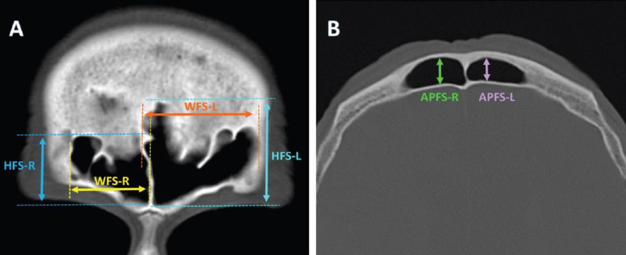
All evaluations and measurements were performed together by 2 scientists in this study with a Digital Imaging Communication in Medicine (DICOM) viewer program. Measurements were expressed in millimeters in the Hospital Information Management Systems and Image Archiving and Management System (HBYS & PACS) program. Volumes were expressed in cubic centimeters. In total, 8 measurements were used in this study: 1) width of the right frontal sinus (WFS-R), 2) width of the left frontal sinus (WFS-L), 3) height of the right frontal sinus (HFS-R), 4) height of the left frontal sinus (HFS-L), 5) anteroposterior length of the right frontal sinus (APFS-R), 6) anteroposterior length of the left frontal sinus (APFS-L), 7) volume of the right frontal sinus (VFS-R), and 8) volume of the left frontal sinus (VFS-L). The width and height of both sinuses were measured on a coronal plane, and the anteroposterior length was measured on an axial plane (Figure 1). Volumes were calculated using the HBYS & PACS program.

A) Measurements of the widths and heights of both sinuses (WFS-R - width of the right frontal sinus, WFS-L - width of the left frontal sinus, HFS-R - height of the right frontal sinus, HFS-L - height of the left frontal sinus) on a coronal plane, and B) the anteroposterior length (APFS-R - anteroposterior length of the right frontal sinus, APFS-L - anteroposterior length of the left frontal sinus) on an axial plane.
This study used CT scans of 180 female and 180 male patients. Cases were divided into 6 age groups according to age for both genders: 20-29, 30-39, 40-49, 50-59, 60-69, and 70-70+. Each group included 30 cases, and the mean age of each group was adjusted for more accurate results, as age was statistically similar to the corresponding group of the other gender except in the 70-70+ group. The mean age could not be adjusted in the 70-70+ age group due to the difficulty of finding cases in this age group.
Descriptive statistics for studied variables were presented as the mean, standard deviation, minimum and maximum values. One-way analysis of variance (ANOVA) (including post hoc tests) were used to compare the age groups and genders.
Statistical significance was considered as p<0.05. The Statistical Package of the Social Science version 13 (SPSS Inc., Chicago, IL, USA) was used for all statistical computations.
The study was approved by the Non-Interventional Clinical Research Ethics Committee of Yuzuncu Yil University, Van, Turkey on February 2016.
Results
Paranasal CT scans of 180 females (mean age: 50.14, standard deviation [SD]: 18.68; median age: 49.50; minimum age: 20, maximum age: 101) and 180 males (mean age: 49.66, SD: 17.53; median ages: 49.50, minimum age: 20, maximum age: 88) (p=0.800) were evaluated in this study.
All of the measurements of the frontal sinus were higher among males than among females. These differences were statistically significant in the WFS-L, HFS-L, APFS-R, APFS-L, VFS-R, and VFS-L (p<0.05), but the differences were not statistically significant in WFS-R and HFS-R (p>0.05) (Table 1).
Comparison of measurements of frontal sinus in females and males.
All the measurements of the frontal sinus were higher at the left side for females, males and the total population. Anteroposterior (AP) length and volume of frontal sinuses were significantly larger on the left side among females and in the total population (p<0.05), whereas other measurements of frontal sinuses were slightly larger on the left side, but the differences were not statistically significant (p>0.05) (Table 2). Of all included cases, 55.6% (n=200) had left sinus dominancy, and 44.4% (n=160) had right sinus dominancy based on volumetric measurements (p<0.05).
Comparison of frontal sinus measurements in right and left sides.
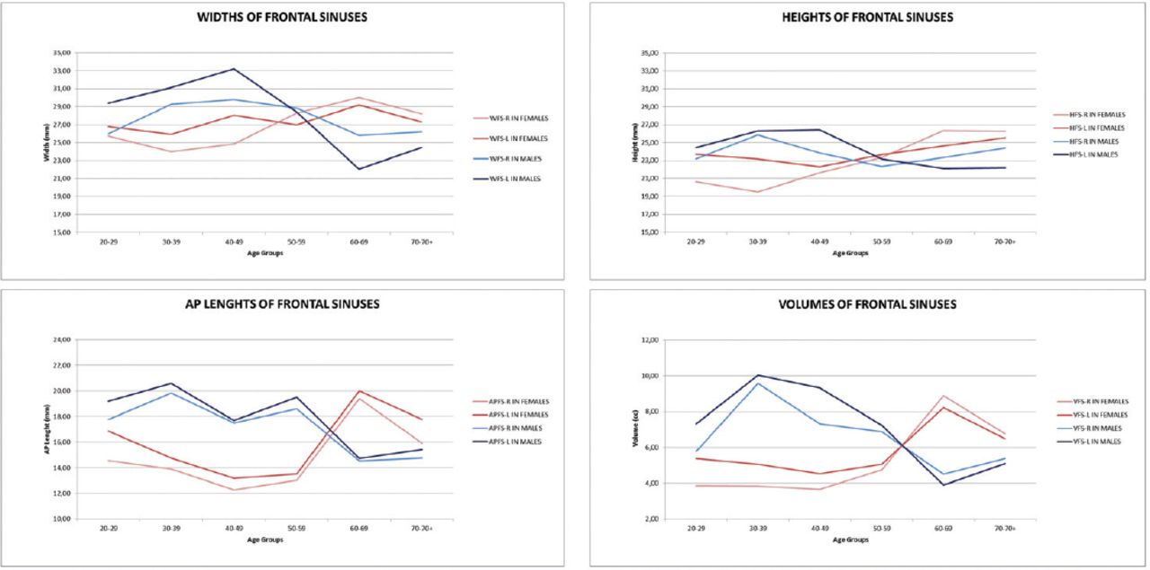
Up to age 50-59 (and up to age 40-49 for widths), the widths, heights, and volumes of the left frontal sinuses were slightly greater than those of the right side. After age 50-59 (after age 40-49 for widths), a crossing occurred, and the widths, heights, and volumes of the right frontal sinuses were found to be slightly greater than the left side. Anteroposterior length was slightly higher in the left frontal sinus than in the right frontal sinus among all age groups for both genders (Figure 2).

Changes of the frontal sinus sizes with age and gender in right and left sides
All measurements of the frontal sinus exhibited different features by age group for both females and males. In females, WFS-R, WFS-L, HFS-R, APFS-R, APFS-L, VFS-R, and VFS-L reached maximum values between the ages of 60 and 69 years old, whereas HFS-L reached a maximum value at over 70 years of age. In males, WFS-R, WFS-L and HFS-L reached maximum value between the ages of 40 to 49 years old; HFS-R, APFS-R, APFS-L, VFS-R, and VFS-L reached maximum value between the ages of 30 and 39 years old (Table 3). When we examined these changes in the right and left frontal sinus for both genders in Figure 2, a second crossing event was observed between frontal sinus sizes of females and males between 60 and 69 years of age in WFS-R, WFS-L, APFS-R, and APFS-L and between 50 and 59 years of age in the HFS-R and HFS-L. The sizes of the frontal sinuses in females increased with age, and the sizes of frontal sinuses in males decreased with age after 50.
Comparison of frontal sinus measurements in right and left sides.
In the overall population, APFS-L reached a maximum value between 20 and 29 years of age; HFS-L, VFS-R and VFS-L reached a maximum value between 30 and 39 years of age; WFS-L reached a maximum value between 40 and 49 years of age; WFR-R reached a maximum value between 50 and 59 years of age; APFS-R reached a maximum value between 60 and 69 years of age; and HFS-R reached a maximum value at over 70 years of age (Table 3).
Discussion
Most previous studies have reported that the frontal sinus was larger in males than in females.8,11,13-16 Only a few authors have reported that the frontal sinus measurements for females and males were not significantly different.3,17,18 In our study, the WFS-L, HFS-L, APFS-R, APFS-L, VFS-R, and VFS-L were significantly larger in males (p<0.05). Other measurements were also slightly larger in males, but the differences were not statistically significant (p>0.05).
In most previous studies, the left frontal sinus was found to be larger than the right frontal sinus, although the cause was not explained.13,15,19 Tatlisumak et al13 stated that the size of the left frontal sinus is more pronounced in both genders. Nevertheless, some authors have reported that there were no statistically significant differences between the right and left frontal sinuses.20 Kanat et al21 reported that the left sinuses were dominant in 50.1% of the cases, the right sinuses were dominant in 35.6% of the cases, and both sinuses were equal in 14.3% of their series.
In the present study, anteroposterior (AP) length and volume of frontal sinuses were significantly larger on the left side in females and in total population (p<0.05), whereas other sizes of frontal sinus were slightly larger on the left side. However, these differences were not statistically significant (p>0.05).
When cases were stratified by age group and gender, interesting results were obtained. Sizes and volumes of left frontal sinuses, except AP lengths, were slightly greater compared to the right side up to 50-59 (40-49 for widths) years of age. However, a crossing occurred after 50-59 (40-49 for widths) years of age, and as a result, the widths, heights and volumes of the right frontal sinuses were found to be larger than those on the left side. Kanat et al21 suggested that lateralization might be one of the causes of human frontal sinus asymmetry. Because we identified a change in the dominant side with age in this study, we cannot agree with this opinion.
We determined that each measurement exhibited different characteristics with aging among both genders. In the total population, we observed a crossing event in frontal sinus sizes between 50 and 59 years of age in WFS-R, WFS-L, APFS-R, APFS-L, VFS-R and VFS-L and between 40 and 49 years of age in HFS-R and HFS-L. In the literature, we did not encounter such findings in any previously conducted studies. In only one article, Tatlisumak et al13 described that most of the measurements of the frontal sinus reached their highest values between 31 and 40 or between 41 and 50 years of age in both genders; a reduction occurred with aging.
All measurements of the frontal sinus after 40-49 years of age tended to increase and continued until 70-70+ years of age in females, at which point a decrease occurred, except in HFS-L. Contrarily, all measurements of frontal sinus tended to decrease after 40-49 years of age in males, until 70-70+ years of age, when the values of the measurements increased again. The changes occurring after 40-49 years of age fit the sex steroid decline that occurs in both genders during this period. McLaughlin et al12 had proposed that growth hormone levels might affect the sizes of frontal sinuses. Further research is required to elucidate whether the levels of sex steroids have effects on diameters of the frontal sinus.
Fatu et al22 reported that frontal sinuses were dynamic structures and that changes in both size and shape continue to occur during adult life. Tatlisumak et al13 determined alterations in the diameters of the frontal sinus in an adult Turkish population. We also defined changes in the sizes of frontal sinuses in a Turkish population from a different region of Turkey. However, all these studies were on local populations with a limited number of subjects.
In conclusion, from past to present, frontal sinuses have been accepted as unique for every person, an indispensable element of forensic identification, and a valuable identification tool that can be successful, even in monozygotic twins. We observed differences of dimensions of frontal sinus in different age groups in a Turkish adult population. We suggest that further studies are required to determine whether frontal sinus dimensions change during adulthood. The results of such studies may affect the opinions of researches working on forensic personal identification using frontal sinus measurements.
Footnotes
Disclosure. Authors have no conflict of interest, and the work was not supported or funded by any drug company.
- Received August 10, 2016.
- Accepted October 13, 2016.
- Copyright: © Saudi Medical Journal
This is an open-access article distributed under the terms of the Creative Commons Attribution-Noncommercial-Share Alike 3.0 Unported, which permits unrestricted use, distribution, and reproduction in any medium, provided the original work is properly cited.
In this issue


Jump to section
Related Articles
Cited By...
- No citing articles found.