
Main menu

  • Home
  • Content
    • Latest
    • Archive
    • home
  • Info for
    • Authors
    • Reviewers
    • Subscribers
    • Institutions
    • Advertisers
    • Join SMJ
  • About Us
    • About Us
    • Editorial Office
    • Editorial Board
  • More
    • Advertising
    • Alerts
    • Feedback
    • Folders
    • Help
  • Other Publications
    • NeuroSciences Journal

User menu

  • My alerts
  • Log in

Search

  • Advanced search
Saudi Medical Journal
  • Other Publications
    • NeuroSciences Journal
  • My alerts
  • Log in
Saudi Medical Journal

Advanced Search

  • Home
  • Content
    • Latest
    • Archive
    • home
  • Info for
    • Authors
    • Reviewers
    • Subscribers
    • Institutions
    • Advertisers
    • Join SMJ
  • About Us
    • About Us
    • Editorial Office
    • Editorial Board
  • More
    • Advertising
    • Alerts
    • Feedback
    • Folders
    • Help
  • Follow psmmc on Twitter
  • Visit psmmc on Facebook
  • RSS
Review ArticleReview Article
Open Access

Middle East respiratory syndrome in children

Dental considerations

Fares S. Al-Sehaibany
Saudi Medical Journal April 2017, 38 (4) 339-343; DOI: https://doi.org/10.15537/smj.2017.4.15777
Fares S. Al-Sehaibany
From the Division of Pediatric Dentistry, Department of Pediatric Dentistry and Orthodontics, College of Dentistry, King Saud University, Riyadh, Kingdom of Saudi Arabia
BDS, DMSc
  • Find this author on Google Scholar
  • Find this author on PubMed
  • Search for this author on this site
  • For correspondence: [email protected]
  • Article
  • Figures & Data
  • eLetters
  • Info & Metrics
  • References
  • PDF
Loading

Abstract

As of January 2016, 1,633 laboratory-confirmed cases of Middle East Respiratory Syndrome Coronavirus (MERS-CoV) infection and 587 MERS-related deaths have been reported by the World Health Organization globally. Middle East Respiratory Syndrome Coronavirus may occur sporadically in communities or may be transmitted within families or hospitals. The number of confirmed MERS-CoV cases among healthcare workers has been increasing. Middle East Respiratory Syndrome Coronavirus may also spread through aerosols generated during various dental treatments, resulting in transmission between patients and dentists. As MERS-CoV cases have also been reported among children, pediatric dentists are at risk of MERS-CoV infection. This review discusses MERS-CoV infection in children and healthcare workers, especially pediatric dentists, and considerations pertaining to pediatric dentistry. Although no cases of MERS-CoV transmission between a patient and a dentist have yet been reported, the risk of MERS-CoV transmission from an infected patient may be high due to the unique work environment of dentists (aerosol generation).

In 2014, Memish et al1 reported the clinical presentation and outcome of Middle East Respiratory Syndrome Coronavirus (MERS-CoV) infection in 11 Saudi pediatric patients. However, MERS-CoV was initially isolated from a 60-year-old Saudi patient in September 2012.2 Middle East Respiratory Syndrome Coronavirus is a novel beta coronavirus of the Coronaviridae family that causes a severe respiratory disease with a high fatality rate.3-6 As of January 2016, 1633 laboratory-confirmed cases of MERS-CoV infection and 587 related deaths have been reported by the World Health Organization (WHO) globally.7 The male-to-female ratio of the affected patients was 2:1, with a median age of 49 years.8 The incubation period for human-to-human transmission ranges from 2-15 days.9

The symptoms of MERS-CoV infection range from being asymptomatic to severe pneumonia, acute respiratory distress syndrome, septic shock, and multi-organ failure, leading to death. The symptomatic patients may have fever, cough, chills, throat soreness, myalgia, arthralgia, vomiting, or diarrhea.10 Furthermore, men over 45 years of age, patients with comorbidities, and healthcare workers have been reported to be high-risk groups for MERS-CoV infection.11 The virus has been detected in respiratory, gastrointestinal and other bodily secretions,12,13 as well as in air samples, which indicates the possibility for airborne transmission.14

Several studies have reported MERS-CoV infections among healthcare workers4,6,15,16 and children.1,17 Viral infections, such as severe acute respiratory syndrome (SARS-CoV), may be transmitted to healthcare workers from infected patients through aerosols.18 Considering that several types of dental equipment, such as handpieces, air-water syringes and ultrasonic scalers, produce considerable amounts of aerosols, the potential for the spread of infections from patients to dentists or dental assistants is high.19 This review is an attempt to discuss MERS-CoV infection among children and those providing dental treatment to them, including precautions and considerations pertaining to the practice of pediatric dentistry.

MERS-CoV infection in healthcare workers

The 3 patterns of MERS-CoV transmission suggested in the literature include the following: a) sporadic cases occurring in communities; b) transmission within families; and c) nosocomial transmission.20 Healthcare-related MERS-CoV transmission is associated with high morbidity, extended use of mechanical ventilation and fatality rates of up to 65%; this type of transmission can be attributed to shortcomings in observing stringent infection control protocols.4,6,15,21 Furthermore, MERS-CoV has been reported to be viable in hospital-like environments for up to 48 hours with a stability that is unaltered during aerosolization.22 Among 200 healthcare workers’ contacts identified in Al-Hassa, Saudi Arabia, MERS-CoV infection was confirmed in 2 cases.4 A Saudi study20 compared healthcare worker and family contact with laboratory-confirmed MERS-CoV patients and reported a lower rate (1.12%) of infection among the healthcare workers than among the families (3.4%).

Healthcare workers could be infected with MERS-CoV through exposure in the community or at their workplace,1,6,20 they could be diagnosed late,1,6 and they could remain asymptomatic or mildly symptomatic.6,8,16 Furthermore, unsuspected cases entering healthcare facilities have been considered the main source of MERS-CoV.4 Considering these aspects and that healthcare workers may continue to work regardless of being symptomatic,15 the possibility of transmitting the infection to their patients is also high.

MERS-CoV infection in children

Memish et al1 reported that the age of 11 Saudi pediatric patients who presented clinically with symptoms of MERS-CoV infection ranged from 2-16 years with a female-to-male ratio of 2.7 to 1. Of the 11 cases, 2 were symptomatic, and 9 were asymptomatic with no underlying comorbidities. Another report by Thabet et al17 concluded that although MERS-CoV infection presents with a wide range of clinical manifestations, the mortality rate in children is lower than that in adults. A review on the effects of Coronavirus infection in children concluded that Human Coronavirus infections may be associated with respiratory symptoms and may also involve central nervous system. The authors suggested that the clinical and genetic traits of Human Coronavirus infection among children should be monitored closely for early prevention.23

Dental considerations. Bioaerosols in dental practice

Bioaerosols are defined as a suspension of biological particles in gaseous media.24 Apart from radiation exposures, dermatitis, eye injuries, and musculoskeletal and respiratory disorders, other occupational health risks exist in dentistry. These risks include percutaneous exposure incidents and exposure to infectious diseases, such as those that may be present in bioaerosols.25 Subgingival scaling of periodontally involved teeth with ultrasonic scalers may produce aerosols containing blood.26 One study reported that ultrasonic scalers and tips produced significantly more aerosol and splatter than a handheld curette, regardless of the scaler type used.27 Miller et al28 studied the characteristics of blood-containing aerosols generated by common powered dental instruments. The author concluded that all the recovered particles could contain the hepatitis B virus and be inhaled and retained (20-100%) in the human respiratory system.

The repeated and chronic exposure to bioaerosols generated during certain dental procedures and the relatively small particle size contribute to an increased risk of infection among dental professionals.29 Furthermore, the protection provided by surgical masks worn by dental professionals may be low for small particles; in addition, these masks may not fit perfectly in clinical practice.30 A study reported that 15-83% of plasma aerosol particles ranging from 0.06-2.5 microns in size passed through the filters of 9 makes of surgical masks used by dentists.28 These factors may play a role in the airborne route of viral infections among dentists and clinical dental auxiliaries.

The likelihood of detecting, reporting, documenting and publishing dentistry-associated infections is relatively less.31 Although no definitive evidence of an extensive public health hazard from exposure to dental unit waterlines has been reported.32 Few case reports have suggested a definitive link between exposure to contaminated dental unit waterlines and Legionella infection.33,34

Infection control in dental practice

A review by Scully and Samaranayake35 on the emerging and fluctuating viral diseases in the new millennium concluded that infection control plays an equally important role in the practice of dentistry as do the understanding of oral manifestations and the diagnosis and management of viral infections. While differences exist in the virology, epidemiology, and clinical outcomes of MERS-CoV and SARS-CoV infections,36 the clinical symptoms of MERS-CoV resemble those of SARS-CoV, apart from acute renal failure.37 Although these results cannot be directly interpreted for a definitive conclusion, the management protocols for different MERS-CoV infection-associated with clinical scenarios for dental professionals may be similar to those of SARS-CoV.

The implications of SARS-CoV for general dental practitioners, the significance of droplets and aerosols in disease transmission, the recommended management protocols for SARS-CoV infection and specific infection control measures have been well described by Li et al.38 A comprehensive review on universal and standard precautions has been published elsewhere39 and is beyond the scope of this article, infection control measures pertaining only to pediatric dental practice in the context of MERS-CoV infection are discussed here.

In pediatric dental practice, effective infection control measures for the prevention or minimization of viral infection transmission can be implemented by a) controlling the gag or cough reflex; b) reducing aerosol/splatter generation; c) managing contaminated air and; d) improving personal protection. The gag or cough reflex may be stimulated by certain procedures, such as posterior intraoral and bite-wing radiographs and taking impressions. Orthopantomographs or oblique lateral views may be considered instead of intraoral radiographs for screening,40 whereas oral mucosa in very sensitive patients may be anesthetized before taking impressions.41 Sedation may also be considered to control gag reflex.42

Varying levels of physical, intellectual, emotional and social development of children and adolescents are major challenges for dentists especially attending to their psychological needs within adequate infection control regimen. Toys provided for pediatric patients may be a potential source of cross-infection. Soft toys are more likely to be contaminated, difficult to disinfect and may re-contaminate quickly compared to hard-surfaced toys. Furthermore, restraining devices used to control movements of pediatric patients such as Velcro fasteners may also be contaminated and should be disinfected accordingly.43 Extra-oral evacuation devices and special aerosol reduction devices may be used in combination with ultrasonic scalers to reduce the amount of aerosol produced.44,45 In high-risk cases, chemomechanical caries removal46 or the atraumatic restorative technique47,48 may be utilized to prevent or reduce aerosol and splatter generation. In addition, high-volume evacuation removes infectious droplets at the source as they are emitted; thereby, minimizing, or preventing their dispersion in the air. To maintain their efficacy, the filters in the suction apparatus should be cleaned every day, and the exhaust air should be vented outside to prevent the recirculation of contaminated air.38

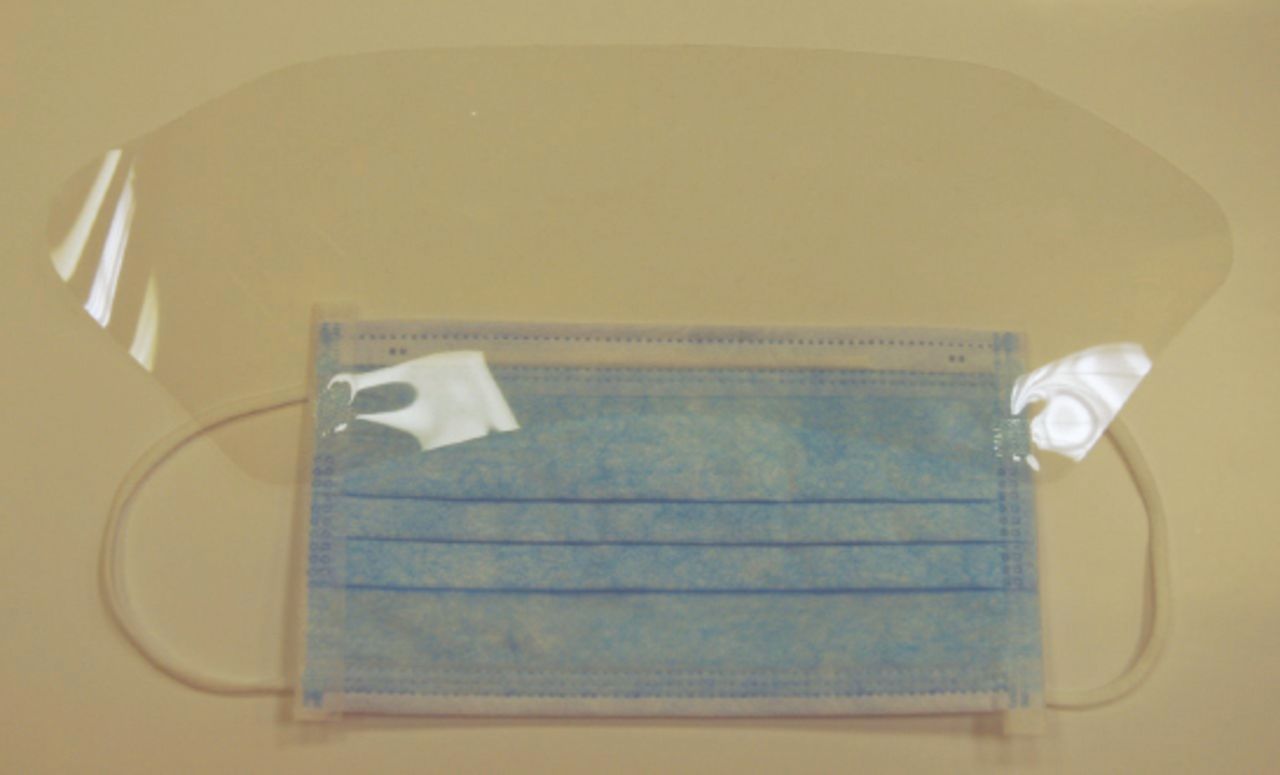
Contaminated air can be managed by improving dental clinic ventilation and/or by disinfecting the air. An ideal airflow pattern combined with a minimum of 3 air changes per hour has been recommended for dental clinics.49-51 Moreover, although its use in dental clinics is unconfirmed, ultraviolet germicidal irradiation may be installed and is effective against fungi, viruses, and bacteria, namely, tubercle bacilli and anthrax.38,49 On the other hand, measures of improving personal protection include washing hands frequently before and after treatment, using disposable barriers, dispensing instruments and materials just before treatment (thereby preventing particles from settling on the surfaces), and sterilizing soiled instruments.38 After each patient visit, surfaces may be disinfected using hospital-grade disinfectants, which are effective against coronavirus.52 Personal protective equipment, such as gowns, hair covers, masks, gloves, shielded face masks and shoe covers, should be used as appropriate.51 A higher level of respiratory protection should be considered, especially with aerosol-generating procedures. The use of N95 respirators offers a certain level of protection against the airborne transmission of SARS-CoV, although the exact level of protection offered by these respirators for individuals may vary.53 Examples of personal protective equipment such as face mask with plastic shield (Figure 1) and N95 respirator (Figure 2). These respirators may also provide some level of protection against MERS-CoV and may be utilized instead of surgical masks in dental clinics. Apart from these measures, vaccination of pediatric dentists and staffs working in pediatric dental clinics against measles, mumps, varicella and rubella may be necessary for their protection from these viral infections.54

Figure 1
  • Download figure
  • Open in new tab
  • Download powerpoint
Figure 1

Face mask with plastic shield.
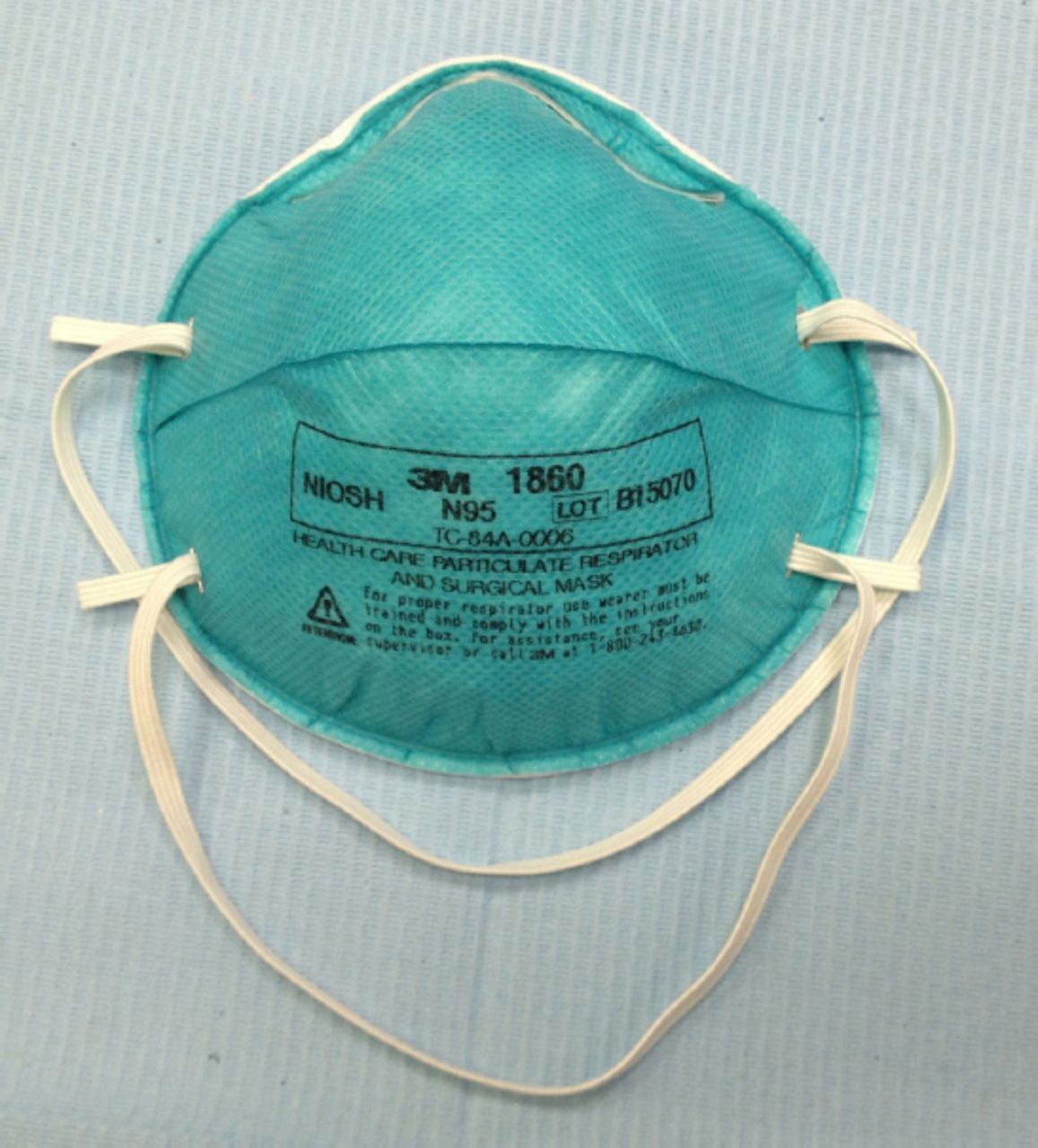
Figure 2
  • Download figure
  • Open in new tab
  • Download powerpoint
Figure 2

The N95 respirator.

In conclusions, considering the unique work environment of dentists, which involves close patient contact and aerosol production, the risk of MERS-CoV transmission from an infected patient is high. Children are also prone to MERS-CoV infection. As the number of MERS-CoV cases may increase in future, pediatric dentists should be well informed and educated about not only the signs and symptoms of the condition but also how to follow stringent infection control measures in these cases.

Acknowledgment

The author would like to extend his sincere gratitude to Prof. Amjad Wyne, Department of Pediatric Dentistry and Orthodontics, College of Dentistry, King Saud University, Riyadh, Saudi Arabia for critically evaluating this review.

Footnotes

  • Disclosure. Authors have no conflict of interest, and the work was not supported or funded by any drug company.

  • Copyright: © Saudi Medical Journal

This is an open-access article distributed under the terms of the Creative Commons Attribution-Noncommercial-Share Alike 3.0 Unported, which permits unrestricted use, distribution, and reproduction in any medium, provided the original work is properly cited.

References

  1. ↵
    1. Memish ZA,
    2. Al-Tawfiq JA,
    3. Assiri A,
    4. AlRabiah FA,
    5. Al Hajjar S,
    6. Albarrak A,
    7. et al.
    (2014) Middle East respiratory syndrome coronavirus disease in children. Pediatr Infect Dis J 33:904–906.
    OpenUrlCrossRefPubMed
  2. ↵
    1. Zaki AM,
    2. van Boheemen S,
    3. Bestebroer TM,
    4. Osterhaus AD,
    5. Fouchier RA
    (2012) Isolation of a novel coronavirus from a man with pneumonia in Saudi Arabia. N Engl J Med 367:1814–1820.
    OpenUrlCrossRefPubMed
  3. ↵
    1. Assiri A,
    2. Al-Tawfiq JA,
    3. Al-Rabeeah AA,
    4. Al-Rabiah FA,
    5. Al-Hajjar S,
    6. Al-Barrak A,
    7. et al.
    (2013) Epidemiological, demographic, and clinical characteristics of 47 cases of Middle East respiratory syndrome coronavirus disease from Saudi Arabia: a descriptive study. Lancet Infect Dis 13:752–761.
    OpenUrlCrossRefPubMed
  4. ↵
    1. Assiri A,
    2. McGeer A,
    3. Perl TM,
    4. Price CS,
    5. Al Rabeeah AA,
    6. Cummings DA,
    7. et al.
    (2013) Hospital outbreak of Middle East respiratory syndrome coronavirus. N Engl J Med 369:407–416.
    OpenUrlCrossRefPubMed
    1. De Groot RJ,
    2. Baker SC,
    3. Baric RS,
    4. Brown CS,
    5. Drosten C,
    6. Enjuanes L,
    7. et al.
    (2013) Middle East respiratory syndrome coronavirus (MERS-CoV): announcement of the Coronavirus Study Group. J Virol 87:7790–7792.
    OpenUrlFREE Full Text
  5. ↵
    1. Arabi YM,
    2. Arifi AA,
    3. Balkhy HH,
    4. Najm H,
    5. Aldawood AS,
    6. Ghabashi A,
    7. et al.
    (2014) Clinical course and outcomes of critically ill patients with Middle East respiratory syndrome coronavirus infection. Ann Intern Med 160:389–397.
    OpenUrlCrossRefPubMed
  6. ↵
    1. World Health Organization
    Middle East respiratory syndrome coronavirus (MERS-CoV) Update 2016, Available at http://www.who.int/csr/don/archive/disease/coronavirus_infections/en/. [Accessed February 2, 2016].
  7. ↵
    1. World Health Organization
    Middle East respiratory syndrome coronavirus (MERS-CoV) summary and literature update-as of 2014, Available at http://www.who.int/csr/disease/coronavirus_infections/MERS_CoV_Update_09_May_2014.pdf?ua=1. [Accessed 2016 February 2].
  8. ↵
    1. Cunha CB,
    2. Opal SM
    (2014) Middle East respiratory syndrome (MERS): a new zoonotic viral pneumonia. Virulence 5:650–654.
    OpenUrlCrossRefPubMed
  9. ↵
    1. WHO MERS-Cov Research Group
    (2013) State of knowledge and data gaps of Middle East respiratory syndrome coronavirus (MERS-CoV) in humans. PLOS Curr, 5.
  10. ↵
    1. Oboho IK,
    2. Tomczyk SM,
    3. Al-Asmari AM,
    4. Banjar AA,
    5. Al-Mugti H,
    6. Aloraini MS,
    7. et al.
    (2015) 2014 MERS-CoV outbreak in Jeddah--a link to health care facilities. N Engl J Med 372:846–854.
    OpenUrlCrossRefPubMed
  11. ↵
    1. Gardner LM,
    2. Rey D,
    3. Heywood AE,
    4. Toms R,
    5. Wood J,
    6. Travis Waller S,
    7. et al.
    (2014) A scenario-based evaluation of the Middle East respiratory syndrome coronavirus and the Hajj. Risk Anal 34:1391–1400.
    OpenUrlCrossRefPubMed
  12. ↵
    1. Lu G,
    2. Hu Y,
    3. Wang Q,
    4. Qi J,
    5. Gao F,
    6. Li Y,
    7. et al.
    (2013) Molecular basis of binding between novel human coronavirus MERS-CoV and its receptor CD26. Nature 500:227–231.
    OpenUrlCrossRefPubMed
  13. ↵
    1. Azhar EI,
    2. Hashem AM,
    3. El-Kafrawy SA,
    4. Sohrab SS,
    5. Aburizaiza AS,
    6. Farraj SA,
    7. et al.
    (2014) Detection of the Middle East respiratory syndrome coronavirus genome in an air sample originating from a camel barn owned by an infected patient. MBio 5:e01450–e01414.
    OpenUrlCrossRefPubMed
  14. ↵
    1. Hijawi B,
    2. Abdallat M,
    3. Sayaydeh A,
    4. Alqasrawi S,
    5. Haddadin A,
    6. Jaarour N,
    7. et al.
    (2013) Novel coronavirus infections in Jordan, April 2012: epidemiological findings from a retrospective investigation. East Mediterr Health J 19(Suppl 1):S12–S18.
    OpenUrlCrossRefPubMed
  15. ↵
    1. Memish ZA,
    2. Zumla AI,
    3. Assiri A
    (2013) Middle East respiratory syndrome coronavirus infections in health care workers. N Engl J Med 369:884–886.
    OpenUrlCrossRefPubMed
  16. ↵
    1. Thabet F,
    2. Chehab M,
    3. Bafaqih H,
    4. Al Mohaimeed S
    (2015) Middle East respiratory syndrome coronavirus in children. Saudi Med J 36:484–486.
    OpenUrlAbstract/FREE Full Text
  17. ↵
    1. Tang JW,
    2. Li Y,
    3. Eames I,
    4. Chan PK,
    5. Ridgway GL
    (2006) Factors involved in the aerosol transmission of infection and control of ventilation in healthcare premises. J Hosp Infect 64:100–114.
    OpenUrlCrossRefPubMed
  18. ↵
    1. Harrel SK,
    2. Molinari J
    (2004) Aerosols and splatter in dentistry: a brief review of the literature and infection control implications. J Am Dent Assoc 135:429–437.
    OpenUrlAbstract/FREE Full Text
  19. ↵
    1. Memish ZA,
    2. Al-Tawfiq JA,
    3. Makhdoom HQ,
    4. Al-Rabeeah AA,
    5. Assiri A,
    6. Alhakeem RF,
    7. et al.
    (2014) Screening for Middle East respiratory syndrome coronavirus infection in hospital patients and their healthcare worker and family contacts: a prospective descriptive study. Clin Microbiol Infect 20:469–474.
    OpenUrlCrossRefPubMed
  20. ↵
    1. Omrani AS,
    2. Matin MA,
    3. Haddad Q,
    4. Al-Nakhli D,
    5. Memish ZA,
    6. Albarrak AM
    (2013) A family cluster of Middle East respiratory syndrome Coronavirus infections related to a likely unrecognized asymptomatic or mild case. Int J Infect Dis 17:e668–e672.
    OpenUrlCrossRefPubMed
  21. ↵
    1. Van Doremalen N,
    2. Bushmaker T,
    3. Munster VJ
    (2013) Stability of Middle East respiratory syndrome coronavirus (MERS-CoV) under different environmental conditions. Euro Surveill 18:1–4.
    OpenUrlPubMed
  22. ↵
    1. Principi N,
    2. Bosis S,
    3. Esposito S
    (2010) Effects of Coronavirus infections in children. Emerg Infect Dis 16:183–188.
    OpenUrlCrossRefPubMed
  23. ↵
    1. Hurst CJ,
    2. Crawford RL,
    3. Garland KL,
    4. Lipson DA,
    5. Mills AL,
    6. Stetzenbach LD
    1. Tanner RS
    (2007) in Manual of environmental microbiology, Cultivation of bacteria and fungi, eds Hurst CJ, Crawford RL, Garland KL, Lipson DA, Mills AL, Stetzenbach LD (ASM Press, Washington (DC)), 3rd ed.
  24. ↵
    1. Leggat PA,
    2. Kedjarune U,
    3. Smith DR
    (2007) Occupational Health problems in modern dentistry: a review. Ind Health 45:611–621.
    OpenUrlCrossRefPubMed
  25. ↵
    1. Barnes JB,
    2. Harrel SK,
    3. Rivera-Hidalgo F
    (1998) Blood contamination of the aerosols produced by in vivo use of ultrasonic scalers. J Periodontol 69:434–438.
    OpenUrlPubMed
  26. ↵
    1. Harrel SK,
    2. Barnes JB,
    3. Rivera-Hidalgo F
    (1998) Aerosol and splatter contamination from the operative site during ultrasonic scaling. J Am Dent Assoc 129:1241–1249.
    OpenUrlAbstract/FREE Full Text
  27. ↵
    1. Miller RL
    (1995) Characteristics of blood-containing aerosols generated by common powered dental instruments. Am Ind Hyg Assoc J 56:670–676.
    OpenUrlCrossRefPubMed
  28. ↵
    1. Dutil S,
    2. Meriaux A,
    3. de Latremoille MC,
    4. Lazure L,
    5. Barbeau J,
    6. Duchaine C
    (2009) Measurement of airborne bacteria and endotoxin generated during dental cleaning. J Occup Environ Hyg 6:121–130.
    OpenUrlCrossRefPubMed
  29. ↵
    1. Checchi L,
    2. Montevecchi M,
    3. Moreschi A,
    4. Graziosi F,
    5. Taddei P,
    6. Violante FS
    (2005) Efficacy of three face masks in preventing inhalation of airborne contaminants in dental practice. J Am Dent Assoc 136:877–882.
    OpenUrlAbstract/FREE Full Text
  30. ↵
    1. Perry JL,
    2. Pearson RD,
    3. Jagger J
    (2006) Infected health care workers and patient safety: a double standard. Am J Infect Control 34:313–319.
    OpenUrlCrossRefPubMed
  31. ↵
    1. Pankhurst CL
    (2003) Risk assessment of dental unit waterline contamination. Prim Dent Care 10:5–10.
    OpenUrlCrossRefPubMed
  32. ↵
    1. Fiore AE,
    2. Butler JC,
    3. Emori TG,
    4. Gaynes RP
    (1999) A survey of methods used to detect nosocomial legionellosis among participants in the National Nosocomial Infections Surveillance System. Infect Control Hosp Epidemiol 20:416–420.
    OpenUrl
  33. ↵
    1. Atlas RM,
    2. Williams JF,
    3. Huntingdon MK
    (1995) Legionella contamination of dental-unit water. Appl Environ Microbiol 61:1208–1213.
    OpenUrlAbstract/FREE Full Text
  34. ↵
    1. Scully C,
    2. Samaranayake LP
    (2015) Emerging and changing viral diseases in the new millennium. Oral Dis 22:171–179.
    OpenUrl
  35. ↵
    1. Banik GR,
    2. Khandaker G,
    3. Rashid H
    (2015) Middle East respiratory syndrome coronavirus “MERS-CoV”: current knowledge gaps. Paediatr Respir Rev 16:197–202.
    OpenUrlCrossRefPubMed
  36. ↵
    1. Eckerle I,
    2. Müller MA,
    3. Kallies S,
    4. Gotthardt DN,
    5. Drosten C
    (2013) In-vitro renal epithelial cell infection reveals a viral kidney tropism as a potential mechanism for acute renal failure during Middle East respiratory syndrome (MERS) Coronavirus infection. Virol J 10:1–5.
    OpenUrlCrossRefPubMed
  37. ↵
    1. Li RW,
    2. Leung KW,
    3. Sun FC,
    4. Samaranayake LP
    (2004) Severe acute respiratory syndrome (SARS) and the GDP. part II: Implications for GDPs. Br Dent J 197:130–134.
    OpenUrlCrossRefPubMed
  38. ↵
    1. Center for Disease Control and Prevention
    (2003) Guidelines for infection control in dental health-Care settings, Available at http://www.cdc.gov/oralhealth/infectioncontrol/guidelines/. [Accessed 2016 February 27].
  39. ↵
    1. Whaites E
    (1996) Essentials of dental radiography and radiography (Churchill-Livingstone, Edinburgh), 2nd ed, pp 107–113.
  40. ↵
    1. Ferguson DB
    (1999) Oral bioscience (Churchill-Livingstone, London (UK)), p 278.
  41. ↵
    1. Robb ND,
    2. Crothers AJ
    (1996) Sedation in dentistry. part 2: Management of the gagging patient. Dent Update 23:182–186.
    OpenUrlPubMed
  42. ↵
    1. Molinari GE
    (2004) Infection control issues related to pediatric dentistry. Compend Contin Educ Dent 25:537–540.
    OpenUrlPubMed
  43. ↵
    1. Klyn SL,
    2. Cummings DE,
    3. Richardson BW,
    4. Davis RD
    (2001) Reduction of bacteria-containing spray produced during ultrasonic scaling. Gen Dent 49:648–652.
    OpenUrlPubMed
  44. ↵
    1. Teanpaisan R,
    2. Taeporamaysamai M,
    3. Rattanachone P,
    4. Poldoung N,
    5. Srisintorn S
    (2001) The usefulness of the modified extra-oral vacuum aspirator (EOVA) from household vacuum cleaner in reducing bacteria in dental aerosols. Int Dent J 51:413–416.
    OpenUrlCrossRefPubMed
  45. ↵
    1. Beeley JA,
    2. Yip HK,
    3. Stevenson AG
    (2000) Chemochemical caries removal: a review of the techniques and latest developments. Br Dent J 188:427–430.
    OpenUrlPubMed
  46. ↵
    1. Cole BO,
    2. Welbury RR
    (2000) The atraumatic restorative treatment (ART) technique: does it have a place in everyday practice? Dent Update 27:118–120.
    OpenUrlPubMed
  47. ↵
    1. Frencken JE,
    2. Pilot T,
    3. Songpaisan Y,
    4. Phantumvanit P
    (1996) Atraumatic restorative treatment (ART): rationale, technique, and development. J Public Health Dent 56:135–140.
    OpenUrlCrossRefPubMed
  48. ↵
    1. Brickner PW,
    2. Vincent RL,
    3. First M,
    4. Nardell E,
    5. Murray M,
    6. Kaufman W
    (2003) The application of ultraviolet germicidal irradiation to control transmission of airborne disease: bioterrorism countermeasure. Public Health Rep 118:99–114.
    OpenUrlCrossRef
    1. Samaranayake LP,
    2. Scheutz F,
    3. Cottone J
    (1991) Infection control for the dental team (Munksgaard, Copenhagen), p 112.
  49. ↵
    1. Woods RG,
    2. Amerena V,
    3. David P,
    4. Fan PL,
    5. Heydt H,
    6. Marianos D
    (1997) Additional precautions for tuberculosis and a self-assessment checklist. FDI World 6:10–17.
    OpenUrlPubMed
  50. ↵
    1. Wenzel RP,
    2. Edmond MB
    (2003) Managing SARS amidst uncertainty. N Engl J Med 348:1947–1948.
    OpenUrlCrossRefPubMed
  51. ↵
    1. Christian MD,
    2. Loutfy M,
    3. McDonald LC,
    4. Martinez KF,
    5. Ofner M,
    6. Wong T,
    7. et al.
    (2004) Possible SARS coronavirus transmission during cardiopulmonary resuscitation. Emerg Infect Dis 10:287–293.
    OpenUrlPubMed
  52. ↵
    1. Fujiwara T
    (2010) Safety management and infection control in pediatric dentistry. Jpn J Pediatr Dent 48:367–373.
    OpenUrl
PreviousNext
Back to top

In this issue

Saudi Medical Journal: 38 (4)
Saudi Medical Journal
Vol. 38, Issue 4
1 Apr 2017
  • Table of Contents
  • Cover (PDF)
  • Index by author
Print
Download PDF
Email Article

Thank you for your interest in spreading the word on Saudi Medical Journal.

NOTE: We only request your email address so that the person you are recommending the page to knows that you wanted them to see it, and that it is not junk mail. We do not capture any email address.

Enter multiple addresses on separate lines or separate them with commas.
Middle East respiratory syndrome in children
(Your Name) has sent you a message from Saudi Medical Journal
(Your Name) thought you would like to see the Saudi Medical Journal web site.
Citation Tools
Middle East respiratory syndrome in children
Fares S. Al-Sehaibany
Saudi Medical Journal Apr 2017, 38 (4) 339-343; DOI: 10.15537/smj.2017.4.15777

Citation Manager Formats

  • BibTeX
  • Bookends
  • EasyBib
  • EndNote (tagged)
  • EndNote 8 (xml)
  • Medlars
  • Mendeley
  • Papers
  • RefWorks Tagged
  • Ref Manager
  • RIS
  • Zotero
Share
Middle East respiratory syndrome in children
Fares S. Al-Sehaibany
Saudi Medical Journal Apr 2017, 38 (4) 339-343; DOI: 10.15537/smj.2017.4.15777
Twitter logo Facebook logo Mendeley logo
  • Tweet Widget
  • Facebook Like
  • Google Plus One
Bookmark this article

Jump to section

  • Article
    • Abstract
    • Acknowledgment
    • Footnotes
    • References
  • Figures & Data
  • eLetters
  • References
  • Info & Metrics
  • PDF

Related Articles

  • No related articles found.
  • PubMed
  • Google Scholar

Cited By...

  • No citing articles found.
  • Google Scholar

More in this TOC Section

  • Harnessing artificial intelligence for infection control and prevention in hospitals
  • Effects of antidiabetic drugs on the level of serum uric acid in patients who have type 2 diabetes
  • The future of personalized medicine in Saudi Arabia
Show more Review Article

Similar Articles

CONTENT

  • home

JOURNAL

  • home

AUTHORS

  • home
Saudi Medical Journal

© 2025 Saudi Medical Journal Saudi Medical Journal is copyright under the Berne Convention and the International Copyright Convention.  Saudi Medical Journal is an Open Access journal and articles published are distributed under the terms of the Creative Commons Attribution-NonCommercial License (CC BY-NC). Readers may copy, distribute, and display the work for non-commercial purposes with the proper citation of the original work. Electronic ISSN 1658-3175. Print ISSN 0379-5284.

Powered by HighWire