
Main menu

  • Home
  • Content
    • Latest
    • Archive
    • home
  • Info for
    • Authors
    • Reviewers
    • Subscribers
    • Institutions
    • Advertisers
    • Join SMJ
  • About Us
    • About Us
    • Editorial Office
    • Editorial Board
  • More
    • Advertising
    • Alerts
    • Feedback
    • Folders
    • Help
  • Other Publications
    • NeuroSciences Journal

User menu

  • My alerts
  • Log in

Search

  • Advanced search
Saudi Medical Journal
  • Other Publications
    • NeuroSciences Journal
  • My alerts
  • Log in
Saudi Medical Journal

Advanced Search

  • Home
  • Content
    • Latest
    • Archive
    • home
  • Info for
    • Authors
    • Reviewers
    • Subscribers
    • Institutions
    • Advertisers
    • Join SMJ
  • About Us
    • About Us
    • Editorial Office
    • Editorial Board
  • More
    • Advertising
    • Alerts
    • Feedback
    • Folders
    • Help
  • Follow psmmc on Twitter
  • Visit psmmc on Facebook
  • RSS
Review ArticleReview Article
Open Access

Review of surgical resection and reconstruction in head and neck cancer

Traditional versus current concepts

Afnan F. Alfouzan
Saudi Medical Journal October 2018, 39 (10) 971-980; DOI: https://doi.org/10.15537/smj.2018.10.22887
Afnan F. Alfouzan
From the Department of Prosthodontics, College of Dentistry, King Saud University, Riyadh, Kingdom of Saudi Arabia
BDS, MS
  • Find this author on Google Scholar
  • Find this author on PubMed
  • Search for this author on this site
  • For correspondence: [email protected] [email protected]
  • Article
  • Figures & Data
  • eLetters
  • Info & Metrics
  • References
  • PDF
Loading

Abstract

This review summarizes the development of head and neck cancer resection and reconstruction. The developments in the treatment of cancer patients are reflected in their surgical outcomes, in addition to functional and aesthetic improvements. New technologies, such as surgical simulation and planning, minimally invasive surgery, and microsurgery have been added to the field to improve surgical resection of the tumor and reconstruction. The field is still growing to optimize the management of head and neck cancer.

Head and neck cancer represent the sixth most frequent malignancies. Worldwide more than 500,000 new cases are diagnosed annually, along with 300,000 deaths. Head and neck cancer predominantly affects men, with a male: female ratio of up to 10:1. There is also an increased risk of developing this type of cancer.1,2 Tobacco smoking, alcohol consumption and human papillomavirus (HPV) infection are the principal risk factors for the development of head and neck cancers.3-5 In the United States, more than 60% of oropharyngeal cancers are associated with HPV infection. Head and neck cancers that are associated with HPV infection usually affect nonsmoking white male patients who are younger than patients with unrelated HPV cancers and who have a higher socioeconomic status and a history of multiple sexual partners.3,6 Most head and neck cancers (more than 90%) are squamous cell carcinomas originating from the epithelium, which are the most common cancer in males and the third most common cancer in females.6,7 This type of cancer may be found on the lips, oral cavity, pharynx, larynx, sinuses, nasal cavity and the salivary glands.8 More than two-thirds of patients present with regional lymph node involvement.9 Early diagnosis of this cancer leads to higher survival rates; the 5-year survival rate drops from 83% to 37% when the diagnosis is made late. Squamous cell carcinoma associated with HPV presents with higher survival rates at 3- and 5-year intervals when compared with squamous cell carcinoma associated with smoking.1

Surgery, chemotherapy, and radiotherapy are the common treatment modalities for head and neck cancer. The purpose of this review was to summarizes the development of surgical resection of head and neck cancer with the reconstruction of the resected human part.

Review

Oral cancer was seldom reported in medical reports before the introduction of tobacco in the 16th century.10 In 1650, Hayes Martin wrote the first report detailing oral cancer treatment.11 Then, in 1664, Marchetti described the first tongue resection procedure as a treatment for cancer.12 Then, in 1712, Joseph de la Charriere recommended cauterization as a treatment for cancer patients.13 After that, the first cancer hospital was established in France in 1740.14 In 1790, the awareness of the importance of lymph node metastasis appeared in the literature.14 In 1860, the dynamics of lymphatic spread were studied.15 In 1846, successful removal of cervical cancer was performed by Dr. Warren, and this was the beginning of major surgical procedures.12 Advances in surgical procedures continued in the 20th century. These advances in surgery and medicine occurred during the first half of the century due to the 2 world wars.12

Surgical resection is the primary treatment modality for oral cancer. Tumor resection involves removal of the lesion with a wide margin of normal tissue; involved cervical lymph nodes also need to be removed.16-18

Head and neck cancer resection

The radical neck dissection for head and neck cancer was first descried in 1906 by Dr. George Crile from the Cleveland Clinic in Ohio.19 His procedure involved the removal of all the lymphatic structures between the mandible and the clavicle. After that, Dr. Hayes reviewed the procedure of Dr. Crill by performing 1450 neck dissections in 1951 at the Memorial Hospital in New York City. His method started with a midline bisection of the lower lip, then by segmental mandibulectomy and in-continuity radical neck dissection.20

Forty percent of head and neck cancer patients present with cervical lymph node metastasis. Cervical lymphadenectomy is a procedure that involves removal of groups of lymph nodes from the neck to decrease disease burden.21,22 Neck dissection can be therapeutic or elective, depending on the presence of clinical or radiographic lymphadenopathy. Elective neck dissection is aimed at ruling out the possibility of cancer metastasis, which may eliminate the need for adjuvant therapy after surgery. Shah23 stratified cervical lymph nodes into 7 group levels; submental (I), submandibular and upper jugular (IIa, IIb), middle jugular (III), lower jugular (IV), posterior triangle (Va, Vb), central compartment (VI), and superior mediastinal (VII).21,23-25

Currently, the classical radical neck dissection is seldom performed for the head and neck cancer patients, where all the lymph nodes in the anterior and posterior triangles, the submandibular salivary gland, and the tail of parotid salivary gland, the spinal accessory nerve, the internal jugular vein and the sternocleidomastoid muscles will be removed. It also involved the removal of the spinal accessory nerve, the internal jugular vein and the sternocleidomastoid muscles. However, the modified radical neck dissection where involved the removal of the cervical lymph nodes in levels I through V without the removal of 3 aforementioned anatomical structures. Currently, cervical lymph nodes removal with the preservation of non-lymphatic structures is more commonly performed through selective neck dissection (levels I-IV) or supraomohyoid neck dissection (levels I-III).21

Conventional head and neck surgery involving neck dissection is performed via a U-shaped or Y-shaped incision through the neck, leavening a visible scar that may have a psychological impact on the patients.26,27 In 2012, Kim et al28 introduced robotic-assisted surgery for lateral neck dissection for squamous cell carcinoma patients that resulted in a good aesthetic appearance and avoided the long scar that was associated with the conventional open technique without compromising the surgical completeness and oncological outcomes.

Surgical treatment modalities for head and neck cancer require complete tumor excision along with excision of the surrounding margins and are usually performed via a transcervical approach and may require a lip-split mandibulotomy or mandibulectomy.29,30

Classical open cancer resection or minimally invasive resection can be performed depending on the specific cancer anatomy and characteristics. Classical open surgery is less favorable in young patients due to the aesthetic impairments caused by the visible scars that this procedure may produce.29,31

Minimally invasive surgeries

In the late 20th century, minimally invasive surgeries were developed. Trans-oral endoscopic head and neck surgery is a minimally invasive surgery for the oropharynx through a trans-oral route that is performed either with conventional instruments or by laser resection.17,32 Trans-oral laser microscopy and trans-oral robotic surgery are the specific approaches used. These can be performed without external skin incisions, so there is no need to gain access through a mandibulotomy or transmandibular approach, which significantly reduces the postoperative morbidity. The surgical time may be shorter than that of the transcervical approach. These techniques provide a highly magnified view of the tumor, which permits confident resection of the tumor.33,34

Trans-oral laser microsurgery (TLM)

The first laser microlaryngoscopy procedure was described in 1971.35 In 2001, Steiner described this approach for more widespread use.36 Surgical resection is performed through direct laryngoscopy using a carbon dioxide (CO2) laser. The laser beam is transferred through the endoscope and is absorbed by water at the tissue-laser interface, which then coverts it into thermal energy and allows precision tissue cutting. This type of resection is performed under a binocular microscope that allows close visualization of the tumor, coupled with flexible microsurgical instruments that enable a better approach to the resected area. This technique requires the surgeons’ understanding of the complete anatomy and extent of the tumor and the surrounding structures.32

Trans-oral robotic surgery (TORS)

In 2009, the United States Food and Drug Administration approved the use of the da Vinci surgical system.37,38 This involves real surgical procedures performed in a virtual environment, where the surgeon sits at a remote console and can manipulate an endoscope and 2 additional instruments that are placed in the patient’s mouth. The surgeon can control every movement of robot’s instruments. The robot’s arms carry interchangeable working instruments, such as grasping forceps and an electrocautery tool. The patient’s mouth is kept open with the help of a suitable oral retractor, and an endoscope or camera is introduced through the mouth. The flexibility of the robotic arms allows suturing of structures in low visibility areas, which is not possible with the standard techniques. Countertraction and suction performed by a bedside assistant are needed throughout the surgery.30

Patients with retrognathia, class II dental occlusion, limited cervical extension and prominent maxillary dentition have limited oropharyngeal access, thus considered not suitable for trans-oral robotic surgery. So, tumors arising from the tonsillar fossa, the lateral pharyngeal wall, the glossopharyngeal sulcus, and the lateral tongue base can be resected with the assistance of trans-oral robotic surgery due to their suitable position for this technique.30

Most surgical resections of head and neck lesions can compromise the patient’s appearance as well as certain functions; thus, reconstruction of the defect area is necessary. Jaw reconstruction restores the continuity of the jaw, separates the oral and nasal cavities, and restores a stable base for the oral cavity and the structures to which muscles are attached, leading to improved mastication, speech, swallowing, tongue function, breathing and definition of the lower third of the face, which affects aesthetics.39-41 In addition, surgical reconstructions of the jaws produces proper phonation and deglutition functions compared with reconstruction with prosthetics.42,43 In 2010, Moreno et al43 compared microvascular free flap reconstruction and palatal obturator reconstruction for maxillectomy defects and concluded that better functional outcomes were associated with free flap reconstruction. Techniques for the reconstructing the defect area vary from the simplest primary closure to the more complex free vascularized flaps.44

History of head and neck reconstruction

Bardenheuer first reported a non-vascularized cortical bone graft in 1892 that used the mandible itself to rebuild its defect. It was used in World War I, harvesting bone from the rib and the tibia. In particular, cancellous bone in a metallic tray or block graft was used.45

Before the 1950s, resected cancer was left without reconstruction. Reconstruction was performed only when no early local recurrence developed.45 In the late 1950s, the first free flap procedure, which involved the removal of tissue from the donor site and transplantation in another site of the body with anastomosis of the vessels, was performed.46,47

Advances in head and neck reconstruction emerged in the 1960s with the introduction of the myocutaneous pedicle flap, and afterward, the description of the pectoralis major myocutaneous flap was described in 1979.48 The transferring and reconstruction of the defects with the flap provided the patient with healthy, vascularized tissue and recovered the resected areas. In the 1990s, free flap reconstruction was the dominant technique for cancer defect reconstruction.45

The traditional method of jaw reconstruction is challenging, particularly with regard to bone graft shaping. Malposition of the bone graft can negatively affect the facial symmetry, appearance, support, occlusal function, masticatory movement and dental rehabilitation. Manual remolding of the graft can prolong the period of ischemia.49

Digital technologies and surgical simulation provide 3-dimensional (3D) virtual models of the resected and grafted areas based on preoperative high-resolution computed tomography (CT) data with specialized printers using rapid prototype modeling technology.49 Three-dimensional printing was first described by Hideo Kodama in 1981 when he started manufacturing 3D plastic models.50 In 1990, Mankovich et al51 described the rapid prototyping technique for medical use, transferring the engineering method to the surgical field. Surgical planning was improved, which facilitated resection, flap harvesting, and graft positioning.

Through surgical planning and simulation, a 3D printed surgical template can be fabricated to guide the procedure of tumor resection, graft harvesting, and shaping and positioning of the graft in the defect area.52,53 It depends on the adaptation of certain anatomical landmarks, and then osteotomies are performed through the metal cutting slots on the surgical template.54

Surgical simulation and planning lead to exact planning and good communication between the resection and reconstruction team, which results in increased accuracy, improved bone to bone contact, decreased operating room time, lower surgeon stress, reduced ischemia time in the microvascular free flap reconstruction and enhanced treatment outcomes.55-67

After the resection of head and neck cancer, more than 50% of the resections require reconstructions of the defect and involve bony and/or soft tissue reconstruction.7 Maxillomandibular resection may involves only an edge (marginal) or complete segment (segmental) reconstruction. A marginal mandibulectomy can be reconstructed with local flaps or skin graft, whereas a segmental mandibulectomy requires an osteocutaneous flap.16,68

Soft tissue reconstruction

Soft tissue reconstructions are usually based on the defect size after the resection of cancer. Primary closure healing or healing by second intention for small defects and skin grafts for larger defects are the simplest options for reconstruction. For larger defects, full or partial thickness skin grafts can be utilized, using different flap methods depending on the defect site and size.16

Several soft tissue flaps can be harvested from several sites of the patient’s body, like the radial forearm, lateral arm, ulnar forearm, anterolateral or lateral thigh, latissimus, dorsi, jejunum, omentum, rectus abdominis, scapula, and temporal parietal fascia.47,69,70 Some axial regional pedicle flap may be preferred because microvascular anastomosis is not necessary and shorter harvesting times are needed for this technique compared with the free flap procedure, such as latissimus dorsi and pectoralis major myocutaneous flaps; however, but the bulkiness and inflexibility of these flaps makes them inferior to the vascularized radial free forearm flap.71

Myocutaneous trapezius flap

The myocutaneous trapezius flap is a thin and large pedicle flap that has been used for intraoral defects and posterior occipital defects. It has a wide arc of rotation, which make it an option for head and neck reconstructions.72,73 In 1980, Baek et al74 reported the use of the trapezius flap for a facial defect pedicle flap reconstruction. It is an alternative reconstructive flap that is suitable for patients who cannot undergo microvascular free flap reconstruction. It can also be used for primary or salvage reconstruction surgery.75

Vascularized radial free forearm flap

In 1983, Soutar et al76 reported the use of the radial free forearm flap for intraoral reconstructions to replace the oral mucosa. This fasciocutaneous flap is now a popular option for soft tissue reconstructions. Because it is usually a mobile, thin, and pliable large paddle of skin without added bulkiness, it can be used for a variety of external skin and intraoral defects, such as defects of the palate, the floor of the mouth and the tongue. The long pedicle flap (up to 20 centimeters) with large caliber vessels facilitates surgery without repositioning of the patient during surgery, which decreases the surgical time.77-79 Full thickness defects of the cheeks or lips can be reconstructed using a folded fasciocutaneous radial forearm free flap.80

Hard tissue reconstruction

Osteocutaneous flaps consist of soft and hard tissue, and bone grafts can be harvested from different donor sites of a patient’s body, such as the fibula, iliac crest, radius and scapula.81-87

Scapular flap

In 1986, Swartz et al88 first reported the use of an osteocutaneous scapular flap for mandibular and maxillary reconstructions. The bone is harvested from the lateral border of the scapula with scapular and parascapular skin paddles. The flap is harvested in the lateral decubitus position and remains vascularized by its pedicle until reconstruction is performed to prevent flap ischemia.89 A 2 team approach is difficult because of the need for intraoperative patient positioning.90 While a limited amount bone of low quality can be harvested and multiple osteotomies cannot be tolerated, this technique does provide large amount of soft tissue. Therefore, this method is an option for mandibular reconstructions in cases were a fibular flap is unsuitable or in cases that require extensive soft tissue reconstruction.91,92

Vascularized fibular free flap

The vascularized fibular free flap is the first option and is the gold standard for reconstructions of the mandible or maxilla due to the shape and quality of the bone, which is suitable for contouring and allows dental rehabilitation through the placement of dental implants.83,93-98 It was described first by Hidalgo in 1989 for mandibular reconstruction, and its successful depends on the surgeon’s skills, decisions and trial and error.82 The surgical plane for resection and reconstruction is dependent on a diagnosis made with 2D images with clinical measurements that may affect the position and the shape of the graft.99,100

The fibular graft has multiple advantages including the following: a large amount of bone and soft tissue are available for harvesting (approximately 20-26 cm); the bone tolerates multiple osteotomies; the graft can be easily shaped; there is a long and anatomically reliable vessel pedicle with a wide vessel diameter; the technique is associated with low donor site morbidity; and the location of the donor site facilitates a 2 team approach without the need to reposition the patient during the surgery.101-103 The site and the length of the graft depend on the defect size.100 The bicortical structure seems to increase the long-term success of dental implants placed in the graft, along with good primary stability of the implants.93

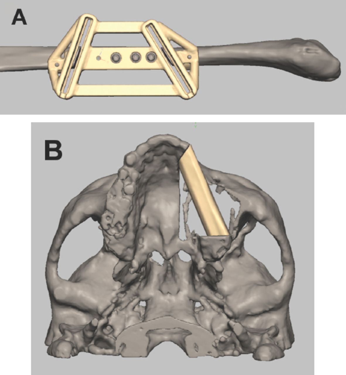
The traditional surgical procedure of transferring the straight fibula into the curved mandible often resulted in imprecise contouring, condylar positioning, and symmetry of the lower third of the face.41,101,104 With advanced surgical technologies, the fibula can be redesigned and reshaped with the help of 3D printed surgical templates, along with the use of osseointegrated dental implants that are placed at the time of the reconstruction (Figures 1A & 1B).63,105

Figure 1
  • Download figure
  • Open in new tab
  • Download powerpoint
Figure 1

Surgical planning and simulation using 3-dimentional printed surgical template for A) fibular cutting guide and B) reconstruction of partial left maxillectomy.

Wang et al41 studied a comparison of virtual planning surgery and conventional surgery for mandibular reconstruction with a vascularized fibular free flap and found that virtual surgical planning produced a more accurate mandibular reconstruction than conventional surgery.

The limitation of the fibula graft is the limited height of the graft (rarely more than 1.5 cm), which is about half of the native mandible, which makes it insufficient to replace the skeletal base and alveolar ridge. Therefore, the iliac crest flap, due to its height and shape, emerged as a good option for bony reconstruction.106

Vascularized iliac crest flap

The iliac crest is commonly used as a source of non-vascularized bone graft.45 In 1979, Taylor first described the use of the vascularized iliac crest flap for mandibular reconstruction. Then, it became widely favored by surgeons due to the large amount of bone volume, rich cancellous blood supply and compact cortex.107-109 Through surgical simulation and planning, the ideal bone graft site inside the iliac crest that is the most similar to the missing bone with regard to the outline and size can be identified thus avoiding the risk of wasting a bone graft.81,110 It is an optimal choice to reconstruct defects related to the inferior orbital rim, the vertical process of the maxilla and the zygomatic arch.111 However, it is not an alternative to the fibular and scapular free flaps due to the difficulty in harvesting the graft and the thin and immobile skin paddle.45

Osteocutaneous radial forearm free flap

In 1979, reconstruction with an osteocutaneous radical forearm free flap was introduced for head and neck reconstruction. This technique is suitable for short bony defects such as defects of the maxilla or the mandible.79 On the other hand, it is not recommended for large head and neck reconstructions due to the limited bone stock and the morbidity of radius harvesting, including the possibility of donor radial fracture.112,113

Pre-bent reconstruction plates

Since the twentieth century, the surgical plates have been used for bone segments stabilization during head and neck reconstruction surgery. Conventionally, surgical plates are available in standard formats and are manually bent by the surgeons during surgery to match the individual bone anatomy.114

The manual plate-bending procedure can be a time intensive and energy consuming technique, particularly for inexperienced surgeons.114 On occasion, surgical plates need to be bent repeatedly, particularly in complicated cases, which may concentrate internal stress within the plate that leads to fatigue under masticatory loading, resulting in complications, such as plate fracture, corrosion, screw loosening and bone resorption.115 To overcome the complications of conventional surgical plates, 3D technologies have facilitated the production of patient-specific pre-bent surgical plates.63,116-119 Pre-bent selective laser 41 sintering (SLS) or milled titanium surgical plates with specific configurations, including the 3D architecture, width, thickness and screw holes, can be individualized for each patient using the patient’s original printed contours.120 This type of plate has threaded screw holes with different angles and can tolerate a greater number of screws than a conventional plate and has a minimum distance between screws of 5.5 mm compared with the 8 mm of a conventional plate.121 In addition, this type of plate can greatly facilitate graft positioning into the resected area, avoiding dental malocclusion due to the accurate contouring of the surgical plate.100,122 Moreover, damage to important structures, such as tooth roots or nerves, can be avoided by determining the locations of each fixation hole.123

Personalized titanium meshes can be used to restore the orbital floor and facial contour during surgical reconstruction after a maxillectomy with orbital floor involvement to avoid displacement of the eyeball and diplopia due to changes in the orbital volume. The titanium mesh is highly biocompatible and can be contoured into the resected areas.49,124,125

Dental rehabilitation

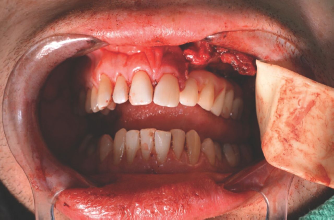
Planning for dental implants and the type of dental prosthesis that is needed to replace missing teeth after a resection is a part of the virtual surgical planning and simulation for maxillary or mandibular resection and the reconstruction procedure. The implants can be placed at the time of jaw reconstruction, which will reduce the time for dental rehabilitation, enhance optimum bone exposure during implant placement and eliminate the need for hyperbaric oxygen after radiotherapy.126 Placing the dental implant within the vascularized bone graft before reconstruction facilitates the correct positioning of the graft within the defects by putting the patient into occlusion using a provisional prosthesis or fitting template for the implants (Figures 2 & 3).53,93

Figure 2
  • Download figure
  • Open in new tab
  • Download powerpoint
Figure 2

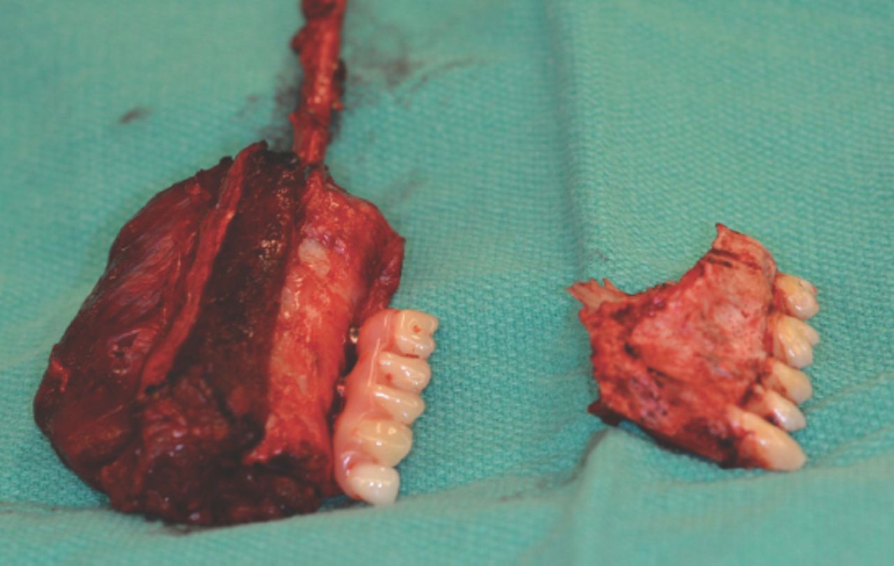
Provisional prosthesis supported by dental implant on fibular graft after partial left maxillectomy.

Figure 3
  • Download figure
  • Open in new tab
  • Download powerpoint
Figure 3

Provisional prosthesis facilitates correct positioning of the graft.

Free flap perfusion assessment and monitoring

The success of the reconstructions depends on the quality of perfusion of the harvested flap and the rapid identification and salvage of a failing flap.127,128 Doppler ultrasonography and angiography have improved surgeons’ abilities to make decisions regarding microvascular reconstructions.129 Laser-assisted fluorescent angiography is now used to monitor perfusion intraoperatively. A perfusion map can be constructed by administering an intravenous injection of indocyanine green (ICG) to the patient, which fluoresces on exposure to a laser light emitted by the machine and is detected by a high-speed imaging system that is sensitive to the ICG wavelength.130 The immediate assessment of vascular anastomosis and venous and arterial flow allows the identification of thrombi and an assessment of the quality of perfusion, which are determinants of early flap necrosis.130 The assessment of tissue oxygenation by near-infrared spectroscopy is one of the advancements in postoperative flap assessment that is a sensitive, specific and non-invasive method.131 Measuring the hemoglobin saturation at the capillary level by visible white light spectroscopy is also an advancement in monitoring. Smartphones can also be used for flap monitoring through infrared thermography cameras.132,133

In the 21st century, new advances in reconstruction of head and neck defects are developing. In 2004, Lendeckel et al134 reconstructed a calvarial defect through the use of autologous stem cells (adipose) combined with the patient’s own cancellous bone and fibrin glue. The resorption of non-vascularized bone grafts or donor site morbidity of vascularized bone flaps during maxillofacial reconstruction can be avoided through the use of stem cells to produce the bone.135 The harvesting of adipose-derived stem cells is easy, where it can be performed by liposuction or lipectomy. The cells can differentiate into different cell lineages, such as adipogenic, osteogenic, chondrogenic, neurogenic, myogenic, cardiomyogenic, angiogenic, tenogenic and periodontogenic cell types.136 After a period of implantation of these cells in a titanium mesh that is seeded with bone marrow stem cells in certain areas of the patient’s muscles, vascularized bone pieces of the size and outline needed to perform reconstruction can be produced. The resultant bone can tolerate dental implants with excellent outcomes.137,138

In conclusion, through the past decades, surgical resection and reconstruction for head and neck cancer have developed significantly. Surgical simulation, 3D printing, microvascular surgery and minimally invasive surgery have improved the surgical outcomes and have led to superior functional and aesthetic outcomes. Undoubtedly, the future will bring more developments in the field to optimize the treatment of head and neck cancer.

Acknowledgment

The author acknowledge the Institute for Reconstructive Sciences in Medicine (iRSM), Alberta, Canada for providing the photos. Also the author acknowledges the American Journal Experts company for the professional editing of the manuscript.

Footnotes

  • Disclosure. Authors have no conflict of interests, and the work was not supported or funded by any drug company.

  • Copyright: © Saudi Medical Journal

This is an open-access article distributed under the terms of the Creative Commons Attribution-Noncommercial-Share Alike 3.0 Unported, which permits unrestricted use, distribution, and reproduction in any medium, provided the original work is properly cited.

References

  1. ↵
    1. Torre LA,
    2. Bray F,
    3. Siegel RL,
    4. Ferlay J,
    5. Lortet-Tieulent J,
    6. Jemal A
    (2015) Global cancer statistics 2012. CA Cancer J Clin 65:87–108.
    OpenUrlCrossRefPubMed
  2. ↵
    1. Vigneswaran N,
    2. Williams MD
    (2014) Epidemiologic trends in head and neck cancer and aids in diagnosis. Oral Maxillofac Surg Clin North Am 26:123–141.
    OpenUrlCrossRef
  3. ↵
    1. Gillison ML,
    2. Chaturvedi AK,
    3. Anderson WF,
    4. Fakhry C
    (2015) Epidemiology of human papillomavirus-positive head and neck squamous cell carcinoma. J Clin Oncol 33:3235–3242.
    OpenUrlAbstract/FREE Full Text
    1. Blot WJ,
    2. McLaughlin JK,
    3. Winn DM,
    4. Austin DF,
    5. Greenberg RS,
    6. Preston-Martin S,
    7. et al.
    (1988) Smoking and drinking in relation to oral and pharyngeal cancer. Cancer Res 48:3282–3287.
    OpenUrlAbstract/FREE Full Text
  4. ↵
    1. Sturgis EM,
    2. Cinciripini PM
    (2007) Trends in head and neck cancer incidence in relation to smoking prevalence: an emerging epidemic of human papillomavirus-associated cancers? Cancer 110:1429–1435.
    OpenUrlCrossRefPubMed
  5. ↵
    1. Chi AC,
    2. Day TA,
    3. Neville BW
    (2015) Oral cavity and oropharyngeal squamous cell carcinoma--an update. CA Cancer J Clin 65:401–421.
    OpenUrlCrossRefPubMed
  6. ↵
    1. Kanazawa T,
    2. Sarukawa S,
    3. Fukushima H,
    4. Takeoda S,
    5. Kusaka G,
    6. Ichimura K
    (2011) Current reconstructive techniques following head and neck cancer resection using microvascular surgery. Ann Vasc Dis 4:189–195.
    OpenUrl
  7. ↵
    1. Vokes EE,
    2. Weichselbaum RR,
    3. Lippman SM,
    4. Hong WK
    (1993) Head and neck cancer. N Engl J Med 328:184–194.
    OpenUrlCrossRefPubMed
  8. ↵
    1. Siegel RL,
    2. Miller KD,
    3. Jemal A
    (2015) Cancer statistics 2015. CA Cancer J Clin 65:5–29.
    OpenUrlCrossRefPubMed
  9. ↵
    1. Nelson WR
    (1987) In search of the first head and neck surgeon. Am J Surg 154:342–346.
    OpenUrlPubMed
  10. ↵
    1. Martin H
    (1951) Richard Wiseman on cancer. Cancer 4:907–912.
    OpenUrlPubMed
  11. ↵
    1. Folz BJ,
    2. Silver CE,
    3. Rinaldo A,
    4. Fagan JJ,
    5. Pratt LW,
    6. Weir N,
    7. et al.
    (2008) An outline of the history of head and neck oncology. Oral Oncol 44:2–9.
    OpenUrlPubMed
  12. ↵
    1. Onuigbo WI
    (1971) False firsts in cancer literature. Oncology 25:163–167.
    OpenUrlPubMed
  13. ↵
    1. Kardinal CG
    (1977) An outline of the history of cancer. Part I. Mo Med 74:662–666.
    OpenUrlPubMed
  14. ↵
    1. Onuigbo WI
    (1972) Historical data on the dynamics of lymphatic metastasis. Oncology 26:505–514.
    OpenUrlPubMed
  15. ↵
    1. Patel SA,
    2. Chang EI
    (2015) Principles and practice of reconstructive surgery for head and neck cancer. Surg Oncol Clin N Am 24:473–489.
    OpenUrl
  16. ↵
    1. Saloura V,
    2. Langerman A,
    3. Rudra S,
    4. Chin R,
    5. Cohen EE
    (2013) Multidisciplinary care of the patient with head and neck cancer. Surg Oncol Clin N Am 22:179–215.
    OpenUrlCrossRefPubMed
  17. ↵
    1. Marur S,
    2. Forastiere AA
    (2016) Head and Neck Squamous Cell Carcinoma: Update on Epidemiology, Diagnosis, and Treatment. Mayo Clin Proc 91:386–396.
    OpenUrlCrossRefPubMed
  18. ↵
    1. Crile G III.
    (1906) On the Technique of Operations upon the Head and Neck. Ann Surg 44:842–850.
    OpenUrlCrossRefPubMed
  19. ↵
    1. Martin H,
    2. Del Valle B,
    3. Ehrlich H,
    4. Cahan WG
    (1951) Neck dissection. Cancer 4:441–499.
    OpenUrlCrossRefPubMed
  20. ↵
    1. Holmes JD
    (2008) Neck dissection: nomenclature, classification, and technique. Oral Maxillofac Surg Clin North Am 20:459–475.
    OpenUrlCrossRefPubMed
  21. ↵
    1. Mendenhall WM,
    2. Million RR,
    3. Cassisi NJ
    (1980) Elective neck irradiation in squamous-cell carcinoma of the head and neck. Head Neck Surg 3:15–20.
    OpenUrlCrossRefPubMed
  22. ↵
    1. Shah JP
    (1990) Patterns of cervical lymph node metastasis from squamous carcinomas of the upper aerodigestive tract. Am J Surg 160:405–409.
    OpenUrlCrossRefPubMed
    1. Candela FC,
    2. Kothari K,
    3. Shah JP
    (1990) Patterns of cervical node metastases from squamous carcinoma of the oropharynx and hypopharynx. Head Neck 12:197–203.
    OpenUrlCrossRefPubMed
  23. ↵
    1. Candela FC,
    2. Shah J,
    3. Jaques DP,
    4. Shah JP
    (1990) Patterns of cervical node metastases from squamous carcinoma of the larynx. Arch Otolaryngol Head Neck Surg 116:432–435.
    OpenUrlCrossRefPubMed
  24. ↵
    1. Hsu DW,
    2. Sayan A,
    3. Ramchandani P,
    4. Ilankovan V
    (2017) Minimally-invasive neck dissection and free flap reconstruction in patients with cancer of the head and neck. Br J Oral Maxillofac Surg 55:46–49.
    OpenUrl
  25. ↵
    1. Mockelmann N,
    2. Lorincz BB,
    3. Knecht R
    (2016) Robotic-assisted selective and modified radical neck dissection in head and neck cancer patients. Int J Surg 25:24–30.
    OpenUrl
  26. ↵
    1. Kim WS,
    2. Lee HS,
    3. Kang SM,
    4. Hong HJ,
    5. Koh YW,
    6. Lee HY,
    7. et al.
    (2012) Feasibility of robot-assisted neck dissections via a transaxillary and retroauricular (“TARA”) approach in head and neck cancer: preliminary results. Ann Surg Oncol 19:1009–1017.
    OpenUrlCrossRefPubMed
  27. ↵
    1. Rumsey N,
    2. Clarke A,
    3. White P
    (2003) Exploring the psychosocial concerns of outpatients with disfiguring conditions. J Wound Care 12:247–252.
    OpenUrlCrossRefPubMed
  28. ↵
    1. Holsinger FC,
    2. Ferris RL
    (2015) Transoral endoscopic head and neck surgery and its role within the multidisciplinary treatment paradigm of oropharynx cancer: Robotics, lasers, and clinical trials. J Clin Oncol 33:3285–3292.
    OpenUrlAbstract/FREE Full Text
  29. ↵
    1. Kofler B,
    2. Laban S,
    3. Busch CJ,
    4. Lorincz B,
    5. Knecht R
    (2014) New treatment strategies for HPV-positive head and neck cancer. Eur Arch Otorhinolaryngol 271:1861–1867.
    OpenUrlCrossRefPubMed
  30. ↵
    1. Li RJ,
    2. Richmon JD
    (2012) Transoral endoscopic surgery: new surgical techniques for oropharyngeal cancer. Otolaryngol Clin North Am 45:823–844.
    OpenUrlCrossRefPubMed
  31. ↵
    1. Kao SS,
    2. Micklem J,
    3. Ofo E,
    4. Edwards S,
    5. Dhatrak D,
    6. Foreman A,
    7. et al.
    A comparison of oncological outcomes between transoral surgical and non-surgical treatment protocols in the management of oropharyngeal squamous cell carcinoma. J Laryngol Otol 2017:1–7.
  32. ↵
    1. Goepfert RP,
    2. Liu C,
    3. Ryan WR
    (2015) Trans-oral robotic surgery and surgeon-performed trans-oral ultrasound for intraoperative location and excision of an isolated retropharyngeal lymph node metastasis of papillary thyroid carcinoma. Am J Otolaryngol 36:710–714.
    OpenUrl
  33. ↵
    1. Strong MS,
    2. Jako GJ
    (1972) Laser surgery in the larynx. Early clinical experience with continuous CO 2 laser. Ann Otol Rhinol Laryngol 81:791–798.
    OpenUrlPubMed
  34. ↵
    1. Steiner W,
    2. Ambrosch P,
    3. Hess CF,
    4. Kron M
    (2001) Organ preservation by transoral laser microsurgery in piriform sinus carcinoma. Otolaryngol Head Neck Surg 124:58–67.
    OpenUrlCrossRefPubMed
  35. ↵
    1. Tanna N,
    2. Joshi AS,
    3. Glade RS,
    4. Zalkind D,
    5. Sadeghi N
    (2006) Da Vinci robot-assisted endocrine surgery: novel applications in otolaryngology. Otolaryngol Head Neck Surg 135:633–635.
    OpenUrlCrossRefPubMed
  36. ↵
    1. Shanti RM,
    2. O'Malley BW Jr.
    (2018) Surgical Management of Oral Cancer. Dent Clin North Am 62:77–86.
    OpenUrl
  37. ↵
    1. Futran ND,
    2. Mendez E
    (2006) Developments in reconstruction of midface and maxilla. Lancet Oncol 7:249–258.
    OpenUrlCrossRefPubMed
    1. Chim H,
    2. Salgado CJ,
    3. Mardini S,
    4. Chen HC
    (2010) Reconstruction of mandibular defects. Semin Plast Surg 24:188–197.
    OpenUrlCrossRefPubMed
  38. ↵
    1. Wang YY,
    2. Zhang HQ,
    3. Fan S,
    4. Zhang DM,
    5. Huang ZQ,
    6. Chen WL,
    7. et al.
    (2016) Mandibular reconstruction with the vascularized fibula flap: comparison of virtual planning surgery and conventional surgery. Int J Oral Maxillofac Surg 45:1400–1405.
    OpenUrl
  39. ↵
    1. Pace-Balzan A,
    2. Shaw RJ,
    3. Butterworth C
    (2011) Oral rehabilitation following treatment for oral cancer. Periodontol 2000 57:102–117.
    OpenUrlPubMed
  40. ↵
    1. Moreno MA,
    2. Skoracki RJ,
    3. Hanna EY,
    4. Hanasono MM
    (2010) Microvascular free flap reconstruction versus palatal obturation for maxillectomy defects. Head Neck 32:860–868.
    OpenUrlPubMed
  41. ↵
    1. de Bree R,
    2. Rinaldo A,
    3. Genden EM,
    4. Suarez C,
    5. Rodrigo JP,
    6. Fagan JJ,
    7. et al.
    (2008) Modern reconstruction techniques for oral and pharyngeal defects after tumor resection. Eur Arch Otorhinolaryngol 265:1–9.
    OpenUrlCrossRefPubMed
  42. ↵
    1. Steel BJ,
    2. Cope MR
    (2015) A brief history of vascularized free flaps in the oral and maxillofacial region. J Oral Maxillofac Surg 73:786e1–e11.
    OpenUrl
  43. ↵
    1. Seidenberg B,
    2. Rosenak SS,
    3. Hurwitt ES,
    4. Som ML
    (1959) Immediate reconstruction of the cervical esophagus by a revascularized isolated jejunal segment. Ann Surg 149:162–171.
    OpenUrlCrossRefPubMed
  44. ↵
    1. Kini E
    (2015) Free Flap Procedures for Reconstruction After Head and Neck Cancer. AORN J 102:644e1–e6.
    OpenUrl
  45. ↵
    1. Ariyan S
    (1979) The pectoralis major myocutaneous flap. A versatile flap for reconstruction in the head and neck. Plast Reconstr Surg 63:73–81.
    OpenUrlCrossRefPubMed
  46. ↵
    1. Zheng GS,
    2. Wang L,
    3. Su YX,
    4. Liao GQ,
    5. Zhang SE,
    6. Lao XM
    (2016) Maxillary reconstruction assisted by preoperative planning and accurate surgical templates. Oral Surg Oral Med Oral Pathol Oral Radiol 121:233–238.
    OpenUrl
  47. ↵
    1. Kodama H
    (1981) Automatic method for fabricating a three-dimensional plastic model with photo-hardening polymer. Review of Scientific Instruments 52:1770–1773.
    OpenUrlCrossRef
  48. ↵
    1. Mankovich NJ,
    2. Cheeseman AM,
    3. Stoker NG
    (1990) The display of three-dimensional anatomy with stereolithographic models. J Digit Imaging 3:200–203.
    OpenUrlCrossRefPubMed
  49. ↵
    1. Zheng GS,
    2. Su YX,
    3. Liao GQ,
    4. Liu HC,
    5. Zhang SE,
    6. Liang LZ
    (2013) Mandibular reconstruction assisted by preoperative simulation and accurate transferring templates: preliminary report of clinical application. J Oral Maxillofac Surg 71:1613–1618.
    OpenUrl
  50. ↵
    1. Rohner D,
    2. Bucher P,
    3. Hammer B
    (2013) Prefabricated fibular flaps for reconstruction of defects of the maxillofacial skeleton: planning, technique, and long-term experience. Int J Oral Maxillofac Implants 28:e221–e229.
    OpenUrl
  51. ↵
    1. Jiang N,
    2. Hsu Y,
    3. Khadka A,
    4. Hu J,
    5. Wang D,
    6. Wang Q,
    7. et al.
    (2012) Total or partial inferior border ostectomy for mandibular contouring: indications and outcomes. J Craniomaxillofac Surg 40:e277–e284.
    OpenUrlPubMed
  52. ↵
    1. D'Urso PS,
    2. Barker TM,
    3. Earwaker WJ,
    4. Bruce LJ,
    5. Atkinson RL,
    6. Lanigan MW,
    7. et al.
    (1999) Stereolithographic biomodelling in cranio-maxillofacial surgery: a prospective trial. J Craniomaxillofac Surg 27:30–37.
    OpenUrlCrossRefPubMed
    1. Logan H,
    2. Wolfaardt J,
    3. Boulanger P,
    4. Hodgetts B,
    5. Seikaly H
    (2013) Exploratory benchtop study evaluating the use of surgical design and simulation in fibula free flap mandibular reconstruction. J Otolaryngol Head Neck Surg 42:42.
    OpenUrl
    1. Foley BD,
    2. Thayer WP,
    3. Honeybrook A,
    4. McKenna S,
    5. Press S
    (2013) Mandibular reconstruction using computer-aided design and computer-aided manufacturing: an analysis of surgical results. J Oral Maxillofac Surg 71:e111–e119.
    OpenUrlCrossRefPubMed
    1. Succo G,
    2. Berrone M,
    3. Battiston B,
    4. Tos P,
    5. Goia F,
    6. Appendino P,
    7. et al.
    (2015) Step-by-step surgical technique for mandibular reconstruction with fibular free flap: application of digital technology in virtual surgical planning. Eur Arch Otorhinolaryngol 272:1491–1501.
    OpenUrl
    1. Hanasono MM,
    2. Skoracki RJ
    (2013) Computer-assisted design and rapid prototype modeling in microvascular mandible reconstruction. Laryngoscope 123:597–604.
    OpenUrl
    1. Zhang L,
    2. Liu Z,
    3. Li B,
    4. Yu H,
    5. Shen SG,
    6. Wang X
    (2016) Evaluation of computer-assisted mandibular reconstruction with vascularized fibular flap compared to conventional surgery. Oral Surg Oral Med Oral Pathol Oral Radiol 121:139–148.
    OpenUrl
    1. Stirling Craig E,
    2. Yuhasz M,
    3. Shah A,
    4. Blumberg J,
    5. Salomon J,
    6. Lowlicht R,
    7. et al.
    (2015) Simulated surgery and cutting guides enhance spatial positioning in free fibular mandibular reconstruction. Microsurgery 35:29–33.
    OpenUrl
    1. Weitz J,
    2. Bauer FJ,
    3. Hapfelmeier A,
    4. Rohleder NH,
    5. Wolff KD,
    6. Kesting MR
    (2016) Accuracy of mandibular reconstruction by three-dimensional guided vascularised fibular free flap after segmental mandibulectomy. Br J Oral Maxillofac Surg 54:506–510.
    OpenUrl
  53. ↵
    1. Roser SM,
    2. Ramachandra S,
    3. Blair H,
    4. Grist W,
    5. Carlson GW,
    6. Christensen AM,
    7. et al.
    (2010) The accuracy of virtual surgical planning in free fibula mandibular reconstruction: comparison of planned and final results. J Oral Maxillofac Surg 68:2824–2832.
    OpenUrlPubMed
    1. Saad A,
    2. Winters R,
    3. Wise MW,
    4. Dupin CL,
    5. St Hilaire H
    (2013) Virtual surgical planning in complex composite maxillofacial reconstruction. Plast Reconstr Surg 132:626–633.
    OpenUrl
    1. Antony AK,
    2. Chen WF,
    3. Kolokythas A,
    4. Weimer KA,
    5. Cohen MN
    (2011) Use of virtual surgery and stereolithography-guided osteotomy for mandibular reconstruction with the free fibula. Plast Reconstr Surg 128:1080–1084.
    OpenUrlCrossRefPubMed
    1. He Y,
    2. Zhu HG,
    3. Zhang ZY,
    4. He J,
    5. Sader R
    (2009) Three-dimensional model simulation and reconstruction of composite total maxillectomy defects with fibula osteomyocutaneous flap flow-through from radial forearm flap. Oral Surg Oral Med Oral Pathol Oral Radiol Endod 108:e6–e12.
    OpenUrlCrossRefPubMed
  54. ↵
    1. Modabber A,
    2. Legros C,
    3. Rana M,
    4. Gerressen M,
    5. Riediger D,
    6. Ghassemi A
    (2012) Evaluation of computer-assisted jaw reconstruction with free vascularized fibular flap compared to conventional surgery: a clinical pilot study. Int J Med Robot 8:215–2120.
    OpenUrlPubMed
  55. ↵
    1. Deleyiannis FW,
    2. Dunklebarger J,
    3. Lee E,
    4. Gastman B,
    5. Lai S,
    6. Ferris R,
    7. et al.
    (2007) Reconstruction of the marginal mandibulectomy defect: an update. Am J Otolaryngol 28:363–366.
    OpenUrlPubMed
  56. ↵
    1. Deschler DG,
    2. Herr MW,
    3. Kmiecik JR,
    4. Sethi R,
    5. Bunting G
    (2015) Tracheoesophageal voice after total laryngopharyngectomy reconstruction. Jejunum versus radial forearm free flap. Laryngoscope 125:2715–2721.
    OpenUrl
  57. ↵
    1. Taylor GI,
    2. Miller GD,
    3. Ham FJ
    (1975) The free vascularized bone graft. A clinical extension of microvascular techniques. Plast Reconstr Surg 55:533–544.
    OpenUrlCrossRefPubMed
  58. ↵
    1. Welz C,
    2. Canis M,
    3. Schwenk-Zieger S,
    4. Spiegel JL,
    5. Weiss BG,
    6. Pilavakis Y
    (2017) Oral Cancer Reconstruction Using the Supraclavicular Artery Island Flap: Comparison to Free Radial Forearm Flap. J Oral Maxillofac Surg 75:2261–2269.
    OpenUrl
  59. ↵
    1. Zenga J,
    2. Sharon JD,
    3. Santiago P,
    4. Nussenbaum B,
    5. Haughey BH,
    6. Fox IK,
    7. et al.
    (2015) Lower Trapezius Flap for Reconstruction of Posterior Scalp and Neck Defects after Complex Occipital-Cervical Surgeries. J Neurol Surg B Skull Base 76:397–408.
    OpenUrl
  60. ↵
    1. Can A,
    2. Orgill DP,
    3. Dietmar Ulrich JO,
    4. Mureau MA
    (2014) The myocutaneous trapezius flap revisited: a treatment algorithm for optimal surgical outcomes based on 43 flap reconstructions. J Plast Reconstr Aesthet Surg 67:1669–1679.
    OpenUrl
  61. ↵
    1. Baek SM,
    2. Biller HF,
    3. Krespi YP,
    4. Lawson W
    (1980) The lower trapezius island myocutaneous flap. Ann Plast Surg 5:108–114.
    OpenUrlCrossRefPubMed
  62. ↵
    1. Patel RS,
    2. McCluskey SA,
    3. Goldstein DP,
    4. Minkovich L,
    5. Irish JC,
    6. Brown DH,
    7. et al.
    (2010) Clinicopathologic and therapeutic risk factors for perioperative complications and prolonged hospital stay in free flap reconstruction of the head and neck. Head Neck 32:1345–1353.
    OpenUrlCrossRefPubMed
  63. ↵
    1. Soutar DS,
    2. Scheker LR,
    3. Tanner NS,
    4. McGregor IA
    (1983) The radial forearm flap: a versatile method for intra-oral reconstruction. Br J Plast Surg 36:1–8.
    OpenUrlPubMed
  64. ↵
    1. Shen Y,
    2. Guo XH,
    3. Sun J,
    4. Li J,
    5. Shi J,
    6. Huang W,
    7. et al.
    (2013) Double-barrel vascularised fibula graft in mandibular reconstruction: a 10-year experience with an algorithm. J Plast Reconstr Aesthet Surg 66:364–371.
    OpenUrl
    1. Fujiki M,
    2. Miyamoto S,
    3. Sakuraba M,
    4. Nagamatsu S,
    5. Hayashi R
    (2013) A comparison of perioperative complications following transfer of fibular and scapular flaps for immediate mandibular reconstruction. J Plast Reconstr Aesthet Surg 66:372–375.
    OpenUrl
  65. ↵
    1. Villaret DB,
    2. Futran NA
    (2003) The indications and outcomes in the use of osteocutaneous radial forearm free flap. Head Neck 25:475–481.
    OpenUrlCrossRefPubMed
  66. ↵
    1. Savant DN,
    2. Patel SG,
    3. Deshmukh SP,
    4. Gujarati R,
    5. Bhathena HM,
    6. Kavarana NM
    (1995) Folded free radial forearm flap for reconstruction of full-thickness defects of the cheek. Head Neck 17:293–296.
    OpenUrlPubMed
  67. ↵
    1. Yu Y,
    2. Zhang WB,
    3. Wang Y,
    4. Liu XJ,
    5. Guo CB,
    6. Peng X
    (2016) A Revised Approach for Mandibular Reconstruction With the Vascularized Iliac Crest Flap Using Virtual Surgical Planning and Surgical Navigation. J Oral Maxillofac Surg 74:1285, e1–e11.
    OpenUrl
  68. ↵
    1. Hidalgo DA
    (1989) Fibula free flap: a new method of mandible reconstruction. Plast Reconstr Surg 84:71–7s9.
    OpenUrlCrossRefPubMed
  69. ↵
    1. Hidalgo DA
    (1994) Fibula free flap mandibular reconstruction. Clin Plast Surg 21:25–35.
    OpenUrlPubMed
    1. Hidalgo DA,
    2. Disa JJ,
    3. Cordeiro PG,
    4. Hu QY
    (1998) A review of 716 consecutive free flaps for oncologic surgical defects: refinement in donor-site selection and technique. Plast Reconstr Surg 102:722–732, discussion 33-34.
    OpenUrlCrossRefPubMed
    1. Cordeiro PG,
    2. Disa JJ,
    3. Hidalgo DA,
    4. Hu QY
    (1999) Reconstruction of the mandible with osseous free flaps: a 10-year experience with 150 consecutive patients. Plast Reconstr Surg 104:1314–1320.
    OpenUrlCrossRefPubMed
    1. Takushima A,
    2. Harii K,
    3. Asato H,
    4. Nakatsuka T,
    5. Kimata Y
    (2001) Mandibular reconstruction using microvascular free flaps: a statistical analysis of 178 cases. Plast Reconstr Surg 108:1555–1563.
    OpenUrlCrossRefPubMed
  70. ↵
    1. Shpitzer T,
    2. Neligan PC,
    3. Gullane PJ,
    4. Boyd BJ,
    5. Gur E,
    6. Rotstein LE,
    7. et al.
    (1999) The free iliac crest and fibula flaps in vascularized oromandibular reconstruction: comparison and long-term evaluation. Head Neck 21:639–647.
    OpenUrlCrossRefPubMed
  71. ↵
    1. Swartz WM,
    2. Banis JC,
    3. Newton ED,
    4. Ramasastry SS,
    5. Jones NF,
    6. Acland R
    (1986) The osteocutaneous scapular flap for mandibular and maxillary reconstruction. Plast Reconstr Surg 77:530–545.
    OpenUrlCrossRefPubMed
  72. ↵
    1. Dassonville O,
    2. Poissonnet G,
    3. Chamorey E,
    4. Vallicioni J,
    5. Demard F,
    6. Santini J,
    7. et al.
    (2008) Head and neck reconstruction with free flaps: a report on 213 cases. Eur Arch Otorhinolaryngol 265:85–95.
    OpenUrlPubMed
  73. ↵
    1. Urken ML,
    2. Weinberg H,
    3. Vickery C,
    4. Buchbinder D,
    5. Lawson W,
    6. Biller HF
    (1991) Oromandibular reconstruction using microvascular composite free flaps. Report of 71 cases and a new classification scheme for bony, soft-tissue, and neurologic defects. Arch Otolaryngol Head Neck Surg 117:733–744.
    OpenUrlCrossRefPubMed
  74. ↵
    1. Coleman JJ 3rd.,
    2. Sultan MR
    (1991) The bipedicled osteocutaneous scapula flap: a new subscapular system free flap. Plast Reconstr Surg 87:682–692.
    OpenUrlCrossRefPubMed
  75. ↵
    1. Bak M,
    2. Jacobson AS,
    3. Buchbinder D,
    4. Urken ML
    (2010) Contemporary reconstruction of the mandible. Oral Oncol 46:71–76.
    OpenUrlCrossRefPubMed
  76. ↵
    1. Rohner D,
    2. Bucher P,
    3. Kunz C,
    4. Hammer B,
    5. Schenk RK,
    6. Prein J
    (2002) Treatment of severe atrophy of the maxilla with the prefabricated free vascularized fibula flap. Clin Oral Implants Res 13:44–52.
    OpenUrlPubMed
    1. Bahr W
    (1996) Maxillary augmentation with a microvascularly anastomosed fibula: a preliminary report. Int J Oral Maxillofac Implants 11:719–727.
    OpenUrlPubMed
    1. Kazaoka Y,
    2. Shinohara A,
    3. Yokou K,
    4. Hasegawa T
    (1999) Functional reconstruction after a total maxillectomy using a fibula osteocutaneous flap with osseointegrated implants. Plast Reconstr Surg 103:1244–1246.
    OpenUrl
    1. Nakayama B,
    2. Matsuura H,
    3. Hasegawa Y,
    4. Ishihara O,
    5. Hasegawa H,
    6. Torii S
    (1994) New reconstruction for total maxillectomy defect with a fibula osteocutaneous free flap. Br J Plast Surg 47:247–249.
    OpenUrlCrossRefPubMed
    1. Yim KK,
    2. Wei FC
    (1994) Fibula osteoseptocutaneous free flap in maxillary reconstruction. Microsurgery 15:353–357.
    OpenUrlCrossRefPubMed
  77. ↵
    1. Wang KH,
    2. Inman JC,
    3. Hayden RE
    (2011) Modern concepts in mandibular reconstruction in oral and oropharyngeal cancer. Curr Opin Otolaryngol Head Neck Surg 19:119–124.
    OpenUrlCrossRefPubMed
  78. ↵
    1. Bell RB,
    2. Weimer KA,
    3. Dierks EJ,
    4. Buehler M,
    5. Lubek JE
    (2011) Computer planning and intraoperative navigation for palatomaxillary and mandibular reconstruction with fibular free flaps. J Oral Maxillofac Surg 69:724–732.
    OpenUrlPubMed
  79. ↵
    1. Liu YF,
    2. Xu LW,
    3. Zhu HY,
    4. Liu SS
    (2014) Technical procedures for template-guided surgery for mandibular reconstruction based on digital design and manufacturing. Biomed Eng Online 13:63.
    OpenUrlCrossRefPubMed
  80. ↵
    1. Thankappan K,
    2. Trivedi NP,
    3. Subash P,
    4. Pullara SK,
    5. Peter S,
    6. Kuriakose MA,
    7. et al.
    (2008) Three-dimensional computed tomography-based contouring of a free fibula bone graft for mandibular reconstruction. J Oral Maxillofac Surg 66:2185–2192.
    OpenUrlPubMed
    1. Disa JJ,
    2. Winters RM,
    3. Hidalgo DA
    (1997) Long-term evaluation of bone mass in free fibula flap mandible reconstruction. Am J Surg 174:503–506.
    OpenUrlPubMed
  81. ↵
    1. Moore EJ,
    2. Hinni ML,
    3. Arce K,
    4. Salinas T
    (2013) Mandibular alveolar reconstruction using three-dimensional planning. Curr Opin Otolaryngol Head Neck Surg 21:328–334.
    OpenUrl
  82. ↵
    1. Rose EH,
    2. Norris MS,
    3. Rosen JM
    (1993) Application of high-tech three-dimensional imaging and computer-generated models in complex facial reconstructions with vascularized bone grafts. Plast Reconstr Surg 91:252–564.
    OpenUrlCrossRefPubMed
  83. ↵
    1. Qaisi M,
    2. Kolodney H,
    3. Swedenburg G,
    4. Chandran R,
    5. Caloss R
    (2016) Fibula Jaw in a Day: State of the Art in Maxillofacial Reconstruction. J Oral Maxillofac Surg 74:1284, e1–e15.
    OpenUrl
  84. ↵
    1. Ortakoglu K,
    2. Suer BT,
    3. Ozyigit A,
    4. Ozen T,
    5. Sencimen M
    (2006) Vertical distraction osteogenesis of fibula transplant for mandibular reconstruction: a case report. Oral Surg Oral Med Oral Pathol Oral Radiol Endod 102:e8–e11.
    OpenUrlPubMed
  85. ↵
    1. Riediger D
    (1988) Restoration of masticatory function by microsurgically revascularized iliac crest bone grafts using enosseous implants. Plast Reconstr Surg 81:861–877.
    OpenUrlCrossRefPubMed
    1. Disa JJ,
    2. Cordeiro PG
    (2000) Mandible reconstruction with microvascular surgery. Semin Surg Oncol 19:226–234.
    OpenUrlCrossRefPubMed
  86. ↵
    1. Shenaq SM,
    2. Klebuc MJ
    (1994) The iliac crest microsurgical free flap in mandibular reconstruction. Clin Plast Surg 21:37–44.
    OpenUrlPubMed
  87. ↵
    1. Juergens P,
    2. Klug C,
    3. Krol Z,
    4. Beinemann J,
    5. Kim H,
    6. Reyes M,
    7. et al.
    (2011) Navigation-guided harvesting of autologous iliac crest graft for mandibular reconstruction. J Oral Maxillofac Surg 69:2915–2923.
    OpenUrlPubMed
  88. ↵
    1. Okay DJ,
    2. Genden E,
    3. Buchbinder D,
    4. Urken M
    (2001) Prosthodontic guidelines for surgical reconstruction of the maxilla: a classification system of defects. J Prosthet Dent 86:352–363.
    OpenUrlCrossRefPubMed
  89. ↵
    1. Thoma A,
    2. Khadaroo R,
    3. Grigenas O,
    4. Archibald S,
    5. Jackson S,
    6. Young JE,
    7. et al.
    (1999) Oromandibular reconstruction with the radial-forearm osteocutaneous flap: experience with 60 consecutive cases. Plast Reconstr Surg 104:368–378, discussion 79-80.
    OpenUrlCrossRefPubMed
  90. ↵
    1. Smith AA,
    2. Bowen CV,
    3. Rabczak T,
    4. Boyd JB
    (1994) Donor site deficit of the osteocutaneous radial forearm flap. Ann Plast Surg 32:372–376.
    OpenUrlCrossRefPubMed
  91. ↵
    1. Marchetti C,
    2. Bianchi A,
    3. Mazzoni S,
    4. Cipriani R,
    5. Campobassi A
    (2006) Oromandibular reconstruction using a fibula osteocutaneous free flap: four different “preplating” techniques. Plast Reconstr Surg 118:643–651.
    OpenUrl
  92. ↵
    1. Katakura A,
    2. Shibahara T,
    3. Noma H,
    4. Yoshinari M
    (2004) Material analysis of AO plate fracture cases. J Oral Maxillofac Surg 62:348–352.
    OpenUrlPubMed
  93. ↵
    1. Modabber A,
    2. Gerressen M,
    3. Stiller MB,
    4. Noroozi N,
    5. Fuglein A,
    6. Holzle F,
    7. et al.
    (2012) Computer-assisted mandibular reconstruction with vascularized iliac crest bone graft. Aesthetic Plast Surg 36:653–359.
    OpenUrlPubMed
    1. Leiggener C,
    2. Messo E,
    3. Thor A,
    4. Zeilhofer HF,
    5. Hirsch JM
    (2009) A selective laser sintering guide for transferring a virtual plan to real time surgery in composite mandibular reconstruction with free fibula osseous flaps. Int J Oral Maxillofac Surg 38:187–192.
    OpenUrlPubMed
    1. Wilde F,
    2. Plail M,
    3. Riese C,
    4. Schramm A,
    5. Winter K
    (2012) Mandible reconstruction with patient-specific pre-bent reconstruction plates: comparison of a transfer key method to the standard method--results of an in vitro study. Int J Comput Assist Radiol Surg 7:57–63.
    OpenUrlPubMed
  94. ↵
    1. Wilde F,
    2. Winter K,
    3. Kletsch K,
    4. Lorenz K,
    5. Schramm A
    (2015) Mandible reconstruction using patient-specific pre-bent reconstruction plates: comparison of standard and transfer key methods. Int J Comput Assist Radiol Surg 10:129–140.
    OpenUrl
  95. ↵
    1. Yang WF,
    2. Choi WS,
    3. Leung YY,
    4. Curtin JP,
    5. Du R,
    6. Zhang CY,
    7. et al.
    (2018) Three-dimensional printing of patient-specific surgical plates in head and neck reconstruction: A prospective pilot study. Oral Oncol 78:31–36.
    OpenUrl
  96. ↵
    1. Wilde F,
    2. Cornelius CP,
    3. Schramm A
    (2014) Computer-Assisted Mandibular Reconstruction using a Patient-Specific Reconstruction Plate Fabricated with Computer-Aided Design and Manufacturing Techniques. Craniomaxillofac Trauma Reconstr 7:158–166.
    OpenUrl
  97. ↵
    1. Mehta RP,
    2. Deschler DG
    (2004) Mandibular reconstruction in 2004: an analysis of different techniques. Curr Opin Otolaryngol Head Neck Surg 12:288–293.
    OpenUrlCrossRefPubMed
  98. ↵
    1. Rana M,
    2. Chin SJ,
    3. Muecke T,
    4. Kesting M,
    5. Groebe A,
    6. Riecke B,
    7. et al.
    (2017) Increasing the accuracy of mandibular reconstruction with free fibula flaps using functionalized selective laser-melted patient-specific implants: A retrospective multicenter analysis. J Craniomaxillofac Surg 45:1212–1219.
    OpenUrl
  99. ↵
    1. Fu K,
    2. Liu Y,
    3. Gao N,
    4. Cai J,
    5. He W,
    6. Qiu W
    (2017) Reconstruction of Maxillary and Orbital Floor Defect With Free Fibula Flap and Whole Individualized Titanium Mesh Assisted by Computer Techniques. J Oral Maxillofac Surg 75:1791, e1–e9.
    OpenUrl
  100. ↵
    1. Reich W,
    2. Seidel D,
    3. Bredehorn-Mayr T,
    4. Eckert AW
    (2014) [Reconstruction of isolated orbital floor fractures with a prefabricated titanium mesh]. Klin Monbl Augenheilkd 231:246–255.
    OpenUrl
  101. ↵
    1. Marunick MT,
    2. Roumanas ED
    (1999) Functional criteria for mandibular implant placement post resection and reconstruction for cancer. J Prosthet Dent 82:107–113.
    OpenUrlPubMed
  102. ↵
    1. Salgado CJ,
    2. Chim H,
    3. Schoenoff S,
    4. Mardini S
    (2010) Postoperative care and monitoring of the reconstructed head and neck patient. Semin Plast Surg 24:281–287.
    OpenUrlPubMed
  103. ↵
    1. Chen KT,
    2. Mardini S,
    3. Chuang DC,
    4. Lin CH,
    5. Cheng MH,
    6. Lin YT,
    7. et al.
    (2007) Timing of presentation of the first signs of vascular compromise dictates the salvage outcome of free flap transfers. Plast Reconstr Surg 120:187–195.
    OpenUrlCrossRefPubMed
  104. ↵
    1. Suchyta M,
    2. Mardini S
    (2017) Innovations and Future Directions in Head and Neck Microsurgical Reconstruction. Clin Plast Surg 44:325–344.
    OpenUrl
  105. ↵
    1. Green JM 3rd.,
    2. Thomas S,
    3. Sabino J,
    4. Howard R,
    5. Basile P,
    6. Dryden S,
    7. et al.
    (2013) Use of intraoperative fluorescent angiography to assess and optimize free tissue transfer in head and neck reconstruction. J Oral Maxillofac Surg 71:1439–1449.
    OpenUrl
  106. ↵
    1. Fox PM,
    2. Zeidler K,
    3. Carey J,
    4. Lee GK
    (2013) White light spectroscopy for free flap monitoring. Microsurgery 33:198–202.
    OpenUrl
  107. ↵
    1. Yamamoto T,
    2. Todokoro T,
    3. Koshima I
    (2012) Handheld thermography for flap monitoring. J Plast Reconstr Aesthet Surg 65:1747–1748.
    OpenUrlPubMed
  108. ↵
    1. Hardwicke JT,
    2. Osmani O,
    3. Skillman JM
    (2016) Detection of Perforators Using Smartphone Thermal Imaging. Plast Reconstr Surg 137:39–41.
    OpenUrl
  109. ↵
    1. Lendeckel S,
    2. Jodicke A,
    3. Christophis P,
    4. Heidinger K,
    5. Wolff J,
    6. Fraser JK,
    7. et al.
    (2004) Autologous stem cells (adipose) and fibrin glue used to treat widespread traumatic calvarial defects: case report. J Craniomaxillofac Surg 32:370–373.
    OpenUrlCrossRefPubMed
  110. ↵
    1. Kozlik M,
    2. Wojcicki P
    (2014) The use of stem cells in plastic and reconstructive surgery. Adv Clin Exp Med 23:1011–1017.
    OpenUrlCrossRefPubMed
  111. ↵
    1. Mizuno H,
    2. Tobita M,
    3. Uysal AC
    (2012) Concise review: Adipose-derived stem cells as a novel tool for future regenerative medicine. Stem Cells 30:804–810.
    OpenUrlCrossRefPubMed
  112. ↵
    1. Warnke PH,
    2. Wiltfang J,
    3. Springer I,
    4. Acil Y,
    5. Bolte H,
    6. Kosmahl M,
    7. et al.
    (2006) Man as living bioreactor: fate of an exogenously prepared customized tissue-engineered mandible. Biomaterials 27:3163–3167.
    OpenUrlCrossRefPubMed
  113. ↵
    1. Mesimaki K,
    2. Lindroos B,
    3. Tornwall J,
    4. Mauno J,
    5. Lindqvist C,
    6. Kontio R,
    7. et al.
    (2009) Novel maxillary reconstruction with ectopic bone formation by GMP adipose stem cells. Int J Oral Maxillofac Surg 38:201–209.
    OpenUrlCrossRefPubMed
PreviousNext
Back to top

In this issue

Saudi Medical Journal: 39 (10)
Saudi Medical Journal
Vol. 39, Issue 10
1 Oct 2018
  • Table of Contents
  • Cover (PDF)
  • Index by author
Print
Download PDF
Email Article

Thank you for your interest in spreading the word on Saudi Medical Journal.

NOTE: We only request your email address so that the person you are recommending the page to knows that you wanted them to see it, and that it is not junk mail. We do not capture any email address.

Enter multiple addresses on separate lines or separate them with commas.
Review of surgical resection and reconstruction in head and neck cancer
(Your Name) has sent you a message from Saudi Medical Journal
(Your Name) thought you would like to see the Saudi Medical Journal web site.
Citation Tools
Review of surgical resection and reconstruction in head and neck cancer
Afnan F. Alfouzan
Saudi Medical Journal Oct 2018, 39 (10) 971-980; DOI: 10.15537/smj.2018.10.22887

Citation Manager Formats

  • BibTeX
  • Bookends
  • EasyBib
  • EndNote (tagged)
  • EndNote 8 (xml)
  • Medlars
  • Mendeley
  • Papers
  • RefWorks Tagged
  • Ref Manager
  • RIS
  • Zotero
Share
Review of surgical resection and reconstruction in head and neck cancer
Afnan F. Alfouzan
Saudi Medical Journal Oct 2018, 39 (10) 971-980; DOI: 10.15537/smj.2018.10.22887
Twitter logo Facebook logo Mendeley logo
  • Tweet Widget
  • Facebook Like
  • Google Plus One
Bookmark this article

Jump to section

  • Article
    • Abstract
    • Acknowledgment
    • Footnotes
    • References
  • Figures & Data
  • eLetters
  • References
  • Info & Metrics
  • PDF

Related Articles

  • No related articles found.
  • PubMed
  • Google Scholar

Cited By...

  • No citing articles found.
  • Google Scholar

More in this TOC Section

  • Harnessing artificial intelligence for infection control and prevention in hospitals
  • Effects of antidiabetic drugs on the level of serum uric acid in patients who have type 2 diabetes
  • The future of personalized medicine in Saudi Arabia
Show more Review Article

Similar Articles

CONTENT

  • home

JOURNAL

  • home

AUTHORS

  • home
Saudi Medical Journal

© 2025 Saudi Medical Journal Saudi Medical Journal is copyright under the Berne Convention and the International Copyright Convention.  Saudi Medical Journal is an Open Access journal and articles published are distributed under the terms of the Creative Commons Attribution-NonCommercial License (CC BY-NC). Readers may copy, distribute, and display the work for non-commercial purposes with the proper citation of the original work. Electronic ISSN 1658-3175. Print ISSN 0379-5284.

Powered by HighWire