


REVIEW ARTICLE
Celiac disease in type 1 diabetes mellitus in the Kingdom of Saudi Arabia
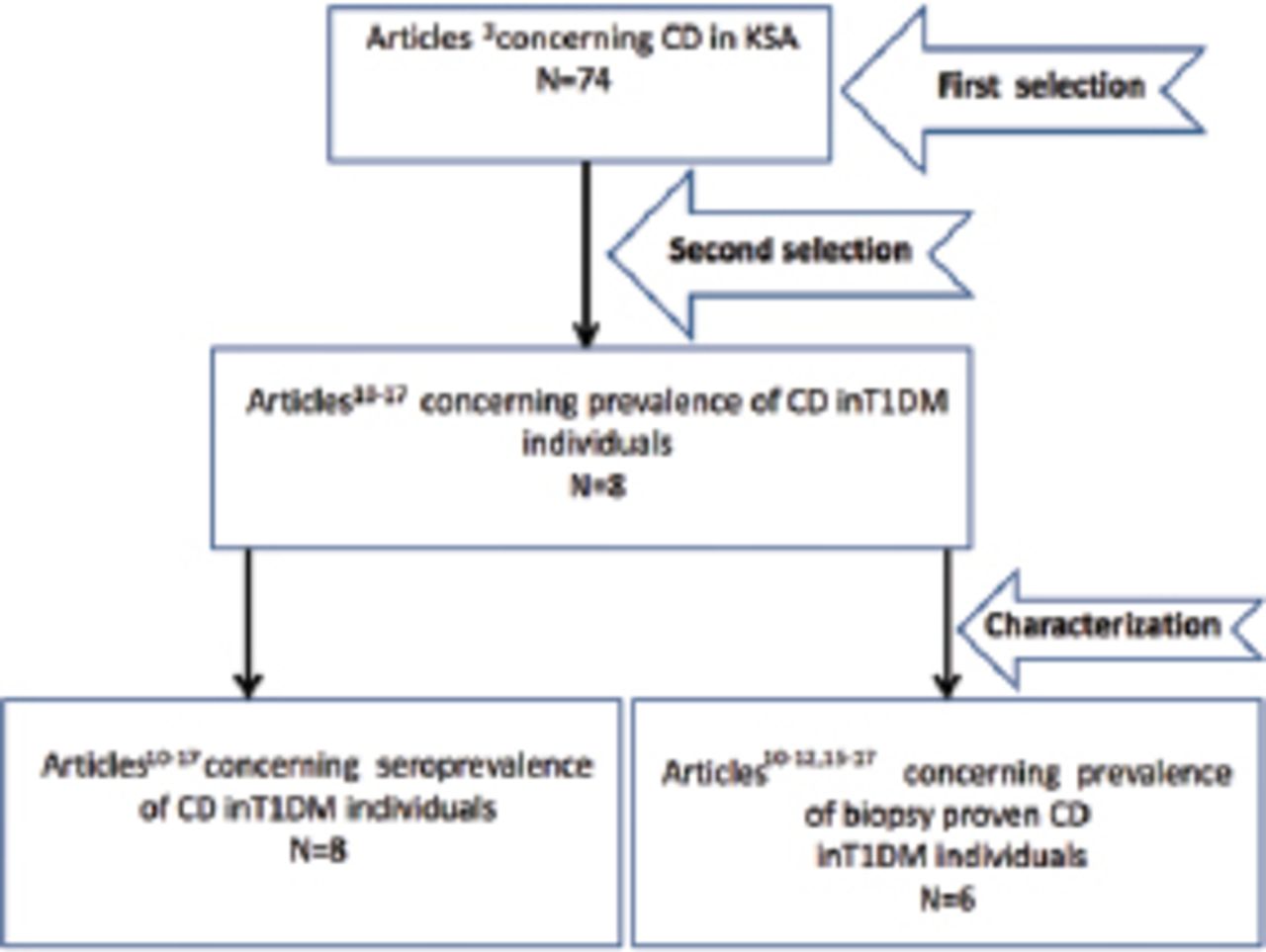
Safi characterize and meta-analyze the pertinent studies concerning celiac disease (CD) among patients with type 1 diabetes mellitus (T1DM) in the Kingdom of Saudi Arabia. The prevalence of seropositive-CD was 15.88% with high heterogeneity, while the prevalence of biopsy-proven CD was 12.0% with high heterogeneity. The prevalence of biopsy-proven CD among T1DM patients in Kingdom of Saudi Arabia (12.0%) was double the global prevalence (6.0%), and much higher than the normal Saudi population (1.4%). The female-to-male ratio (2:1) of CD patients in T1DM was the same as in the normal population in Kingdom of Saudi Arabia.

PRISMA flow-diagram showing the selection process of the pertinent studies
see page 647
ORIGINAL ARTICLES
The evaluation of leukocyte-platelet rich fibrin as an anti-inflammatory autologous biological additive. A novel in vitro study
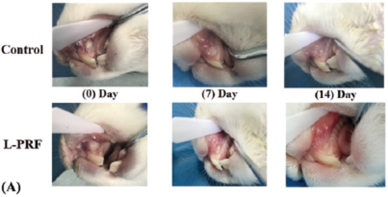
Mudalal et al investigate the use of leukocyte-platelet rich fibrin on suppressing the porphyromonas gingivalis (PG-LPS)-induced secretion of proinflammatory cytokines. The quantitative experimental study successfully established a modified technique for the production of HGFCs culture. One µg/mL PG-LPS was the recommended concentration to inhibit fibroblast proliferation. The expression of the pro-inflammatory cytokines messenger ribnucleic acid was notably raised at 3 and 6 hours post-PG-LPS treatment. The cumulative release of growth factors peaked during the first 24 hours and the production continued for 10 days. However, the fibroblast expression of cytokines was significantly suppressed after treatment with leucocyte- and platelet-rich fibrin (L-PRF). The study provided a novel way of obtaining HGFCs and greater understanding of the clinical impacts through the assessment of the anti-inflammatory properties of L-PRF in vitro.

Macroscopic observation showing the comparison of the ulcerated area between the L-PRF-treated group and control group. It show the more superior healing progress for the L-PRF group.
see page 657
Adaptation and validation of the Arabic version of self-efficacy scale for mammography. A report on psychometric properties
Al Zalabani confirm that the scale’s Arabic version has good psychometric properties, using reliability analysis, confirmatory factor analysis, and extreme groups validation. The scale is likely to be useful for evaluating interventional studies aimed at improving mammography screening participation rates. The scale showed a good internal consistency. The confirmatory factor analysis supported the scale’s single-factor structure and the goodness-of-fit indices confirmed the model’s good fit. Women who had a mammogram in the last 2 years scored significantly higher on the scale than did women who had never received a mammogram.
Item summary and internal consistency of the Arabic version of the mammography-specific self-efficacy scale.
see page 707
CASE REPORT
Parry-Romberg syndrome in Kuwait: neurological manifestations in 2 children. A case report and literature review
Zakkiriah et al presented 2 cases of progressive hemifacial atrophy (Parry-Romberg syndrome) with different neurological manifestations from Kuwait. The first case was a 14-year-old boy who initially presented with recurrent transient stroke-like episodes followed by focal seizures and hemifacial atrophy. Magnetic resonance imaging showed significant white matter changes and cerebral hemiatrophy. The second case was a 7-year-old girl who presented with complex partial seizures and hemifacial atrophy, her magnetic resonance imaging scan showed minimal changes in the hemiatrophy of the temporal cerebral lobe. Both patients’ disease activity was well controlled with immunosuppressive therapy and anticonvulsant.

Hemiatrophy of the left side with abnormal signal in the subcortical left temporal region and left periventricular area.
see page 721
- Copyright: © Saudi Medical Journal
This is an open-access article distributed under the terms of the Creative Commons Attribution-Noncommercial-Share Alike 3.0 Unported, which permits unrestricted use, distribution, and reproduction in any medium, provided the original work is properly cited.
In this issue


Jump to section
Related Articles
Cited By...
- No citing articles found.