


Article Figures & Data
Figures
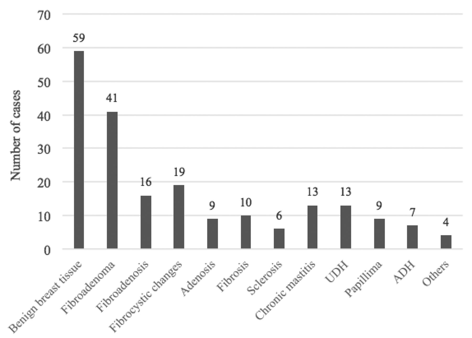
- Figure 1
- Pathological distribution of benign lesions found (n=206). UDH: usual ductal hyperplasia, ADH: atypical ductal hyperplasia
- Figure 2
- Pathological distribution of malignant lesions found (n=41). IDC: invasive ductal carcinoma, ILC: invasive lobular carcinoma
- Appendix 1
- Data handling, imaging, and biopsy of the raters.
- Appendix 2
- Mammography and sonography of a 65-year-old woman presented with clear right nipple discharge. A) Standard craniocaudal and B) mediolateral oblique views were obtained. Focal asymmetry in the right inner lower anterior to middle third (arrows) was detected. C-D) Sonography. It showed an avascular branching mass with posterior acoustic enhancement measuring 2x1.7x3 cm at 4 O’clock. Bilateral Abnormal axillary lymph nodes with cortical thickening approximately 0.4 cm. The histology of US-guided biopsy revealed intraductal papilloma.
- Appendix 3
- Mammography and sonography of 43-year-old women. A) Standard craniocaudal and B) mediolateral oblique views were obtained. Focal asymmetry was detected in the upper outer quadrant left breast (arrows). C) Sonography. It showed a heterogenous mass with an indistinct border measuring 7mm at 2 O’clock (arrow). The histology of the US-guided biopsy revealed benign breast tissue.


Tables
Variables n (%) Age (years), mean±SD 44.47±14.35 Age (years), median (IQR) 44.5 (35.0-54.0) Ultrasound Yes 246 (99.6) No 1 (0.4) Mammography Yes 197 (79.8) No 50 (20.2) Affected side Left 127 (51.4) Right 120 (48.6) Lesion size (cm), mean±SD 2.25±1.80 Lesion size (cm), median (IQR) 1.5 (1.0-2.8) Pathology Benign 199 (80.6) Malignant 41 (16.6) Hyperplasia 7 (2.8) Imaging findings Mass 164 (66.4) Mass with calcification 14 (5.7) Calcification only 5 (2.0) Asymmetry 39 (15.8) Architectural distortion 26 (10.5) Intraductal mass 37 (15.0) Complex mass 18 (7.3) Skin thickening 6 (2.4) Nipple retraction 2 (0.8) Mammography breast density (n=197) A 28 (14.4) B 84 (43.0) C 75 (38.5) D 8 (4.1) Lesion location Retroareolar 57 (23.1) Upper outer quadrant 133 (53.8) Lower outer quadrant 22 (8.9) Upper inner quadrant 20 (8.1) Lower inner quadrant 15 (6.1) Values are presented as numbers and percentages (%), or mean ± standard deviation (SD), or median interquartile range (IQR).
BIRADS Pathology P-value* Benign Malignant Rater 1 4A (n=135) 133 (98.5) 2 (1.5) <0.001 4B (n=68) 63 (92.6) 5 (7.4) 4C (n=44) 10 (22.7) 34 (77.3) Rater 2 4A (n=151) 149 (98.7) 2 (1.3) <0.001 4B (n=48) 44 (91.7) 4 (8.3) 4C (n=48) 13 (27.1) 35 (72.9) Values are presented as numbers and percentages (%).
BIRADS: breast-imaging reporting and data system
↵* Chi-square test for proportions was used to estimate the p-values.
- Table 3
- Comparison of positive predictive value of breast imaging-reporting and data system 4 subcategories with reference value.
Rater BI-RADS subcategory Total Benign Malignant PPV for malignancy BIRADS PPV Actual (95% CI) Reference Rater 1 4A 135 133 (98.5) 2 (1.5) 1.5 (0.2-5.2) >2 - ≤10 4B 68 63 (92.6) 5 (7.4) 7.4 (2.4-16.3) >10 - ≤50 4C 44 10 (22.7) 34 (77.3) 77.3 (62.2-88.5) >50 - <95 Total 247 206 (83.4) 41 (16.6) >2 - <95 Rater 2 4A 151 149 (98.7) 2 (1.3) 1.3 (0.2-4.7) >2 - ≤10 4B 48 44 (91.7) 4 (8.3) 8.3 (2.3-20.0) >10 - ≤50 4C 48 13 (27.1) 35 (72.9) 72.9 (58.2-84.7) >50 - <95 Total 247 206 (83.4) 41 (16.6) >2 - <95 Values are presented as numbers and percentages (%).
BI-RADS: breast-imaging reporting and data system, PPV: positive predictive value, CI: confidence interval
Inter-rater Rater 1 K-values* Rater 2 BIRADS Observer 2 4A 4B 4C BIRADS 4A 121 (89.6) 30 (44.1) 0 (0.0) 0.79 (0.73-0.85) BIRADS 4B 12 (8.9) 30 (44.1) 6 (13.6) BIRADS 4C 2 (1.5) 8 (11.8) 38 (86.4) p<0.001 Total 135 68 44 Values are presented as numbers and percentages (%).
BIRADS: breast-imaging reporting and data system
↵* Weighted Cohen’s Kappa (K) value and 95% confidence interval (CI).
- Table 5
- Issues of differences in the feature assignment reported By Rater 1 and Rater 2.
Mammography Rater (n=52) P-values Rater 1 Rater 2 Density Hypo- 1 (1.9) 0 (0.0) Iso- 42 (77.8) 31 (58.5) 0.025* Hyper- 11 (20.4) 22 (41.5) Shape (n=24) Oval 7 (29.2) 13 (54.2) 0.041** Round 11 (45.8) 4 (16.7) 0.044** Irregular 6 (25.0) 7 (29.1) 0.124** Ultrasound (n=54) Echo Hypo- 36 (66.7) 43 (76.8) Iso- 1 (1.9) 3 (7.1) 0.264* Mixed 17 (31.5) 11 (16.1) Vascularity (n=24) 20 (83.3) 3 (12.5) 0.017** Borders (n=54) Circumscribed 13 (24.1) 24 (44.4) 0.046** Indistinct 13 (24.1) 18 (33.3) 0.102** Angular 23 (42.6) 1 (1.9) 0.015*** Micro-lobular 29 (53.7) 21 (38.9) 0.048** Spiculated 2 (3.7) 1 (1.9) 0.214*** Anti-parallel 7 (13.9) 3 (5.6) 0.084*** Values are presented as numbers and percentages (%).
In this issue


Jump to section
Related Articles
Cited By...
- No citing articles found.