
Main menu

  • Home
  • Content
    • Latest
    • Archive
    • home
  • Info for
    • Authors
    • Reviewers
    • Subscribers
    • Institutions
    • Advertisers
    • Join SMJ
  • About Us
    • About Us
    • Editorial Office
    • Editorial Board
  • More
    • Advertising
    • Alerts
    • Feedback
    • Folders
    • Help
  • Other Publications
    • NeuroSciences Journal

User menu

  • My alerts
  • Log in
  • Log out

Search

  • Advanced search
Saudi Medical Journal
  • Other Publications
    • NeuroSciences Journal
  • My alerts
  • Log in
  • Log out
Saudi Medical Journal

Advanced Search

  • Home
  • Content
    • Latest
    • Archive
    • home
  • Info for
    • Authors
    • Reviewers
    • Subscribers
    • Institutions
    • Advertisers
    • Join SMJ
  • About Us
    • About Us
    • Editorial Office
    • Editorial Board
  • More
    • Advertising
    • Alerts
    • Feedback
    • Folders
    • Help
  • Follow psmmc on Twitter
  • Visit psmmc on Facebook
  • RSS
Review ArticleReview Article
Open Access

Dental pulp stem cells

Biology and use for periodontal tissue engineering

Nahid Y. Ashri, Sumaiah A. Ajlan and Abdullah M. Aldahmash
Saudi Medical Journal December 2015, 36 (12) 1391-1399; DOI: https://doi.org/10.15537/smj.2015.12.12750
Nahid Y. Ashri
From the Department of Periodontics and Community Dentistry (Ashri, Ajlan), College of Dentistry, the Stem Cell Unit, Department of Anatomy (Aldahmash), King Saud University, Riyadh, Kingdom of Saudi Arabia, and the Department of Endocrinology (Aldahmash), Odense University Hospital and University of Southern Denmark, Odense, Denmark
BDS, MSc
  • Find this author on Google Scholar
  • Find this author on PubMed
  • Search for this author on this site
Sumaiah A. Ajlan
From the Department of Periodontics and Community Dentistry (Ashri, Ajlan), College of Dentistry, the Stem Cell Unit, Department of Anatomy (Aldahmash), King Saud University, Riyadh, Kingdom of Saudi Arabia, and the Department of Endocrinology (Aldahmash), Odense University Hospital and University of Southern Denmark, Odense, Denmark
BDS, MSc
  • Find this author on Google Scholar
  • Find this author on PubMed
  • Search for this author on this site
  • For correspondence: [email protected]
Abdullah M. Aldahmash
From the Department of Periodontics and Community Dentistry (Ashri, Ajlan), College of Dentistry, the Stem Cell Unit, Department of Anatomy (Aldahmash), King Saud University, Riyadh, Kingdom of Saudi Arabia, and the Department of Endocrinology (Aldahmash), Odense University Hospital and University of Southern Denmark, Odense, Denmark
MSc, PhD
  • Find this author on Google Scholar
  • Find this author on PubMed
  • Search for this author on this site
  • Article
  • Figures & Data
  • eLetters
  • Info & Metrics
  • References
  • PDF
Loading

Abstract

Inflammatory periodontal disease is a major cause of loss of tooth-supporting structures. Novel approaches for regeneration of periodontal apparatus is an area of intensive research. Periodontal tissue engineering implies the use of appropriate regenerative cells, delivered through a suitable scaffold, and guided through signaling molecules. Dental pulp stem cells have been used in an increasing number of studies in dental tissue engineering. Those cells show mesenchymal (stromal) stem cell-like properties including self-renewal and multilineage differentiation potentials, aside from their relative accessibility and pleasant handling properties. The purpose of this article is to review the biological principles of periodontal tissue engineering, along with the challenges facing the development of a consistent and clinically relevant tissue regeneration platform. This article includes an updated review on dental pulp stem cells and their applications in periodontal regeneration, in combination with different scaffolds and growth factors.

Chronic inflammation and loss of the tooth-supporting structures are distinct features of chronic periodontal diseases. Preservation and enhancement of the regeneration of periodontal structures are the main goals of periodontal treatment. However, the periodontium is a complex structure as it contains a minimum of 6 distinct tissue types including: the gingival epithelium, the gingival connective tissue, the periodontal ligament, the tooth root surface cementum, the alveolar bone and the corresponding vasculature. All these tissues are affected during chronic inflammation and restoration of their normal status is important for allowing periodontal regeneration to occur.1,2 Different periodontal surgical procedures concerning root conditioning, autografts, allografts, xenografts, and/or barrier membranes for guided tissue regeneration have been employed to enhance periodontal tissue regeneration.3 While histological evidence of tissue regeneration has been observed in some studies of regenerative therapies, complete periodontal tissue regeneration is still difficult to obtain.4-6 In a previous study,7 we provided adequate description on tissue engineering and the involvement of mesenchymal (stromal) stem cell (MSC) with and without scaffold. Briefly, tissue engineering represents a novel approach for regeneration of damaged tissues and organs. Tissue engineering is based on establishing the essential conditions that support the natural regenerative potential of tissues, and where each functional stage of reconstruction is based on a biologically enhanced process. By employing the conceptual framework of tissue engineering, it may be possible to obtain complete periodontal tissue regeneration. The purpose of this article is to review the biological principles of periodontal tissue engineering, along with the challenges facing the development of a consistent and clinically relevant tissue regeneration platform.

Components of periodontal tissue engineering

The essential components of periodontal tissue engineering are a) cells including stem cells, b) scaffold materials, and c) appropriate signals like morphogens/growth factors. Each one of these components plays an important role in the regenerative process. The cells define the nature of the tissue to be regenerated, morphogens and growth factors are required to direct the proliferation and the differentiation of cells to specific tissue fate, and scaffolds are used to provide a 3-dimensional micro-environment to facilitate 3-dimensional-tissue formation and enhancement of lineage differentiation. These 3 components are the focus of studies of periodontal tissue engineering.8

Stem cells

Stem cells are defined as undifferentiated cells that exhibit self-renewal and multi-lineage differentiation capacity. Stem cells can be classified into pluripotent (embryonic) or induced-pluripotent stem cells, and adult (also known as tissue-specific) stem cells.7,9 Recently, a number of adult stem cell types have been isolated from dental tissues, including dental pulp stem cells (DPSCs),10-13 exfoliated deciduous teeth (SHED),14-16 periodontal ligament (PDLSCs),17,18 apical papilla (SCAP),19-21 and dental follicle progenitor cells (DFPCs).22,23 In addition, putative stem cells have been isolated from inflamed pulpal,24,25 and periodontal26 tissues.

Dental pulp progenitor cells are the most attractive cells for periodontal tissue engineering based on their good growth and differentiation capacity in ex vivo cultures. Dental pulp progenitor cells are derived from mesodermal tissues and have been originally described by Gronthos et al.11 They are closely related to mesenchymal (stromal) stem cells (MSC) that are present in the stromal compartment of different tissues including bone marrow. A Gene expression profile of DPSCs has been reported to be similar to that of bone marrow MSC.27 Those cells are derived from embryonic neural crest cells and exhibit self-renewal and multilineage differentiation potentials.10,11,28,29 Dental pulp progenitor cells can differentiate into a number of mesodermal and non-mesodermal tissue cells that include osteoblast,13,28,30 adipocytestes,28,29 chondrocytes,28,29 and myocytes,28,29 as well as neuronal,28,31 and endothelial cells,30,32 hepatocytes,33 melanocytes,34 in addition to the dentin forming odontoblasts.35 It is not known whether DPSC cultures contain a homogenous population with respect to differentiation or contain subpopulations with different lineage specific differentiation potentials. In support of the later hypothesis, CD34+ subpopulations of DPSCs has been reported to be committed to bone formation evidenced by formation of mineralized nodules and bone tissue.36,37 Further subpopulations with different characteristics are also being studied.38,39 Overall, DPSC represent a unique cell population with potential for dental tissue engineering.

Isolation and characterization of DPSC

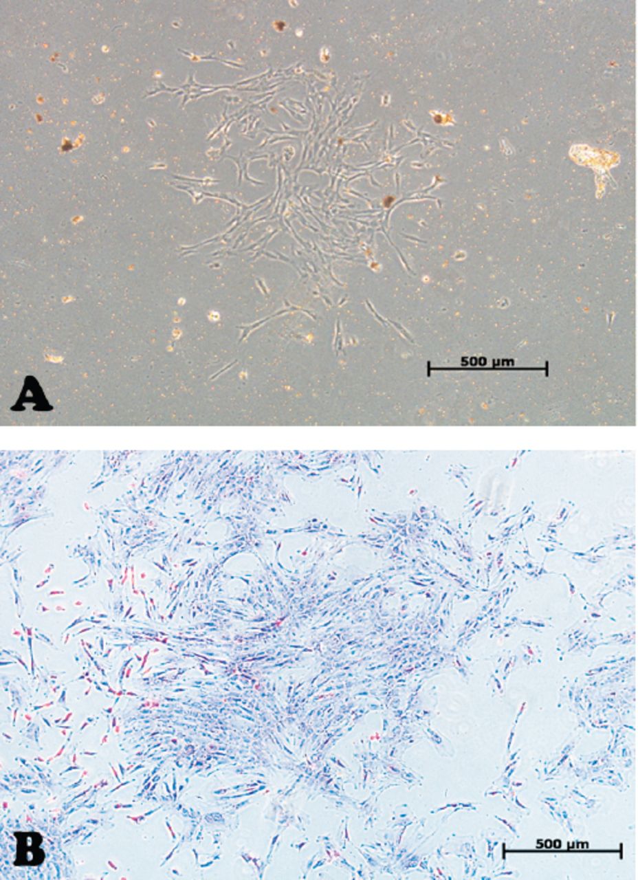
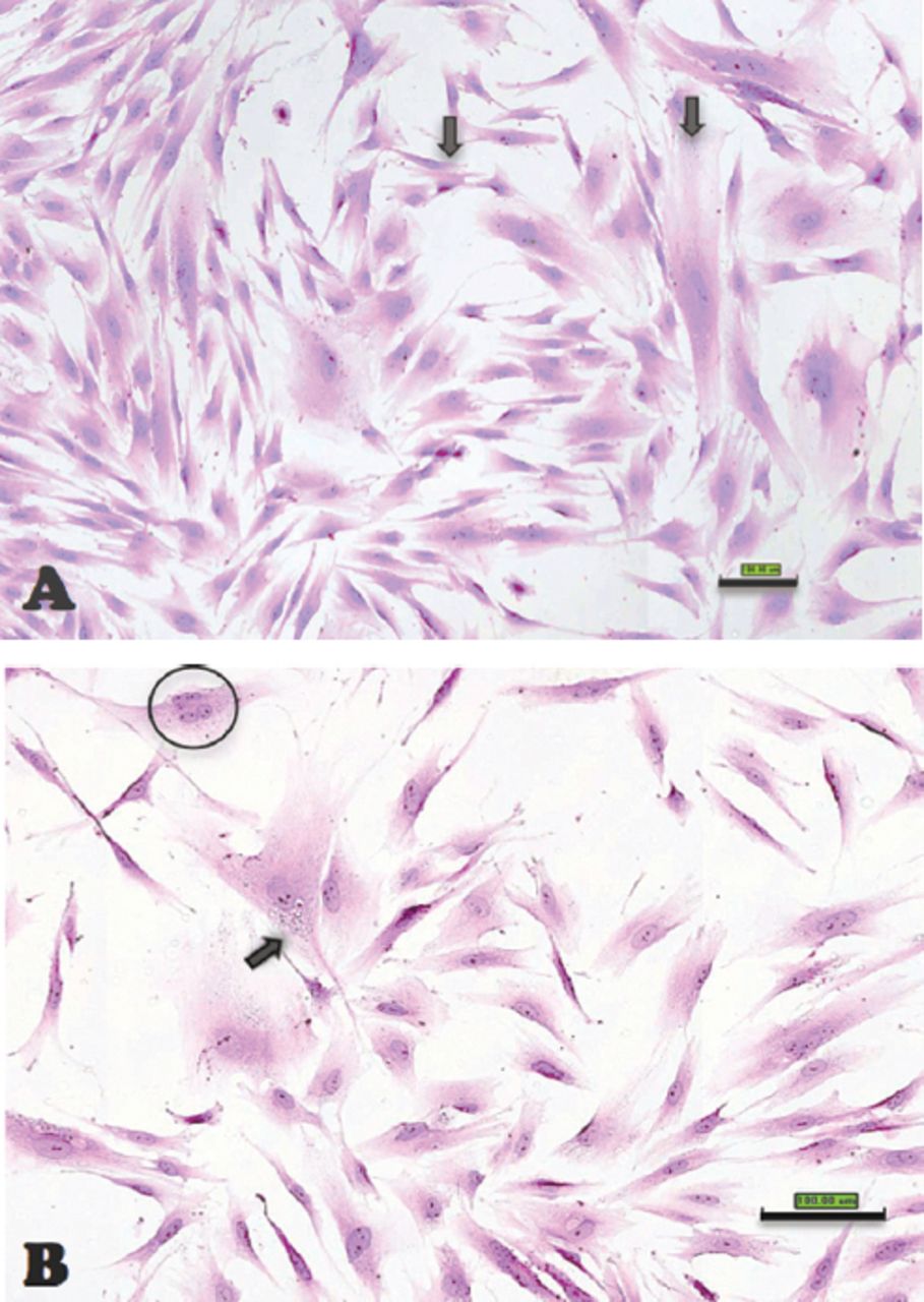
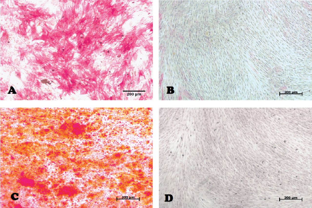
In our laboratory, we established DPSCs, which were isolated from the pulp tissue of extracted human third molar teeth. The teeth were vital, free from caries or periodontal diseases, or infections. Pulp tissue was exposed to enzymatic digestion and resultant cells were cultured in standard medium. Dental pulp stem cells initially formed multiple colonies similar to the MSC-derived colony forming unit fibroblast (CFU-F) (Figure 1). The cells exhibit heterogeneity in size and shape. They contained pale, round or oval central nucleus with multiple nucleoli, which indicated active DNA transcription and RNA synthesis. Multiple cytoplasmic vacuoles represented cellular secretory vesicles (Figure 2). These morphological characteristics are similar to MSC obtained from bone marrow and other tissues.11,40 Similar to MSC, DPSCs were positive to CD 105, CD 90, CD 70 and showed weak or negative expression of the common hematopoietic markers CD 34, CD 45, CD 31, CD 14, and HLA-DR.41 (Figure 3). Dental pulp progenitor cells differentiated readily into osteoblast-like cells in vitro as shown by presence of alkaline phosphatase activity and ability to form mineralized matrix (Figure 4). When implanted in vivo (subcutaneously) in immune deficient mice/rabbits, DPSCs formed dentin-pulp-like complex.10,42 These characterizations and differentiation capacities were confirmed with different tissues in our laboratory.

Figure 1
  • Download figure
  • Open in new tab
  • Download powerpoint
Figure 1

Inverted light microscopic images showing: A) Primary culture of dental pulp stem cells. B) The morphology of a dental pulp stem cells’ colony passage 2. Original magnification 5x. Scale bar 500µm

Figure 2
  • Download figure
  • Open in new tab
  • Download powerpoint
Figure 2

Scanoscope images representing different morphologic features of dental pulp stem cells (scale bar 1 mm) A) Cells with difference in size (black arrows) B) A multinucleated cell (nucleus circled with black), and a cell with cytoplasmic vacuoles (black arrow).

Figure 3
  • Download figure
  • Open in new tab
  • Download powerpoint
Figure 3

Fluorescence activated cell sorting (FACS) analysis results of a representative dental pulp stem cell line.

Figure 4
  • Download figure
  • Open in new tab
  • Download powerpoint
Figure 4

Inverted light microscopic image representing dental pulp stem cells 14 days post osteogenic induction after staining with: Alkaline phosphatase (ALP) A) test (red color represent ALP) B) control, and alizarin red-S (ARS) C) test (red color represent mineralized nodules), and d) control original magnification 10x, scale bar 200µm.

Scaffolds

For achieving periodontal regeneration, scaffolds are required to guide the 3-dimensional growth of tissues. Scaffolds should be able to support cell growth and preferably contain growth factors needed for induction of cell differentiation. In addition, the scaffold should be able to permit the transportation of oxygen, nutrients and waste products and should be bio-degreadable to be replaced by the periodontal tissues and should be highly porous with suitable pore size that support the above mentioned functions.43

Scaffold materials

Several scaffolds are currently available for periodontal tissue regeneration. Scaffolds are usually classified as either biologically derived polymers isolated from the extra-cellular matrix of animal tissues, from plants, or seaweed; for example, collagen type I or fibronectin, alginate from brown algae, chitosan from shells, and coralline materials, or synthetic; for example, hydroxyapatite (HA), tri-calcium phosphate (TCP) ceramics, polylactide and polyglycolide and a combination of these in the form of poly DL-lactic-co-glycolic acid (PLGA).44 Several examples have been employed in periodontal tissue regeneration. Biocorals are among the famous natural scaffolds used for that purpose.45 In addition to being resorbable and biocompatible, those scaffolds have interconnected porous structure that enhance cellular seeding, bone ingrowth, and withstand stresses.46 Bone grafts and bone substitutes supply a structure for the development of the regenerative tissues.47-49 Autologous platelet-rich plasma (PRP) and platelet rich fibrin (PRF) are easy to obtain during the surgical procedure and are rich in bone stimulating growth factors. In addition, platelet concentrates are slowly degradable and can provide a 3D-fibrin matrix formation that can enhance regeneration.50-52 Another scaffold used in periodontal regeneration is mineral trioxide aggregate (MTA),53,54 which is composed of dicalcium and tricalcium silicate, tricalcium aluminate, gypsum, tetra-calcium-alumino-ferrite, and bismuth oxide.55 Mineral trioxide aggregate is biocompatible and exhibit osteoconductive effects on bone cells.56 Tani-Ishii et al57 reported that, in osteoblasts, faster growth of cells and increased mineralized matrix gene expression has been observed in the presence of MTA. Also, Perinpanayagam & Al-Rabeah58 have reported that MTA supports cell attachment and expression of Runt-related transcription factor 2 (RUNX2), the osteoblast lineage master transcriptional factor. In addition, some studies have evaluated the use of different titanium alloys combined with implant surfaces bio-modifications to function as stem cell carrier for enhancement of osteointegration.59,60

Morphogen/growth factors

Growth factors and morphogens are employed to enhance proliferation and differentiation of stem cells into specific tissue type and to stimulate stem cells to synthesize and secrete mineralized matrix. A variety of growth factors have successfully been identified for periodontal regeneration. Enamel matrix derivative (EMD) has been reported to support periodontal regeneration.61,62 It contains a mixture of low molecular weight proteins that get absorbed into hydroxyapatite and collagen fibers of the root surface and induce cementum formation and periodontal regeneration.63 On cellular level, EMD exerts stimulatory effects on periodontal cells.63-66 Other growth factors known to enhance periodontal regeneration have also been employed.67,68 An example is platelet-derived growth factor (PDGF), which is a strong regulatory growth factor in bone that stimulates differentiation of osteoprogenitor cells and enhance vascularization.69 Histological studies showed significant increase in bone and cementum formation upon PDGF treatment.70,71 On cellular level, PDGF increases the number of collagen-synthesizing cells,72 and stimulates cell maturation evidenced by increased bone sialoprotein transcription.73 Another example is bone morphogenetic proteins (BMPs). They were discovered in bone matrix based on their ability for osteoinduction.74 Bone morphogenetic proteins are family of differentiation factors and each is capable of inducing the formation of new bone tissue when administered in vivo. Bone morphogenetic proteins produced by osteoblast, stored in bone matrix and released from bone matrix by osteoclastic bone resorption. The content and diffusibility of BMPs mediate the enhanced bone formation effects of demineralized bone extract when implanted in soft tissues or in osseous tissues.75 Blum et al,76 Pietrzack et al,77 and Wildemann et al78 evaluated the concentration of BMPs in demineralized free-dried bone allografts (DFDBA), and found it to vary with donor age and the source of bone extract, and associated their concentration with osteoinductive ability of the graft.

Combination of bone grafts with growth factors results in efficient tissue regeneration.79 Recombinant human (rh) BMP-2 added to the DFDBA has resulted in increased osteoinduction by almost 50%.80 Jones et al81 reported a higher percentage of defect fill and new bone contact at implant sites treated with rhBMP-2. In addition to the newly formed bone and new cementum, a connective tissue attachment was formed demonstrating the ability of rhBMP-2 application to induce periodontal tissue regeneration.82,83

Another approach involves the use of different histone deacetylase (HDAC) inhibitors to enhance cellular osteogenic differentiation. An example of such material is valporic acid. Although Schroeder and Westendorf84 reported the material to enhance osteogenic maturation, Paino et al85 reported such material may enhance early differentiation events, without inducing terminal differentiation stages.

Examples of periodontal tissue engineering

In cell-based replacement of a functional periodontium, the challenge is to form a physiological attachment between the dental apparatus and the hosting bone, with a cushion effect that absorb and distribute the mechanical load during function. A novel approach for periodontal tissue regeneration is to employ different populations of dental stem cells in order to recapitulate key events during periodontal tissue development; thus, allowing healing to occur in a synchronized way to regenerate the periodontium.86,87 As a corollary, the ideal positioning of cells in a tissue engineering context would include insertion of osteoblastic cells to maintain and repair bone together with periodontal ligament cells supporting a network of vascular and nerve cells. A conceptually simpler approach to periodontal regeneration methods involves engineered cell sheets to facilitate human periodontal ligament cell transplantation together with bone grafting materials and growth factors to regenerate complete periodontal tissues.88,89 However, maintaning the ability of reconstituted periodontium to preserve integrity and function during mastication over long periods of time is currently an area of intensive investigation.

Since alveolar bone loss is a major complication of chronic periodontal diseases, the use of DPSCs for alveolar bone regeneration is clinically relevant. D’Aquino et al,90 implanted DPSCs enriched collagen sponge at the extraction defect of third molar teeth. Radiographic evaluation revealed formation of significant amount of new well organized lamellar bone that was histologically verified at 3 months follow up. There was also more attachment gain next to the adjacent second molar. Interestingly, after 3 years, the regenerated tissue formed complete compact bone.91 Brunelli et al92 tried to utilize DPSCs to perform sinus elevation, and reported successful results as more bone formed during 4 months follow up.

More advanced protocols have demonstrated possible periodontal and craniofacial tissue engineering using different combinations of MSC-like cells from dental and non-dental origins together in combination with growth factors.93-95 Yamada et al,93,94 achieved significant bone regeneration around dental implants using DPSCs combined with platelet rich plasma (PRP) compared to PRP alone and similar results have been reported by Ito et al.95 At experimental levels, a more complex approach has been described by Liu et al96 where the group have reported a successful use of DPSCs in a functionalized hydroxyapatite/collagen/poly (L-lactide) (nHAC/PLA) scaffold using rhBMP-2; to reconstruct critical-size alveolar bone defects in rabbits. The feasibility of utilization of these cell lines in human remains to be determined.

Is whole tooth regeneration possible?

Tooth-like tissues have been generated by seeding a number of cell types on biodegradable scaffolds. The simplest described technique has been to harvest, expand and differentiate cells in vitro, followed by seeding them onto scaffolds, and finally implanting them in vivo. In some cases, the scaffolds were re-implanted into an extracted tooth socket or in the jaw. Ikeda et al97 have successfully regenerated and implanted a fully functioning tooth in an adult mouse, through transplantation of bioengineered tooth germ cells into the alveolar bone of the lost tooth region. The bioengineered tooth was fully erupted and exhibited normal tooth structure, hard-mineralized tissues and adequate response to noxious stimulations such as mechanical stress and pain. Xu et al98 reported seeding rat tooth bud cells on scaffolds fabricated from silk fibroin with 2 different pore sizes. The scaffolds were used as fabricated or after functionalization with an integerin Arg-Gly-Asp (RGD) binding peptide. The scaffolds were placed in the omenta of immune deficient athymic adult rats for 20 weeks. Histological analysis of tissue formed in both scaffolds revealed the presence of mineralized osteodentin-like tissues. Although most dental tissues were regenerated, the success rate was only 15-20% for achieving the correct arrangement of a natural tooth. Further studies are required to achieve replicable reconstituted and structurally sound teeth suitable for human use.

Future perspectives

Despite the impressive progress in tissue engineering approaches for enhancing the regeneration of periodontal tissues, several challenges remain to be solved. Although several animal studies were conducted, more human trials are required to evaluate the efficacy of those procedures in regenerating true periodontal defects. There is a need to evaluate the ability of those transplanted cells to survive, function and integrate into the recipient site in the presence of all other cellular and molecular components, including inflammatory mediators. In preparation for further clinical applications, emphasis on development of clear cell handling protocols to reach good manufacturing practice (GMP) is needed.46 This practice aims at standardization of the cells used in tissue engineering studies, as well as enhancing cell proliferation and differentiation. The production of vascularized scaffolds is a key requirement for enhancing tissue growth.43 Technologies for functionalization of scaffolds with specific signaling peptides that enhance angiogenesis are being developed and tested for their ability for mediating efficient periodontal tissue engineering. Finally, comprehensive interdisciplinary research programs that include cell biologist, material scientists and clinicians are prerequisite for the development of successful and clinically suitable regenerative periodontal tissues.

Copyright

Whenever a manuscript contains material (tables, figures, etc.) which is protected by copyright (previously published), it is the obligation of the author to obtain written permission from the holder of the copyright (usually the publisher) to reproduce the material in Saudi Medical Journal. This also applies if the material is the authors own work. Please submit copies of the material from the source in which it was first published.

Footnotes

  • Disclosure. This work was supported by the National Plan for Sciences and Technology Program, King Saud University, Riyadh, Kingdom of Saudi Arabia (Grant (No. 10-BIO1304-02).

  • Received July 9, 2015.
  • Accepted September 22, 2015.
  • Copyright: © Saudi Medical Journal

This is an open-access article distributed under the terms of the Creative Commons Attribution-Noncommercial-Share Alike 3.0 Unported, which permits unrestricted use, distribution, and reproduction in any medium, provided the original work is properly cited.

References

  1. ↵
    1. Zander HA,
    2. Polson AM,
    3. Heijl LC
    (1976) Goals of periodontal therapy. J Periodontol 47:261–266.
    OpenUrlPubMed
  2. ↵
    (1995) The potential role of growth and differentiation factors in periodontal regeneration. J Periodontol 66:506–510.
    OpenUrlPubMed
  3. ↵
    1. Garrett S
    (1996) Periodontal regeneration around natural teeth. Ann Periodontol 1:621–666.
    OpenUrlCrossRefPubMed
  4. ↵
    1. Bowers GM,
    2. Chadroff B,
    3. Carnevale R,
    4. Mellonig J,
    5. Corio R,
    6. Emerson J,
    7. et al.
    (1989) Histologic evaluation of new attachment apparatus formation in humans. Part III. J Periodontol 60:683–693.
    OpenUrlCrossRefPubMed
    1. Bowers GM,
    2. Chadroff B,
    3. Carnevale R,
    4. Mellonig J,
    5. Corio R,
    6. Emerson J,
    7. et al.
    (1989) Histologic evaluation of new attachment apparatus formation in humans. Part II. J Periodontol 60:675–682.
    OpenUrlPubMed
  5. ↵
    1. Bowers GM,
    2. Chadroff B,
    3. Carnevale R,
    4. Mellonig J,
    5. Corio R,
    6. Emerson J,
    7. et al.
    (1989) Histologic evaluation of new attachment apparatus formation in humans. Part I. J Periodontol 60:664–674.
    OpenUrlPubMed
  6. ↵
    1. Vishnubalaji R,
    2. Al-Nbaheen M,
    3. Kadalmani B,
    4. Aldahmash A,
    5. Ramesh T
    (2012) Skin-derived multipotent stromal cells--an archrival for mesenchymal stem cells. Cell Tissue Res 350:1–12.
    OpenUrlPubMed
  7. ↵
    1. Bianco P,
    2. Robey PG
    (2001) Stem cells in tissue engineering. Nature 414:118–121.
    OpenUrlCrossRefPubMed
  8. ↵
    1. Friedenstein AJ,
    2. Chailakhyan RK,
    3. Gerasimov UV
    (1987) Bone marrow osteogenic stem cells: in vitro cultivation and transplantation in diffusion chambers. Cell Tissue Kinet 20:263–272.
    OpenUrlPubMed
  9. ↵
    1. Gronthos S,
    2. Brahim J,
    3. Li W,
    4. Fisher LW,
    5. Cherman N,
    6. Boyde A,
    7. et al.
    (2002) Stem cell properties of human dental pulp stem cells. J Dent Res 81:531–535.
    OpenUrlCrossRefPubMed
  10. ↵
    1. Gronthos S,
    2. Mankani M,
    3. Brahim J,
    4. Robey PG,
    5. Shi S
    (2000) Postnatal human dental pulp stem cells (DPSCs) in vitro and in vivo. Proc Natl Acad Sci U S A 97:13625–13630.
    OpenUrlAbstract/FREE Full Text
    1. Kraft DC,
    2. Bindslev DA,
    3. Melsen B,
    4. Abdallah BM,
    5. Kassem M,
    6. Klein-Nulend J
    (2010) Mechanosensitivity of dental pulp stem cells is related to their osteogenic maturity. Eur J Oral Sci 118:29–38.
    OpenUrlCrossRefPubMed
  11. ↵
    1. Alkhalil M,
    2. Smajilagic A,
    3. Redzic A
    (2015) Human dental pulp mesenchymal stem cells isolation and osteoblast differentiation. Med Glas (Zenica) 12:27–32.
    OpenUrl
  12. ↵
    1. Telles PD,
    2. Machado MA,
    3. Sakai VT,
    4. Nor JE
    (2011) Pulp tissue from primary teeth: new source of stem cells. J Appl Oral Sci 19:189–194.
    OpenUrlPubMed
    1. Kashyap R
    (2015) SHED - Basic Structure for Stem Cell Research. J Clin Diagn Res 9:ZE07–ZE09.
    OpenUrl
  13. ↵
    1. Miura M,
    2. Gronthos S,
    3. Zhao M,
    4. Lu B,
    5. Fisher LW,
    6. Robey PG,
    7. et al.
    (2003) SHED: stem cells from human exfoliated deciduous teeth. Proc Natl Acad Sci U S A 100:5807–5812.
    OpenUrlAbstract/FREE Full Text
  14. ↵
    1. Seo BM,
    2. Miura M,
    3. Gronthos S,
    4. Bartold PM,
    5. Batouli S,
    6. Brahim J,
    7. et al.
    (2004) Investigation of multipotent postnatal stem cells from human periodontal ligament. Lancet 364:149–155.
    OpenUrlCrossRefPubMed
  15. ↵
    1. Zhu W,
    2. Liang M
    (2015) Periodontal ligament stem cells: current status, concerns, and future prospects. Stem Cells International 2015:972313.
    OpenUrl
  16. ↵
    1. Prateeptongkum E,
    2. Klingelhoffer C,
    3. Morsczeck C
    (2015) The influence of the donor on dental apical papilla stem cell properties. Tissue Cell 47:382–388.
    OpenUrl
    1. Ikeda E,
    2. Hirose M,
    3. Kotobuki N,
    4. Shimaoka H,
    5. Tadokoro M,
    6. Maeda M,
    7. et al.
    (2006) Osteogenic differentiation of human dental papilla mesenchymal cells. Biochem Biophys Res Commun 342:1257–1262.
    OpenUrlCrossRefPubMed
  17. ↵
    1. Sonoyama W,
    2. Liu Y,
    3. Yamaza T,
    4. Tuan RS,
    5. Wang S,
    6. Shi S,
    7. et al.
    (2008) Characterization of the apical papilla and its residing stem cells from human immature permanent teeth: a pilot study. J Endod 34:166–171.
    OpenUrlCrossRefPubMed
  18. ↵
    1. Kemoun P,
    2. Laurencin-Dalicieux S,
    3. Rue J,
    4. Farges JC,
    5. Gennero I,
    6. Conte-Auriol F,
    7. et al.
    (2007) Human dental follicle cells acquire cementoblast features under stimulation by BMP-2/-7 and enamel matrix derivatives (EMD) in vitro. Cell Tissue Res 329:283–294.
    OpenUrlCrossRefPubMed
  19. ↵
    1. Morsczeck C,
    2. Gotz W,
    3. Schierholz J,
    4. Zeilhofer F,
    5. Kuhn U,
    6. Mohl C,
    7. et al.
    (2005) Isolation of precursor cells (PCs) from human dental follicle of wisdom teeth. Matrix Biol 24:155–165.
    OpenUrlCrossRefPubMed
  20. ↵
    1. Alongi DJ,
    2. Yamaza T,
    3. Song Y,
    4. Fouad AF,
    5. Romberg EE,
    6. Shi S,
    7. et al.
    (2010) Stem/progenitor cells from inflamed human dental pulp retain tissue regeneration potential. Regen Med 5:617–631.
    OpenUrlCrossRefPubMed
  21. ↵
    1. Kim J,
    2. Park JC,
    3. Kim SH,
    4. Im GI,
    5. Kim BS,
    6. Lee JB,
    7. et al.
    (2014) Treatment of FGF-2 on stem cells from inflamed dental pulp tissue from human deciduous teeth. Oral Dis 20:191–204.
    OpenUrlCrossRefPubMed
  22. ↵
    1. Park JC,
    2. Kim JM,
    3. Jung IH,
    4. Kim JC,
    5. Choi SH,
    6. Cho KS,
    7. et al.
    (2011) Isolation and characterization of human periodontal ligament (PDL) stem cells (PDLSCs) from the inflamed PDL tissue: in vitro and in vivo evaluations. J Clin Periodontol 38:721–731.
    OpenUrlCrossRefPubMed
  23. ↵
    1. Shi S,
    2. Robey PG,
    3. Gronthos S
    (2001) Comparison of human dental pulp and bone marrow stromal stem cells by cDNA microarray analysis. Bone 29:532–539.
    OpenUrlCrossRefPubMed
  24. ↵
    1. Zhang W,
    2. Walboomers XF,
    3. Shi S,
    4. Fan M,
    5. Jansen JA
    (2006) Multilineage differentiation potential of stem cells derived from human dental pulp after cryopreservation. Tissue Eng 12:2813–2823.
    OpenUrlCrossRefPubMed
  25. ↵
    1. Yang X,
    2. Zhang W,
    3. van den Dolder J,
    4. Walboomers XF,
    5. Bian Z,
    6. Fan M,
    7. et al.
    (2007) Multilineage potential of STRO-1+rat dental pulp cells in vitro. J Tissue Eng Regen Med 1:128–135.
    OpenUrlCrossRefPubMed
  26. ↵
    1. d’Aquino R,
    2. Graziano A,
    3. Sampaolesi M,
    4. Laino G,
    5. Pirozzi G,
    6. De Rosa A,
    7. et al.
    (2007) Human postnatal dental pulp cells co-differentiate into osteoblasts and endotheliocytes: a pivotal synergy leading to adult bone tissue formation. Cell Death Differ 14:1162–1171.
    OpenUrlCrossRefPubMed
  27. ↵
    1. Karaoz E,
    2. Demircan PC,
    3. Saglam O,
    4. Aksoy A,
    5. Kaymaz F,
    6. Duruksu G
    (2011) Human dental pulp stem cells demonstrate better neural and epithelial stem cell properties than bone marrow-derived mesenchymal stem cells. Histochem Cell Biol 136:455–473.
    OpenUrlCrossRefPubMed
  28. ↵
    1. Li X,
    2. Hou J,
    3. Wu B,
    4. Chen T,
    5. Luo A
    (2014) Effects of platelet-rich plasma and cell coculture on angiogenesis in human dental pulp stem cells and endothelial progenitor cells. J Endod 40:1810–1814.
    OpenUrl
  29. ↵
    1. Cho YA,
    2. Noh K,
    3. Jue SS,
    4. Lee SY,
    5. Kim EC
    (2015) Melatonin promotes hepatic differentiation of human dental pulp stem cells: clinical implications for the prevention of liver fibrosis. J Pineal Res 58:127–135.
    OpenUrlCrossRefPubMed
  30. ↵
    1. Paino F,
    2. Ricci G,
    3. De Rosa A,
    4. D’Aquino R,
    5. Laino L,
    6. Pirozzi G,
    7. et al.
    (2010) Ecto-mesenchymal stem cells from dental pulp are committed to differentiate into active melanocytes. Eur Cell Mater 20:295–305.
    OpenUrlCrossRefPubMed
  31. ↵
    1. Tran HL,
    2. Doan VN
    (Feb, 2015) Human dental pulp stem cells cultured onto dentin derived scaffold can regenerate dentin-like tissue in vivo. Cell Tissue Bank, doi.10.1007/s10561-015-9503-z.
  32. ↵
    1. Graziano A,
    2. d’Aquino R,
    3. Laino G,
    4. Proto A,
    5. Giuliano MT,
    6. Pirozzi G,
    7. et al.
    (2008) Human CD34+stem cells produce bone nodules in vivo. Cell Prolif 41:1–11.
    OpenUrlCrossRefPubMed
  33. ↵
    1. Laino G,
    2. Carinci F,
    3. Graziano A,
    4. d’Aquino R,
    5. Lanza V,
    6. De Rosa A,
    7. et al.
    (2006) In vitro bone production using stem cells derived from human dental pulp. J Craniofac Surg 17:511–515.
    OpenUrlCrossRefPubMed
  34. ↵
    1. Horibe H,
    2. Murakami M,
    3. Iohara K,
    4. Hayashi Y,
    5. Takeuchi N,
    6. Takei Y,
    7. et al.
    (2014) Isolation of a stable subpopulation of mobilized dental pulp stem cells (MDPSCs) with high proliferation, migration, and regeneration potential is independent of age. PLoS One 9:e98553.
    OpenUrl
  35. ↵
    1. Kawanabe N,
    2. Fukushima H,
    3. Ishihara Y,
    4. Yanagita T,
    5. Kurosaka H,
    6. Yamashiro T
    (2015) Isolation and characterization of SSEA-4-positive subpopulation of human deciduous dental pulp cells. Clin Oral Investig 19:363–371.
    OpenUrlCrossRefPubMed
  36. ↵
    1. Kuo MY,
    2. Lan WH,
    3. Lin SK,
    4. Tsai KS,
    5. Hahn LJ
    (1992) Collagen gene expression in human dental pulp cell cultures. Arch Oral Biol 37:945–952.
    OpenUrlCrossRefPubMed
  37. ↵
    1. Dominici M,
    2. Le Blanc K,
    3. Mueller I,
    4. Slaper-Cortenbach I,
    5. Marini F,
    6. Krause D,
    7. et al.
    (2006) Minimal criteria for defining multipotent mesenchymal stromal cells. The International Society for Cellular Therapy position statement. Cytotherapy 8:315–317.
    OpenUrlCrossRefPubMed
  38. ↵
    1. El-Backly RM,
    2. Massoud AG,
    3. El-Badry AM,
    4. Sherif RA,
    5. Marei MK
    (2008) Regeneration of dentine/pulp-like tissue using a dental pulp stem cell/poly(lactic-co-glycolic) acid scaffold construct in New Zealand white rabbits. Aust Endod J 34:52–67.
    OpenUrlPubMed
  39. ↵
    1. Liu Y,
    2. Lim J,
    3. Teoh SH
    (2013) Review: development of clinically relevant scaffolds for vascularised bone tissue engineering. Biotechnol Adv 31:688–705.
    OpenUrlCrossRefPubMed
  40. ↵
    1. Chaudhary C,
    2. Garg T
    (2015) Scaffolds: A novel carrier and potential wound healer. Crit Rev Ther Drug Carrier Syst 32:277–321.
    OpenUrl
  41. ↵
    1. Mangano C,
    2. Paino F,
    3. d’Aquino R,
    4. De Rosa A,
    5. Iezzi G,
    6. Piattelli A,
    7. et al.
    (2011) Human dental pulp stem cells hook into biocoral scaffold forming an engineered biocomplex. PLoS One 6:e18721.
    OpenUrlCrossRefPubMed
  42. ↵
    1. La Noce M,
    2. Paino F,
    3. Spina A,
    4. Naddeo P,
    5. Montella R,
    6. Desiderio V,
    7. et al.
    (2014) Dental pulp stem cells: state of the art and suggestions for a true translation of research into therapy. J Dent 42:761–768.
    OpenUrlCrossRefPubMed
  43. ↵
    1. Reynolds MA,
    2. Aichelmann-Reidy ME,
    3. Branch-Mays GL
    (2010) Regeneration of periodontal tissue: bone replacement grafts. Dent Clin North Am 54:55–71.
    OpenUrlCrossRefPubMed
    1. Hanes PJ
    (2007) Bone replacement grafts for the treatment of periodontal intrabony defects. Oral Maxillofac Surg Clin North Am 19:499–512.
    OpenUrl
  44. ↵
    1. Minsk L
    (2005) Bone replacement grafts for periodontal regeneration. Compend Contin Educ Dent 26:676, 678, 680 passim.
    OpenUrl
  45. ↵
    1. Del Corso M,
    2. Vervelle A,
    3. Simonpieri A,
    4. Jimbo R,
    5. Inchingolo F,
    6. Sammartino G,
    7. et al.
    (2012) Current knowledge and perspectives for the use of platelet-rich plasma (PRP) and platelet-rich fibrin (PRF) in oral and maxillofacial surgery part 1: Periodontal and dentoalveolar surgery. Curr Pharm Biotechnol 13:1207–1230.
    OpenUrlPubMed
    1. Simonpieri A,
    2. Del Corso M,
    3. Vervelle A,
    4. Jimbo R,
    5. Inchingolo F,
    6. Sammartino G,
    7. et al.
    (2012) Current knowledge and perspectives for the use of platelet-rich plasma (PRP) and platelet-rich fibrin (PRF) in oral and maxillofacial surgery part 2: Bone graft, implant and reconstructive surgery. Curr Pharm Biotechnol 13:1231–1256.
    OpenUrlCrossRefPubMed
  46. ↵
    1. Dohan Ehrenfest DM,
    2. Bielecki T,
    3. Mishra A,
    4. Borzini P,
    5. Inchingolo F,
    6. Sammartino G,
    7. et al.
    (2012) In search of a consensus terminology in the field of platelet concentrates for surgical use: platelet-rich plasma (PRP), platelet-rich fibrin (PRF), fibrin gel polymerization and leukocytes. Curr Pharm Biotechnol 13:1131–1137.
    OpenUrlCrossRefPubMed
  47. ↵
    1. Naik RM,
    2. Pudakalkatti PS,
    3. Hattarki SA
    (2014) Can MTA be: Miracle trioxide aggregate? J Indian Soc Periodontol 18:5–8.
    OpenUrl
  48. ↵
    1. Katsamakis S,
    2. Slot DE,
    3. Van der Sluis LW,
    4. Van der Weijden F
    (2013) Histological responses of the periodontium to MTA: a systematic review. J Clin Periodontol 40:334–344.
    OpenUrl
  49. ↵
    1. Camilleri J,
    2. Pitt Ford TR
    (2006) Mineral trioxide aggregate: a review of the constituents and biological properties of the material. Int Endod J 39:747–754.
    OpenUrlCrossRefPubMed
  50. ↵
    1. Chen CL,
    2. Huang TH,
    3. Ding SJ,
    4. Shie MY,
    5. Kao CT
    (2009) Comparison of calcium and silicate cement and mineral trioxide aggregate biologic effects and bone markers expression in MG63 cells. J Endod 35:682–685.
    OpenUrlCrossRefPubMed
  51. ↵
    1. Tani-Ishii N,
    2. Hamada N,
    3. Watanabe K,
    4. Tujimoto Y,
    5. Teranaka T,
    6. Umemoto T
    (2007) Expression of bone extracellular matrix proteins on osteoblast cells in the presence of mineral trioxide. J Endod 33:836–839.
    OpenUrlPubMed
  52. ↵
    1. Perinpanayagam H,
    2. Al-Rabeah E
    (2009) Osteoblasts interact with MTA surfaces and express Runx2. Oral Surg Oral Med Oral Pathol Oral Radiol Endod 107:590–596.
    OpenUrlPubMed
  53. ↵
    1. Zhang W,
    2. Li Z,
    3. Huang Q,
    4. Xu L,
    5. Li J,
    6. Jin Y,
    7. et al.
    (2013) Effects of a hybrid micro/nanorod topography-modified titanium implant on adhesion and osteogenic differentiation in rat bone marrow mesenchymal stem cells. Int J Nanomedicine 8:257–265.
    OpenUrl
  54. ↵
    1. Cordoba A,
    2. Monjo M,
    3. Hierro-Oliva M,
    4. Gonzalez-Martin ML,
    5. Ramis JM
    (2015) Bioinspired Quercitrin Nanocoatings: A fluorescence-based method for their surface quantification, and their effect on stem cell adhesion and differentiation to the osteoblastic lineage. ACS Appl Mater Interfaces 7:16857–16864.
    OpenUrl
  55. ↵
    1. Hammarstrom L,
    2. Heijl L,
    3. Gestrelius S
    (1997) Periodontal regeneration in a buccal dehiscence model in monkeys after application of enamel matrix proteins. J Clin Periodontol 24(9 Pt 2):669–677.
    OpenUrlCrossRefPubMed
  56. ↵
    1. Correa MG,
    2. Gomes Campos ML,
    3. Marques MR,
    4. Bovi Ambrosano GM,
    5. Casati MZ,
    6. Nociti FH Jr.,
    7. et al.
    (2014) Outcome of enamel matrix derivative treatment in the presence of chronic stress: histometric study in rats. J Periodontol 85:e259–e267.
    OpenUrl
  57. ↵
    1. Stout BM,
    2. Alent BJ,
    3. Pedalino P,
    4. Holbrook R,
    5. Gluhak-Heinrich J,
    6. Cui Y,
    7. et al.
    (2014) Enamel matrix derivative: protein components and osteoinductive properties. J Periodontol 85:e9–e17.
    OpenUrl
    1. Miron RJ,
    2. Chandad F,
    3. Buser D,
    4. Sculean A,
    5. Cochran DL,
    6. Zhang Y
    (2015) Effect of enamel matrix derivative (EMD)-liquid on osteoblast and periodontal ligament cell proliferation and differentiation. J Periodontol 3:1–14.
    OpenUrl
    1. Bosshardt DD
    (2008) Biological mediators and periodontal regeneration: a review of enamel matrix proteins at the cellular and molecular levels. J Clin Periodontol 35(8 Suppl):87–105.
    OpenUrlCrossRefPubMed
  58. ↵
    1. Kemoun P,
    2. Gronthos S,
    3. Snead ML,
    4. Rue J,
    5. Courtois B,
    6. Vaysse F,
    7. et al.
    (2011) The role of cell surface markers and enamel matrix derivatives on human periodontal ligament mesenchymal progenitor responses in vitro. Biomaterials 32:7375–7388.
    OpenUrlCrossRef
  59. ↵
    1. Smith PC,
    2. Martinez C,
    3. Caceres M,
    4. Martinez J
    (2015) Research on growth factors in periodontology. Periodontol 2000 67:234–250.
    OpenUrl
  60. ↵
    1. Darby IB,
    2. Morris KH
    (2013) A systematic review of the use of growth factors in human periodontal regeneration. J Periodontol 84:465–476.
    OpenUrlCrossRefPubMed
  61. ↵
    1. Shah P,
    2. Keppler L,
    3. Rutkowski J
    (2014) A review of platelet derived growth factor playing pivotal role in bone regeneration. J Oral Implantol 40:330–340.
    OpenUrl
  62. ↵
    1. Al-Hazmi BA,
    2. Al-Hamdan KS,
    3. Al-Rasheed A,
    4. Babay N,
    5. Wang HL,
    6. Al-Hezaimi K
    (2013) Efficacy of using PDGF and xenograft with or without collagen membrane for bone regeneration around immediate implants with induced dehiscence-type defects: a microcomputed tomographic study in dogs. J Periodontol 84:371–378.
    OpenUrl
  63. ↵
    1. Nevins M,
    2. Kao RT,
    3. McGuire MK,
    4. McClain PK,
    5. Hinrichs JE,
    6. McAllister BS,
    7. et al.
    (2013) Platelet-derived growth factor promotes periodontal regeneration in localized osseous defects: 36-month extension results from a randomized, controlled, double-masked clinical trial. J Periodontol 84:456–564.
    OpenUrlCrossRefPubMed
  64. ↵
    1. Canalis E,
    2. McCarthy TL,
    3. Centrella M
    (1989) Effects of platelet-derived growth factor on bone formation in vitro. J Cell Physiol 140:530–537.
    OpenUrlCrossRefPubMed
  65. ↵
    1. Mezawa M,
    2. Araki S,
    3. Takai H,
    4. Sasaki Y,
    5. Wang S,
    6. Li X,
    7. et al.
    (2009) Regulation of human bone sialoprotein gene transcription by platelet-derived growth factor-BB. Gene 435:80–87.
    OpenUrl
  66. ↵
    1. Diaz-Sanchez RM,
    2. Yanez-Vico RM,
    3. Fernandez-Olavarria A,
    4. Mosquera-Perez R,
    5. Iglesias-Linares A,
    6. Torres-Lagares D
    (2015) Current approaches of bone morphogenetic proteins in dentistry. J Oral Implantol 41:337–342.
    OpenUrl
  67. ↵
    1. Lohmann CH,
    2. Andreacchio D,
    3. Koster G,
    4. Carnes DL Jr.,
    5. Cochran DL,
    6. Dean DD,
    7. et al.
    (2001) Tissue response and osteoinduction of human bone grafts in vivo. Arch Orthop Trauma Surg 121:583–590.
    OpenUrl
  68. ↵
    1. Blum B,
    2. Moseley J,
    3. Miller L,
    4. Richelsoph K,
    5. Haggard W
    (2004) Measurement of bone morphogenetic proteins and other growth factors in demineralized bone matrix. Orthopedics 27(1 Suppl):s161–s165.
    OpenUrlPubMed
  69. ↵
    1. Pietrzak WS,
    2. Woodell-May J,
    3. McDonald N
    (2006) Assay of bone morphogenetic protein-2, -4, and -7 in human demineralized bone matrix. J Craniofac Surg 17:84–90.
    OpenUrlCrossRefPubMed
  70. ↵
    1. Wildemann B,
    2. Kadow-Romacker A,
    3. Haas NP,
    4. Schmidmaier G
    (2007) Quantification of various growth factors in different demineralized bone matrix preparations. J Biomed Mater Res A 81:437–442.
    OpenUrlPubMed
  71. ↵
    1. Del Rosario C,
    2. Rodriguez-Evora M,
    3. Reyes R,
    4. Delgado A,
    5. Evora C
    (2015) BMP-2, PDGF-BB, and bone marrow mesenchymal cells in a macroporous beta-TCP scaffold for critical-size bone defect repair in rats. Biomed Mater 10:045008.
    OpenUrlCrossRefPubMed
  72. ↵
    1. Boyan BD,
    2. Ranly DM,
    3. Schwartz Z
    (2006) Use of growth factors to modify osteoinductivity of demineralized bone allografts: lessons for tissue engineering of bone. Dent Clin North Am 50:217–212.
    OpenUrlCrossRefPubMed
  73. ↵
    1. Jones AA,
    2. Buser D,
    3. Schenk R,
    4. Wozney J,
    5. Cochran DL
    (2006) The effect of rhBMP-2 around endosseous implants with and without membranes in the canine model. J Periodontol 77:1184–1193.
    OpenUrlCrossRefPubMed
  74. ↵
    1. King GN,
    2. Hughes FJ
    (2001) Bone morphogenetic protein-2 stimulates cell recruitment and cementogenesis during early wound healing. J Clin Periodontol 28:465–475.
    OpenUrlCrossRefPubMed
  75. ↵
    1. Oda S,
    2. Kinoshita A,
    3. Higuchi T,
    4. Shizuya T,
    5. Ishikawa I
    (1997) Ectopic bone formation by biphasic calcium phosphate (BCP) combined with recombinant human bone morphogenetic protein-2 (rhBMP-2). J Med Dent Sci 44:53–62.
    OpenUrlPubMed
  76. ↵
    1. Schroeder TM,
    2. Westendorf JJ
    (2005) Histone deacetylase inhibitors promote osteoblast maturation. J Bone Miner Res 20:2254–2263.
    OpenUrlCrossRefPubMed
  77. ↵
    1. Paino F,
    2. La Noce M,
    3. Tirino V,
    4. Naddeo P,
    5. Desiderio V,
    6. Pirozzi G,
    7. et al.
    (2014) Histone deacetylase inhibition with valproic acid downregulates osteocalcin gene expression in human dental pulp stem cells and osteoblasts: evidence for HDAC2 involvement. Stem Cells 32:279–289.
    OpenUrlCrossRefPubMed
  78. ↵
    1. Bartold PM,
    2. Shi S,
    3. Gronthos S
    (2006) Stem cells and periodontal regeneration. Periodontol 2000 40:164–172.
    OpenUrlCrossRefPubMed
  79. ↵
    1. Lin NH,
    2. Gronthos S,
    3. Bartold PM
    (2008) Stem cells and periodontal regeneration. Aust Dent J 53:108–121.
    OpenUrlCrossRefPubMed
  80. ↵
    1. Hasegawa M,
    2. Yamato M,
    3. Kikuchi A,
    4. Okano T,
    5. Ishikawa I
    (2005) Human periodontal ligament cell sheets can regenerate periodontal ligament tissue in an athymic rat model. Tissue Eng 11:469–478.
    OpenUrlCrossRefPubMed
  81. ↵
    1. Gao H,
    2. Li B,
    3. Zhao L,
    4. Jin Y
    (2015) Influence of nanotopography on periodontal ligament stem cell functions and cell sheet based periodontal regeneration. Int J Nanomedicine 10:4009–4027.
    OpenUrl
  82. ↵
    1. d’Aquino R,
    2. De Rosa A,
    3. Lanza V,
    4. Tirino V,
    5. Laino L,
    6. Graziano A,
    7. et al.
    (2009) Human mandible bone defect repair by the grafting of dental pulp stem/progenitor cells and collagen sponge biocomplexes. Eur Cell Mater 18:75–83.
    OpenUrlCrossRefPubMed
  83. ↵
    1. Giuliani A,
    2. Manescu A,
    3. Langer M,
    4. Rustichelli F,
    5. Desiderio V,
    6. Paino F,
    7. et al.
    (2013) Three years after transplants in human mandibles, histological and in-line holotomography revealed that stem cells regenerated a compact rather than a spongy bone: biological and clinical implications. Stem Cells Transl Med 2:316–324.
    OpenUrlCrossRefPubMed
  84. ↵
    1. Brunelli G,
    2. Motroni A,
    3. Graziano A,
    4. D’Aquino R,
    5. Zollino I,
    6. Carinci F
    (2013) Sinus lift tissue engineering using autologous pulp micro-grafts: A case report of bone density evaluation. Journal of Indian Society of Periodontology 17:644–647.
    OpenUrl
  85. ↵
    1. Yamada Y,
    2. Ito K,
    3. Nakamura S,
    4. Ueda M,
    5. Nagasaka T
    (2011) Promising cell-based therapy for bone regeneration using stem cells from deciduous teeth, dental pulp, and bone marrow. Cell Transplant 20:1003–1013.
    OpenUrlCrossRefPubMed
  86. ↵
    1. Yamada Y,
    2. Nakamura S,
    3. Ito K,
    4. Sugito T,
    5. Yoshimi R,
    6. Nagasaka T,
    7. et al.
    (2010) A feasibility of useful cell-based therapy by bone regeneration with deciduous tooth stem cells, dental pulp stem cells, or bone-marrow-derived mesenchymal stem cells for clinical study using tissue engineering technology. Tissue Eng Part A 16:1891–1900.
    OpenUrlCrossRefPubMed
  87. ↵
    1. Ito K,
    2. Yamada Y,
    3. Nakamura S,
    4. Ueda M
    (2011) Osteogenic potential of effective bone engineering using dental pulp stem cells, bone marrow stem cells, and periosteal cells for osseointegration of dental implants. Int J Oral Maxillofac Implants 26:947–954.
    OpenUrlPubMed
  88. ↵
    1. Liu HC,
    2. E LL,
    3. Wang DS,
    4. Su F,
    5. Wu X,
    6. Shi ZP,
    7. et al.
    (2011) Reconstruction of alveolar bone defects using bone morphogenetic protein 2 mediated rabbit dental pulp stem cells seeded on nano-hydroxyapatite/collagen/poly(L-lactide). Tissue Eng Part A 17:2417–24133.
    OpenUrlCrossRefPubMed
  89. ↵
    1. Ikeda E,
    2. Morita R,
    3. Nakao K,
    4. Ishida K,
    5. Nakamura T,
    6. Takano-Yamamoto T,
    7. et al.
    (2009) Fully functional bioengineered tooth replacement as an organ replacement therapy. Proc Natl Acad Sci U S A 106:13475–13480.
    OpenUrlAbstract/FREE Full Text
  90. ↵
    1. Xu WP,
    2. Zhang W,
    3. Asrican R,
    4. Kim HJ,
    5. Kaplan DL,
    6. Yelick PC
    (2008) Accurately shaped tooth bud cell-derived mineralized tissue formation on silk scaffolds. Tissue Eng Part A 14:549–557.
    OpenUrlCrossRefPubMed
PreviousNext
Back to top

In this issue

Saudi Medical Journal: 36 (12)
Saudi Medical Journal
Vol. 36, Issue 12
1 Dec 2015
  • Table of Contents
  • Cover (PDF)
  • Index by author
Print
Download PDF
Email Article

Thank you for your interest in spreading the word on Saudi Medical Journal.

NOTE: We only request your email address so that the person you are recommending the page to knows that you wanted them to see it, and that it is not junk mail. We do not capture any email address.

Enter multiple addresses on separate lines or separate them with commas.
Dental pulp stem cells
(Your Name) has sent you a message from Saudi Medical Journal
(Your Name) thought you would like to see the Saudi Medical Journal web site.
Citation Tools
Dental pulp stem cells
Nahid Y. Ashri, Sumaiah A. Ajlan, Abdullah M. Aldahmash
Saudi Medical Journal Dec 2015, 36 (12) 1391-1399; DOI: 10.15537/smj.2015.12.12750

Citation Manager Formats

  • BibTeX
  • Bookends
  • EasyBib
  • EndNote (tagged)
  • EndNote 8 (xml)
  • Medlars
  • Mendeley
  • Papers
  • RefWorks Tagged
  • Ref Manager
  • RIS
  • Zotero
Share
Dental pulp stem cells
Nahid Y. Ashri, Sumaiah A. Ajlan, Abdullah M. Aldahmash
Saudi Medical Journal Dec 2015, 36 (12) 1391-1399; DOI: 10.15537/smj.2015.12.12750
Twitter logo Facebook logo Mendeley logo
  • Tweet Widget
  • Facebook Like
  • Google Plus One
Bookmark this article

Jump to section

  • Article
    • Abstract
    • Footnotes
    • References
  • Figures & Data
  • eLetters
  • References
  • Info & Metrics
  • PDF

Related Articles

  • No related articles found.
  • PubMed
  • Google Scholar

Cited By...

  • No citing articles found.
  • Google Scholar

More in this TOC Section

  • Harnessing artificial intelligence for infection control and prevention in hospitals
  • Effects of antidiabetic drugs on the level of serum uric acid in patients who have type 2 diabetes
  • The future of personalized medicine in Saudi Arabia
Show more Review Article

Similar Articles

CONTENT

  • home

JOURNAL

  • home

AUTHORS

  • home
Saudi Medical Journal

© 2025 Saudi Medical Journal Saudi Medical Journal is copyright under the Berne Convention and the International Copyright Convention.  Saudi Medical Journal is an Open Access journal and articles published are distributed under the terms of the Creative Commons Attribution-NonCommercial License (CC BY-NC). Readers may copy, distribute, and display the work for non-commercial purposes with the proper citation of the original work. Electronic ISSN 1658-3175. Print ISSN 0379-5284.

Powered by HighWire