
Main menu

  • Home
  • Content
    • Latest
    • Archive
    • home
  • Info for
    • Authors
    • Reviewers
    • Subscribers
    • Institutions
    • Advertisers
    • Join SMJ
  • About Us
    • About Us
    • Editorial Office
    • Editorial Board
  • More
    • Advertising
    • Alerts
    • Feedback
    • Folders
    • Help
  • Other Publications
    • NeuroSciences Journal

User menu

  • My alerts
  • Log in

Search

  • Advanced search
Saudi Medical Journal
  • Other Publications
    • NeuroSciences Journal
  • My alerts
  • Log in
Saudi Medical Journal

Advanced Search

  • Home
  • Content
    • Latest
    • Archive
    • home
  • Info for
    • Authors
    • Reviewers
    • Subscribers
    • Institutions
    • Advertisers
    • Join SMJ
  • About Us
    • About Us
    • Editorial Office
    • Editorial Board
  • More
    • Advertising
    • Alerts
    • Feedback
    • Folders
    • Help
  • Follow psmmc on Twitter
  • Visit psmmc on Facebook
  • RSS
Research ArticleOriginal Article
Open Access

The impact of viability assessment using cardiac MRI and echocardiogram on the outcome of revascularization in patients with multi-vessels coronary artery disease and moderate to severe ischemic cardiomyopathy

Atif Alzahrani, Hani Mufti, Anas Alswat, Bsaim Altirkistani, Mohammed Aljehani, Ahmed Jazzar, Fahad Alutaibi, Amir Abushouk, Jamilah Al Rahimi, Wail Al Kashkari and Mohammed Althobaiti
Saudi Medical Journal April 2023, 44 (4) 373-378; DOI: https://doi.org/10.15537/smj.2023.44.4.20220133
Atif Alzahrani
From King Faisal Cardiac Center (Alzahrani, Mufti, Al Rahimi, Al Kashkari), King Abdulaziz Medical City, Ministry of National Guard Health Affairs, Jeddah; from the Department of Radiology (Althobaiti), King Abdulaziz Medical City, Ministry of National Guard Health Affairs, Jeddah, from King Abdullah International Medical Research Center, (Alzahrani, Mufti, Alswat, Altirkistani, Aljehani, Jazzar, Alutaibi, Abushouk, Rahimi, Kashkari, Althobaiti), Jeddah, Kingdom of Saudi Arabia; from the College of Medicine (Alzahrani, Mufti, Alsawat, Altirkistani, Aljehani, Jazzar, Alutaibi, Abushouk, Al Rahimi, Al Kashkari, Althobaiti), King Saud Bin Aldulaziz University for Health Sciences, Jeddah, Kingdom of Saudi Arabia.
MD
  • Find this author on Google Scholar
  • Find this author on PubMed
  • Search for this author on this site
  • For correspondence: [email protected]
Hani Mufti
From King Faisal Cardiac Center (Alzahrani, Mufti, Al Rahimi, Al Kashkari), King Abdulaziz Medical City, Ministry of National Guard Health Affairs, Jeddah; from the Department of Radiology (Althobaiti), King Abdulaziz Medical City, Ministry of National Guard Health Affairs, Jeddah, from King Abdullah International Medical Research Center, (Alzahrani, Mufti, Alswat, Altirkistani, Aljehani, Jazzar, Alutaibi, Abushouk, Rahimi, Kashkari, Althobaiti), Jeddah, Kingdom of Saudi Arabia; from the College of Medicine (Alzahrani, Mufti, Alsawat, Altirkistani, Aljehani, Jazzar, Alutaibi, Abushouk, Al Rahimi, Al Kashkari, Althobaiti), King Saud Bin Aldulaziz University for Health Sciences, Jeddah, Kingdom of Saudi Arabia.
MD
  • Find this author on Google Scholar
  • Find this author on PubMed
  • Search for this author on this site
Anas Alswat
From King Faisal Cardiac Center (Alzahrani, Mufti, Al Rahimi, Al Kashkari), King Abdulaziz Medical City, Ministry of National Guard Health Affairs, Jeddah; from the Department of Radiology (Althobaiti), King Abdulaziz Medical City, Ministry of National Guard Health Affairs, Jeddah, from King Abdullah International Medical Research Center, (Alzahrani, Mufti, Alswat, Altirkistani, Aljehani, Jazzar, Alutaibi, Abushouk, Rahimi, Kashkari, Althobaiti), Jeddah, Kingdom of Saudi Arabia; from the College of Medicine (Alzahrani, Mufti, Alsawat, Altirkistani, Aljehani, Jazzar, Alutaibi, Abushouk, Al Rahimi, Al Kashkari, Althobaiti), King Saud Bin Aldulaziz University for Health Sciences, Jeddah, Kingdom of Saudi Arabia.
MS
  • Find this author on Google Scholar
  • Find this author on PubMed
  • Search for this author on this site
Bsaim Altirkistani
From King Faisal Cardiac Center (Alzahrani, Mufti, Al Rahimi, Al Kashkari), King Abdulaziz Medical City, Ministry of National Guard Health Affairs, Jeddah; from the Department of Radiology (Althobaiti), King Abdulaziz Medical City, Ministry of National Guard Health Affairs, Jeddah, from King Abdullah International Medical Research Center, (Alzahrani, Mufti, Alswat, Altirkistani, Aljehani, Jazzar, Alutaibi, Abushouk, Rahimi, Kashkari, Althobaiti), Jeddah, Kingdom of Saudi Arabia; from the College of Medicine (Alzahrani, Mufti, Alsawat, Altirkistani, Aljehani, Jazzar, Alutaibi, Abushouk, Al Rahimi, Al Kashkari, Althobaiti), King Saud Bin Aldulaziz University for Health Sciences, Jeddah, Kingdom of Saudi Arabia.
MS
  • Find this author on Google Scholar
  • Find this author on PubMed
  • Search for this author on this site
Mohammed Aljehani
From King Faisal Cardiac Center (Alzahrani, Mufti, Al Rahimi, Al Kashkari), King Abdulaziz Medical City, Ministry of National Guard Health Affairs, Jeddah; from the Department of Radiology (Althobaiti), King Abdulaziz Medical City, Ministry of National Guard Health Affairs, Jeddah, from King Abdullah International Medical Research Center, (Alzahrani, Mufti, Alswat, Altirkistani, Aljehani, Jazzar, Alutaibi, Abushouk, Rahimi, Kashkari, Althobaiti), Jeddah, Kingdom of Saudi Arabia; from the College of Medicine (Alzahrani, Mufti, Alsawat, Altirkistani, Aljehani, Jazzar, Alutaibi, Abushouk, Al Rahimi, Al Kashkari, Althobaiti), King Saud Bin Aldulaziz University for Health Sciences, Jeddah, Kingdom of Saudi Arabia.
MS
  • Find this author on Google Scholar
  • Find this author on PubMed
  • Search for this author on this site
Ahmed Jazzar
From King Faisal Cardiac Center (Alzahrani, Mufti, Al Rahimi, Al Kashkari), King Abdulaziz Medical City, Ministry of National Guard Health Affairs, Jeddah; from the Department of Radiology (Althobaiti), King Abdulaziz Medical City, Ministry of National Guard Health Affairs, Jeddah, from King Abdullah International Medical Research Center, (Alzahrani, Mufti, Alswat, Altirkistani, Aljehani, Jazzar, Alutaibi, Abushouk, Rahimi, Kashkari, Althobaiti), Jeddah, Kingdom of Saudi Arabia; from the College of Medicine (Alzahrani, Mufti, Alsawat, Altirkistani, Aljehani, Jazzar, Alutaibi, Abushouk, Al Rahimi, Al Kashkari, Althobaiti), King Saud Bin Aldulaziz University for Health Sciences, Jeddah, Kingdom of Saudi Arabia.
MS
  • Find this author on Google Scholar
  • Find this author on PubMed
  • Search for this author on this site
Fahad Alutaibi
From King Faisal Cardiac Center (Alzahrani, Mufti, Al Rahimi, Al Kashkari), King Abdulaziz Medical City, Ministry of National Guard Health Affairs, Jeddah; from the Department of Radiology (Althobaiti), King Abdulaziz Medical City, Ministry of National Guard Health Affairs, Jeddah, from King Abdullah International Medical Research Center, (Alzahrani, Mufti, Alswat, Altirkistani, Aljehani, Jazzar, Alutaibi, Abushouk, Rahimi, Kashkari, Althobaiti), Jeddah, Kingdom of Saudi Arabia; from the College of Medicine (Alzahrani, Mufti, Alsawat, Altirkistani, Aljehani, Jazzar, Alutaibi, Abushouk, Al Rahimi, Al Kashkari, Althobaiti), King Saud Bin Aldulaziz University for Health Sciences, Jeddah, Kingdom of Saudi Arabia.
MS
  • Find this author on Google Scholar
  • Find this author on PubMed
  • Search for this author on this site
Amir Abushouk
From King Faisal Cardiac Center (Alzahrani, Mufti, Al Rahimi, Al Kashkari), King Abdulaziz Medical City, Ministry of National Guard Health Affairs, Jeddah; from the Department of Radiology (Althobaiti), King Abdulaziz Medical City, Ministry of National Guard Health Affairs, Jeddah, from King Abdullah International Medical Research Center, (Alzahrani, Mufti, Alswat, Altirkistani, Aljehani, Jazzar, Alutaibi, Abushouk, Rahimi, Kashkari, Althobaiti), Jeddah, Kingdom of Saudi Arabia; from the College of Medicine (Alzahrani, Mufti, Alsawat, Altirkistani, Aljehani, Jazzar, Alutaibi, Abushouk, Al Rahimi, Al Kashkari, Althobaiti), King Saud Bin Aldulaziz University for Health Sciences, Jeddah, Kingdom of Saudi Arabia.
PhD
  • Find this author on Google Scholar
  • Find this author on PubMed
  • Search for this author on this site
Jamilah Al Rahimi
From King Faisal Cardiac Center (Alzahrani, Mufti, Al Rahimi, Al Kashkari), King Abdulaziz Medical City, Ministry of National Guard Health Affairs, Jeddah; from the Department of Radiology (Althobaiti), King Abdulaziz Medical City, Ministry of National Guard Health Affairs, Jeddah, from King Abdullah International Medical Research Center, (Alzahrani, Mufti, Alswat, Altirkistani, Aljehani, Jazzar, Alutaibi, Abushouk, Rahimi, Kashkari, Althobaiti), Jeddah, Kingdom of Saudi Arabia; from the College of Medicine (Alzahrani, Mufti, Alsawat, Altirkistani, Aljehani, Jazzar, Alutaibi, Abushouk, Al Rahimi, Al Kashkari, Althobaiti), King Saud Bin Aldulaziz University for Health Sciences, Jeddah, Kingdom of Saudi Arabia.
MD
  • Find this author on Google Scholar
  • Find this author on PubMed
  • Search for this author on this site
Wail Al Kashkari
From King Faisal Cardiac Center (Alzahrani, Mufti, Al Rahimi, Al Kashkari), King Abdulaziz Medical City, Ministry of National Guard Health Affairs, Jeddah; from the Department of Radiology (Althobaiti), King Abdulaziz Medical City, Ministry of National Guard Health Affairs, Jeddah, from King Abdullah International Medical Research Center, (Alzahrani, Mufti, Alswat, Altirkistani, Aljehani, Jazzar, Alutaibi, Abushouk, Rahimi, Kashkari, Althobaiti), Jeddah, Kingdom of Saudi Arabia; from the College of Medicine (Alzahrani, Mufti, Alsawat, Altirkistani, Aljehani, Jazzar, Alutaibi, Abushouk, Al Rahimi, Al Kashkari, Althobaiti), King Saud Bin Aldulaziz University for Health Sciences, Jeddah, Kingdom of Saudi Arabia.
MD
  • Find this author on Google Scholar
  • Find this author on PubMed
  • Search for this author on this site
Mohammed Althobaiti
From King Faisal Cardiac Center (Alzahrani, Mufti, Al Rahimi, Al Kashkari), King Abdulaziz Medical City, Ministry of National Guard Health Affairs, Jeddah; from the Department of Radiology (Althobaiti), King Abdulaziz Medical City, Ministry of National Guard Health Affairs, Jeddah, from King Abdullah International Medical Research Center, (Alzahrani, Mufti, Alswat, Altirkistani, Aljehani, Jazzar, Alutaibi, Abushouk, Rahimi, Kashkari, Althobaiti), Jeddah, Kingdom of Saudi Arabia; from the College of Medicine (Alzahrani, Mufti, Alsawat, Altirkistani, Aljehani, Jazzar, Alutaibi, Abushouk, Al Rahimi, Al Kashkari, Althobaiti), King Saud Bin Aldulaziz University for Health Sciences, Jeddah, Kingdom of Saudi Arabia.
MD
  • Find this author on Google Scholar
  • Find this author on PubMed
  • Search for this author on this site
  • Article
  • Figures & Data
  • eLetters
  • Info & Metrics
  • References
  • PDF
Loading

Abstract

Objectives: To investigate the influence of viability assessment in the management of patients with ischemic cardiomyopathy (ICM).

Methods: This retrospective cohort study included all patients with ICM with moderate to severely reduced left ventricular ejection fraction (LVEF) who underwent viability assessment using cardiac magnetic resonance imaging (MRI) and echocardiogram as modalities of imaging. In addition, LVEF, modality of choice, and treatment plans were all extracted as main variables from the electronic database. One hundred 6 patients who met the inclusion criteria from December 2014 to December 2019 were included.

Results: Posttreatment LVEF improved by 5% in the viable group compared to the nonviable group (p=0.016). Regardless of the treatment received, 6 (8.8%) patients in the viable group died due to cardiac causes after an 18-month follow-up period; in contrast, 7 (18.4%) patients died due to cardiac causes in the nonviable group. However, despite that difference, this was not statistically significant (p=0.153). Medical therapy alone was observed in 32 (84.2%) patients in the nonviable group compared to 32 (47.1%) in the viable group (p<0.001). Although the reduction in hospitalization for cardiac reasons was not statistically significant, the viable arm had 50% fewer hospitalizations than the nonviable arm (p=0.051).

Conclusion: Patients with viable myocardium had better outcomes in which LVEF significantly improved posttreatment. Additionally, there was a reduction in the number of hospitalizations for cardiac reasons in the viable group compared to the nonviable group, even though the difference was not statistically significant. However, further studies with a larger number of patients are needed to determine a definite conclusion.

Keywords:
  • revascularization
  • viability assessment
  • cardiac mri
  • echo
  • ischemic cardiomyopathy

Ischemic cardiomyopathy (ICM) is when the left ventricular systolic function is impaired due to significant coronary artery disease. Ischemic cardiomyopathy results from the reduction of blood supply to the myocardium that is caused by coronary artery diseases (CAD). The reduction of the myocardial blood flow can be acute secondary to myocardial infarction with ventricular remodeling or chronic by progressive narrowing of coronary arteries, which will reduce the ventricular contractile reserve and lead to a condition called hibernating myocardium.1-3

Ischemic cardiomyopathy is considered the most common cause of death worldwide. Moreover, CAD are the major contributor of deaths in ischemic cardiomyopathy, killing more than 370,000 people worldwide annually. In the United States, ICM accounts for 1 in every 4 deaths, killing one person every 36 seconds.4 Thus, 103 per 100,000 patients is the rate of death resulted from CAD.1,4 Regionally, 42% of deaths in Saudi Arabia are related to cardiac causes.5 The Prevalence of CAD in Saudi Arabia is estimated to be 5.5% in the whole population, with more Prevalence in males than females.6

When dealing with CAD, treatment aims to re-establish the blood supply and restore the ventricular function of the heart, either by revascularization using coronary artery bypass grafting (CABG), percutaneous coronary intervention (PCI) or by conventional medical therapy alone when the revascularization is not possible. When discussing revascularization, the term myocardial viability is defined as the myocardium in which the contractility of the heart is predicted to be improved or in which remolding or necrosis can be averted when the blood flow returns.7 Viability assessment of the myocardium is used to determine the extent of damage caused by the ischemic event; by predicting any improvement in the left ventricle ejection fraction (LVEF). Positron emission tomography (PET) is considered the gold standard for detecting myocardial viability; however, other modalities like cardiac MRI (CMR) and dobutamine echocardiography can also be used for the same purpose. The popularity of CMR and dobutamine echocardiography is mainly due to their availability and low cost compared to PET.7,8 According to the literature, some researchers stated that myocardial viability in patients with ischemic cardiomyopathy who underwent CABG has no predictive benefits, especially in the long term.9 However, other researches found that the viability assessment is valuable and beneficial as it decreases the cardiac event (myocardial infarction, cardiac death) and decreases the re-hospitalization for cardiac causes within one year.10-12 Since this topic is controversial, there is a need for more studies on myocardial viability assessment and whether it is beneficial for patients with ischemic cardiomyopathy in short and long-term aspects.

Our study measured the impact of viability assessment on outcomes in patients with multi-vessel coronary artery disease and ischemic cardiomyopathy. The primary objectives of this study were to determine the effect of viability assessment using cardiac MRI or echocardiography on reducing mortality, reducing hospitalization, and increasing the left ventricular ejection fraction (LVEF) within 18 months as a follow-up period.

Methods

A retrospective chart review study with a cohort design was conducted at King Faisal Cardiac Center (KFCC), King Abdulaziz Medical City, Jeddah, Saudi Arabia. The inclusion criteria includes adult patients with CAD who had done viability assessment from December 2014 to December 2019. In addition, patients with mildly reduced LVEF (LVEF >/=45%) and patients under the age of 18 years were all excluded from the study as per exclusion criteria. Using the electronic hospital medical records system, there were 106 patients from which the data was collected, using a consecutive sampling technique.

The data was obtained from the hospital’s electronic records using the Best Care 2.0 System. A data collection sheet that contained the study variables was used. These variables included demographics data such as age, gender, height, weight, and nationality. Other variables included pre and post-assessments of LVEF as percentages, state of living, number of hospitalizations for cardiac-related causes, results of viability assessment, LV size, modality of imaging. They were it either CMR or echocardiography, other cardiac risk factors and medications usage, and lastly, the plan of treatment and whether it was PCI, CABG, or medical therapy alone. As the study was conducted retrospectively, along with the sampling technique used, which was a consecutive one, there was no need for informed consent; therefore, Institutional Review Board approval was used instead. Then, these data were collected and entered manually on the excel sheet by the research team. The validity and quality of the collected data were ensured by the principal investigator of the research.

Statistical analysis

Characteristics of patients who underwent viability, based on the above-mentioned inclusion criteria, were evaluated. Because of the small sample size, non-parametric measures of central tendency and variance were used. Continuous variables were reported using the median and interquartile range and were compared using the Wilcoxon rank-sum test. Categorical variables were reported as frequencies and percentages and were analyzed by Chi-square or Fisher’s exact test as appropriate (Fisher’s exact test when more than 20% of cells have expected frequencies <5. The Kruskal-Wallis test was used for ordinal attributes. All statistical tests were 2-tailed, and p-values of <0.05 were considered significant. Statistical analyses were performed using R software, version 4.0.2. (R Foundation for Statistical Computing, Vienna, Austria).

Results

A total of 106 subjects who met the inclusion criteria were included in the study. Out of these, viable myocardium and non-viable myocardium were respectively reported to be 68 (64.2%) vs. 38 (35.8%). The median age of the viable group was 66 years (interquartile range 58-74 years), while 68 years (inter-quartile range 60-73 years) was the median for the non-viable group. Myocardial infarction, as a history of cardiac pathology, was presented in 21 (55.3%) of the non-viable group, which was significantly higher than the viable group 21 (30.9%) (p=0.013). The detailed demographic characteristics are reported in Table 1.

View this table:
  • View inline
  • View popup
Table 1

- Patients demographic characteristics (N=106).

In the viable group, when it comes to therapeutic modalities, medical therapy alone was considered in 32 (47.1%) of patients, CABG was presented in 22 (32.4%), and only 14 (20.6%) underwent PCI. In the non-viable group, on the contrary, medical therapy alone was reported in 32 (84.2%) of cases (p<0.001). Collectively, CABG, and PCI represented only 6 (15.8%) of the patients. The survival rate after 18 months of follow-up was higher in viable myocardium compared to the non-viable (56 [82.4%] vs. 25 [65.8%]) even though this difference was not statistically significant (p=0.153). Cardiac-related outcomes are summarized in Table 2.

View this table:
  • View inline
  • View popup
Table 2

- Imaging modalities, treatment and cardiac outcomes based on the myocardial viability status.

When medical therapy alone was considered as a therapeutic modality, 7 (21.9%) of the non-viable group died due to cardiac causes compared to the viable group 2 (6.2%). Furthermore, the viable group had a median of 0.5 visits due to cardiac reasons; on the other hand, a median of 1 visit was in the non-viable group. Thus, the viable group reported fewer visits by at least 50% during the follow-up period even though this reduction was not statistically significant (p=0.532). Characteristics of the medical therapy group are presented in Table 3.

View this table:
  • View inline
  • View popup
Table 3

- Characteristics and outcome of patients who received medical therapy alone based on viability status.

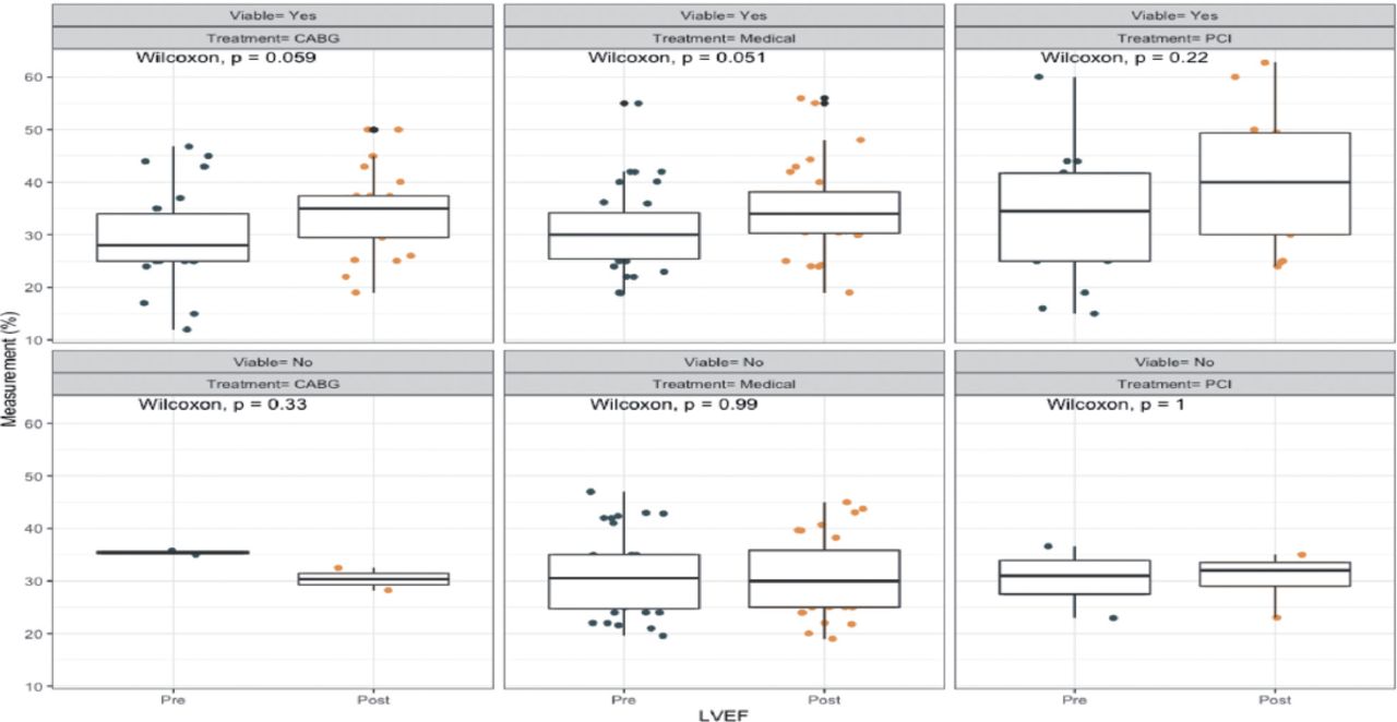
For those who had viable myocardium, overall LVEF was improved and significantly increased by 5% (p=0.016) post-treatment regardless of which therapy was performed. However, in patients in the non-viable group, therapeutic modalities did not play a positive role in terms of LVEF improvement. More demonstrations regarding LVEF with different treatment modalities are presented in (Figure 1).

Figure 1
  • Download figure
  • Open in new tab
  • Download powerpoint
Figure 1

- Left ventricular ejection fraction by treatment modality and viability.

Discussion

When planning coronary revascularization, the primary purpose is to restore the blood supply to the ischemic myocardium.13 Multiple imaging modalities have been used to detect viable myocardium and predict functional recovery, including echocardiography, cardiac MRI, and PET.7,14 Given that there are controversies surrounding the current utility of viability assessment, this study aimed to evaluate the impact of viability assessment on the outcomes of revascularization in patients with CAD-ICM.

Our results showed that patients with viable myocardium underwent more revascularization strategies with CABG or PCI when compared to the non-viable group and showed relatively fewer cardiac-related deaths, events, and hospitalization, even though this difference was not statistically significant. Moreover, LVEF improved by 5% in patients with viable myocardium in comparison with the non-viable. One of the possible explanations is that viable but dysfunctional myocardium has the ability to recover when blood flow is restored. Also, revascularization has been shown to improve survival in viable myocardium.15,16 On the other hand, non-viable myocardium is associated with poorer outcomes, and any further reduction in blood supply or increased oxygen demand in these non-viable regions will eventually lead to re-infarction or fatal arrhythmia, increasing the mortality in non-viable patients, regardless of which treatment plan was chosen.16,17 In patients with non-viable myocardium, more previous myocardial infarctions were reported. These prior infarcts could leave the myocardium in an irreversibly damaged state and the formation of myocardial scars in the non-viable group.16

In comparison to another study, similar results were implicated, suggesting that the presence of myocardial infarction is associated more with non-viable myocardium.18 After assessing the myocardial viability by the 3 used imaging modalities, it was suggestive that going for medical treatment alone did not show any significant effect on the mortality rate of both the viable and non-viable groups. Yet, this finding contradicted prior studies, which state that medical therapy alone has a significant effect on increasing the mortality rate in patients with viable myocardium by 158% rather than both.19 It appears that there is no clear beneficial outcome when comparing between the pre and post-treatment effect of using medical treatment, CABG, or PCI as a route of treatment on the LVEF for the non-viable myocardium; nevertheless, a significant increase in the LVEF on the viable group was present regardless what therapy the patients were on. Also, choosing revascularization as a treatment route resulted in a better predicting outcome of the post-treatment effect on the LVEF with the patients who were in the viable group (Figure 1), which would support the results of prior studies stating that there is a better overall outcome of revascularization on the patients with viable myocardium.10,17,19 Adding to a previous study, which states that there is a relative increase of hospitalization for cardiac causes in terms of going for medical therapy as a route of treatment and in this study, it expands it to elucidate that patient with viable myocardium and using medical therapy showed a decreased incidence of hospitalization contrary to the non-viable group, but it is still not statistically significant.20

Study limitations

Determining the need for viability assessment has been a challenge for cardiac specialists when discussing patients with ischemic cardiomyopathy. There were some limitations in this study. First, this study is a single-center study, and it only involved patients at KFCC in Jeddah. The second limitation, this study is a retrospective electronic chart review. A third limitation is that the number of patients in this study is small, making it difficult to find statistically significant differences between the 2 groups of patients. Given our sample size and with the assumption of a moderate effect size of myocardial viability on mortality (calculated effect size=0.57388), we calculated our study power of 80.8% at a significance level of 0.05 that is adequate for such study design.21 Additional research with a prospective design involving multi centers with a larger sample size is needed to shed light on viability assessment utility.

In conclusion, the current study suggests that patients with viable myocardium could have relatively less mortality and hospitalization. However, significant LVEF improvement can be observed if they adhere to the viability assessment recommendations. Conversely, patients with non-viable myocardium generally tend to have worse outcomes regardless of the treatment given, implicating the need to balance each treatment modality’s risks and benefits in those patients. Further research with a larger sample size is needed to investigate the viability assessment and its role in managing ischemic cardiomyopathy patients.

Acknowledgment

We would like to thank the data collectors for their valuable contributions, especially during these challenging circumstances, the Covid-19 pandemic. Also, we would like to acknowledge Dr. Mohammad Khan and Dr. Alaa Althobiti for their appreciated effort in reviewing and editing the manuscript.

Footnotes

  • Disclosure. Authors have no conflict of interests, and the work was not supported or funded by any drug company.

  • Received December 11, 2022.
  • Accepted February 18, 2023.
  • Copyright: © Saudi Medical Journal

This is an Open Access journal and articles published are distributed under the terms of the Creative Commons Attribution-NonCommercial License (CC BY-NC). Readers may copy, distribute, and display the work for non-commercial purposes with the proper citation of the original work.

References

  1. 1.↵
    1. Bhandari B,
    2. Quintanilla Rodriguez BS,
    3. Masood W. Ischemic Cardiomyopathy
    . Treasure Island (FL): StatPearls Publishing; 2022.
  2. 2.
    1. Cleaveland Clinic
    . Ischemic Cardiomyopathy What is ischemic cardiomyopathy? [Updated 2018 July 12; Accessed 2021 Jul 6]. Available at: https://my.clevelandclinic.org/health/diseases/17145-ischemic-cardiomyopathy
  3. 3.↵
    Mayo Clinic. Coronary artery disease - symptoms and causes. Overview. [Updated 2022 May 25; Accessed 2021 Jul 6]. Available at: https://www.mayoclinic.org/diseases-conditions/coronary-artery-disease/symptoms-causes/syc-20350613
  4. 4.↵
    Centers for Disease Control and Prevention. Heart Disease Facts. [Updated 2022 October 14; Accessed 2021 Jul 6]. Available at: https://www.cdc.gov/heartdisease/facts.htm
  5. 5.↵
    1. Aljefree N,
    2. Ahmed F.
    Prevalence of cardiovascular disease and associated risk factors among adult population in the gulf region: A systematic review. Adv Public Health 2015; 2015.
  6. 6.↵
    1. Al-Nozha MM,
    2. Arafah MR,
    3. Al-Mazrou YY,
    4. Al-Maatouq MA,
    5. Khan NB,
    6. Khalil MZ, et al.
    Coronary artery disease in Saudi Arabia. Saudi Med J 2004; 25: 1165–1171.
    OpenUrlPubMed
  7. 7.↵
    1. Jiménez Borreguero LJ,
    2. Ruiz-Salmerón R.
    Assessment of Myocardial Viability in Patients Before Revascularization. Rev Esp Cardiol 2003; 56: 721–733.
    OpenUrlPubMed
  8. 8.↵
    1. Hunold P,
    2. Jakob H,
    3. Erbel R,
    4. Barkhausen J,
    5. Heilmaier C.
    Accuracy of myocardial viability imaging by cardiac MRI and PET depending on left ventricular function. World J Cardiol2018; 10: 110–118.
    OpenUrlPubMed
  9. 9.↵
    1. Panza JA,
    2. Ellis AM,
    3. Al-Khalidi HR,
    4. Holly TA,
    5. Berman DS,
    6. Oh JK, et al.
    Myocardial viability and long-term outcomes in ischemic cardiomyopathy. N Engl J Med 2019; 381: 739–748.
    OpenUrlPubMed
  10. 10.↵
    1. Beanlands RS,
    2. Nichol G,
    3. Huszti E,
    4. Humen D,
    5. Racine N,
    6. Freeman M, et al.
    F-18-fluorodeoxyglucose positron emission tomography imaging-assisted management of patients with severe left ventricular dysfunction and suspected coronary disease: a randomized, controlled trial (PARR-2). J Am Coll Cardiol 2007; 50: 2002–2012.
    OpenUrlFREE Full Text
  11. 11.
    1. Haas F,
    2. Haehnel CJ,
    3. Picker W,
    4. Nekolla S,
    5. Martinoff S,
    6. Meisner H, et al.
    Preoperative positron emission tomographic viability assessment and perioperative and postoperative risk in patients with advanced ischemic heart disease. J Am Coll Cardiol 1997; 30: 1693–1700.
    OpenUrlFREE Full Text
  12. 12.↵
    1. Eitzman D,
    2. al-Aouar Z,
    3. Kanter HL,
    4. vom Dahl J,
    5. Kirsh M,
    6. Deeb GM, et al.
    Clinical outcome of patients with advanced coronary artery disease after viability studies with positron emission tomography. J Am Coll Cardiol 1992; 20: 559–565.
    OpenUrlFREE Full Text
  13. 13.↵
    Diabetes & Revascularization Procedures to Restore Blood Flow. Society for cardiovascular angiography and intervention. [Updated 2014 Mar 9; Accessed 2021 Aug 4]. Available at: http://www.secondscount.org/treatments/treatments-detail-2/diabetes-revascularization-procedures-to-restore-b#.YHHwF8rXehD
  14. 14.↵
    1. Patel H,
    2. Mazur W,
    3. Williams Sr KA,
    4. Kalra DK.
    Myocardial viability–state of the art: is it still relevant and how to best assess it with imaging? Trends Cardiovasc Med 2018; 28: 24–37.
    OpenUrl
  15. 15.↵
    1. Kandolin RM,
    2. Wiefels CC,
    3. Mesquita CT,
    4. Chong AY,
    5. Boland P,
    6. Glineur D, et al.
    The current role of viability imaging to guide revascularization and therapy decisions in patients with heart failure and reduced left ventricular function. Can J Cardiol 2019; 35: 1015–1029.
    OpenUrl
  16. 16.↵
    1. Löffler AI,
    2. Kramer CM.
    Myocardial viability testing to guide coronary revascularization. Interv Cardiol Clin 2018; 7: 355–365.
    OpenUrl
  17. 17.↵
    1. Di Carli MF,
    2. Maddahi J,
    3. Rokhsar S,
    4. Schelbert HR,
    5. Bianco-Batlles D,
    6. Brunken RC, et al.
    Long-term survival of patients with coronary artery disease and left ventricular dysfunction: implications for the role of myocardial viability assessment in management decisions. J Thorac Cardiovasc Surg 1998; 116: 997–1004.
    OpenUrlCrossRefPubMed
  18. 18.↵
    1. Auerbach MA,
    2. Schoder H,
    3. Hoh C,
    4. Gambhir SS,
    5. Yaghoubi S,
    6. Sayre JW, et al.
    Prevalence of myocardial viability as detected by positron emission tomography in patients with ischemic cardiomyopathy. Circulation 1999; 99: 2921–2926.
    OpenUrlAbstract/FREE Full Text
  19. 19.↵
    1. Allman KC,
    2. Shaw LJ,
    3. Hachamovitch R,
    4. Udelson JE.
    Myocardial viability testing and impact of revascularization on prognosis in patients with coronary artery disease and left ventricular dysfunction: a meta-analysis. J Am Coll Cardiol 2002; 39: 1151–1158.
    OpenUrlFREE Full Text
  20. 20.↵
    1. Howlett JG,
    2. Stebbins A,
    3. Petrie MC,
    4. Jhund PS,
    5. Castelvecchio S,
    6. Cherniavsky A, et al.
    CABG improves outcomes in patients with ischemic cardiomyopathy: 10-year follow-up of the STICH trial. JACC Heart Fail 2019; 7: 878–887.
    OpenUrlAbstract/FREE Full Text
  21. 21.↵
    1. Serdar CC,
    2. Cihan M,
    3. Yücel D,
    4. Serdar MA.
    Sample size, power and effect size revisited: simplified and practical approaches in pre-clinical, clinical and laboratory studies. Biochem Med (Zagreb) 2021; 31: 010502.
    OpenUrl
PreviousNext
Back to top

In this issue

Saudi Medical Journal: 44 (4)
Saudi Medical Journal
Vol. 44, Issue 4
1 Apr 2023
  • Table of Contents
  • Cover (PDF)
  • Index by author
Print
Download PDF
Email Article

Thank you for your interest in spreading the word on Saudi Medical Journal.

NOTE: We only request your email address so that the person you are recommending the page to knows that you wanted them to see it, and that it is not junk mail. We do not capture any email address.

Enter multiple addresses on separate lines or separate them with commas.
The impact of viability assessment using cardiac MRI and echocardiogram on the outcome of revascularization in patients with multi-vessels coronary artery disease and moderate to severe ischemic cardiomyopathy
(Your Name) has sent you a message from Saudi Medical Journal
(Your Name) thought you would like to see the Saudi Medical Journal web site.
Citation Tools
The impact of viability assessment using cardiac MRI and echocardiogram on the outcome of revascularization in patients with multi-vessels coronary artery disease and moderate to severe ischemic cardiomyopathy
Atif Alzahrani, Hani Mufti, Anas Alswat, Bsaim Altirkistani, Mohammed Aljehani, Ahmed Jazzar, Fahad Alutaibi, Amir Abushouk, Jamilah Al Rahimi, Wail Al Kashkari, Mohammed Althobaiti
Saudi Medical Journal Apr 2023, 44 (4) 373-378; DOI: 10.15537/smj.2023.44.4.20220133

Citation Manager Formats

  • BibTeX
  • Bookends
  • EasyBib
  • EndNote (tagged)
  • EndNote 8 (xml)
  • Medlars
  • Mendeley
  • Papers
  • RefWorks Tagged
  • Ref Manager
  • RIS
  • Zotero
Share
The impact of viability assessment using cardiac MRI and echocardiogram on the outcome of revascularization in patients with multi-vessels coronary artery disease and moderate to severe ischemic cardiomyopathy
Atif Alzahrani, Hani Mufti, Anas Alswat, Bsaim Altirkistani, Mohammed Aljehani, Ahmed Jazzar, Fahad Alutaibi, Amir Abushouk, Jamilah Al Rahimi, Wail Al Kashkari, Mohammed Althobaiti
Saudi Medical Journal Apr 2023, 44 (4) 373-378; DOI: 10.15537/smj.2023.44.4.20220133
Twitter logo Facebook logo Mendeley logo
  • Tweet Widget
  • Facebook Like
  • Google Plus One
Bookmark this article

Jump to section

  • Article
    • Abstract
    • Methods
    • Results
    • Discussion
    • Acknowledgment
    • Footnotes
    • References
  • Figures & Data
  • eLetters
  • References
  • Info & Metrics
  • PDF

Related Articles

  • No related articles found.
  • PubMed
  • Google Scholar

Cited By...

  • No citing articles found.
  • Google Scholar

More in this TOC Section

  • Exploring communication challenges with children and parents among pharmacists in Saudi Arabia
  • Exploring hypothyroidism’s effects on lipid profiles
  • Assessment of asthma control levels in a tertiary hospital
Show more Original Article

Similar Articles

Keywords

  • revascularization
  • viability assessment
  • cardiac mri
  • echo
  • ischemic cardiomyopathy

CONTENT

  • home

JOURNAL

  • home

AUTHORS

  • home
Saudi Medical Journal

© 2025 Saudi Medical Journal Saudi Medical Journal is copyright under the Berne Convention and the International Copyright Convention.  Saudi Medical Journal is an Open Access journal and articles published are distributed under the terms of the Creative Commons Attribution-NonCommercial License (CC BY-NC). Readers may copy, distribute, and display the work for non-commercial purposes with the proper citation of the original work. Electronic ISSN 1658-3175. Print ISSN 0379-5284.

Powered by HighWire