


Abstract
Myiasis is the infestation of live vertebrates by dipterous larvae. Cutaneous myiasis is the most common form, although many organs can be infected by these larvae. Cutaneous myiasis is divided into 3 forms: localized furuncular, migratory, and wound myiasis, which have a worldwide distribution, but tropical and subtropical countries have a heavier burden of the disease. Herein, we report a case of scalp wound myiasis in a patient with pemphigus vulgaris caused by Muscidae domestica (M. domestica) in Riyadh, Saudi Arabia. Cases of M. domestica myiasis are limited in the literature. We would like to raise awareness regarding the possibility of cutaneous myiasis in M. domestica in Riyadh, Saudi Arabia.
Myiasis is a term used to describe parasitic infection of humans and other vertebrates by dipteran larvae. The term myiasis is derived from the Greek word “muia,” which means, “fly”. It occurs by either direct deposition of eggs in open wounds or body orifices, ingestion of eggs or larvae, or through phoresis.1 Generally, 4 families of flies’ cause myiasis: Calliphoridae, Sarcophagidae, Oestridae and Muscidae.2 The incidence of myasis is related to low socioeconomic status, unsanitary conditions, fly population density, hot climates, and increased travel to exotic destinations. In addition, it is more commonly observed in individuals with psychological and mental disorders who have poor self-hygiene.2 Myiasis can be divided into primary (obligate), secondary (facultative), and tertiary (accidental) types. Primary obligate flies initiate wounds, whereas secondary and tertiary flies ingest existing wounds. Furthermore, these categories can be subdivided according to the infested body region into cutaneous, oral, nasal, aural, ophthalmomyiasis, enteric, urogenital, and rectal. Of these types, cutaneous myiasis is the most common and can be divided into furuncular, migratory, and wound myiasis, depending on the type of larvae and clinical presentation. Furuncular myiasis presents as a furuncle-like nodule and is most commonly caused by Dermatobia hominis and Cordylobia anthropophaga. In migratory myiasis, maggots creep through burrows in the skin and are mostly caused by Gasterophilus (horse bot fly) and Hypoderma (cattle bot fly). Finally, wound myiasis presents with maggots in an already existing wound and is most commonly caused by the obligate flies Cochliomyia hominivorax, Chrysomya bezziana, and Wohlfahrtia magnifica. Few cases of wound myiasis have been caused by the facultative Muscidae domestica (M. domestica) “house fly”. These include myiasis of tracheostomy wounds, bedsores, diabetic ulcers, ulcers in a patient with leprosy, and scalp wounds in a healthy child and an indigent man.
Case Report
A 62-year-old female visited the emergency department (ER) of Prince Sultan Military Medical City in Riyadh, Saudi Arabia. She complained of a moderately painful scalp wound that was first noticed 7 days before her visit. She was known to have type 2 diabetes which is being treated with insulin, hypertension, and pemphigus vulgaris (PV). She was on the following medications prescribed by her primary dermatologist: prednisolone 5 mg once daily, azathioprine 100 mg twice daily, and received 2 sessions of rituximab infusion.
Clinical findings
Upon examination, the patient was conscious, alert, and stable. A skin examination revealed a single vegetating plaque with a peripheral ulcer on the right temporoparietal scalp. The ulcer was discharging puss and had multiple moving maggots (Figure 1 & 2). The patient was mentally stable and lived with her husband and children in Riyadh, Saudi Arabia. There was no history of travel, animal contact, or farms in the area of residence. According to the patient, the mass was present for almost 2 weeks, but the ulcer started to develop only a week prior. In addition, she started to feel movement under her skin in the scalp area a few days before her ER visit.
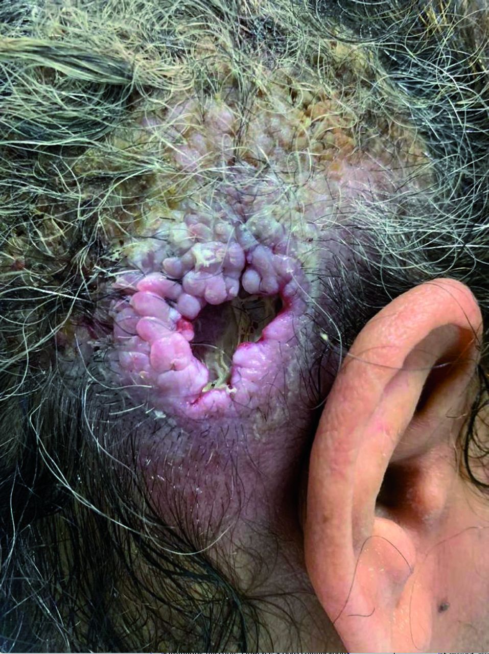
- Single vegetating verrucous plaque with a peripheral ulcer discharging puss with multiple moving maggots on the right temporoparietal scalp.

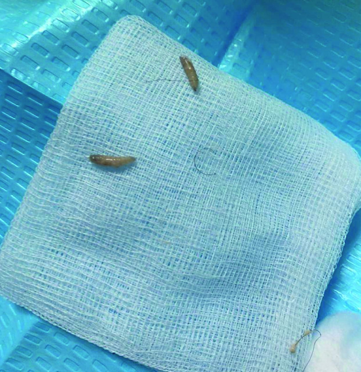
- Multiple moving maggots extracted from the ulcer.
Diagnostic assessment
The patient was admitted with contact precautions. An incisional skin tissue biopsy was obtained from the borders of the ulcer for histopathology and fungal and bacterial cultures. No fungal growth or acid-fast bacilli were detected, but a light growth of Staphylococcus aureus methicillin-sensitive bacteria was present. Histopathology with hematoxylin and eosin (H&E) staining showed acanthosis and a supra-basal cleft with acantholysis, suggestive of PV with no evidence of malignancy. Furthermore, head computed tomography without contrast showed right temporal, periauricular, and occipital/suboccipital subcutaneous infective processes with associated signs of osteomyelitis. No involvement of the mastoid bone, external auditory canal, or intracranial invasion was observed. Complete blood count and differential, liver function test, urea, and electrolytes were within normal limits, whereas C-reactive protein (119 mg/L) and erythrocyte sedimentation rate (46 mm/h) were elevated.
Therapeutic intervention
Wound cleaning with hydrogen peroxide and occlusion with petroleum jelly were carried out twice daily. The maggots were slowly pulled out using forceps to prevent tearing.
Follow-up and outcomes
The patient improved and was discharged after 3 days.
Discussion
Cutaneous myiasis is a benign, self-limiting infection. However, serious complications and even death can occur in certain types that cause local destruction and invasion of deep tissue, such as obligate myiasis. Clinically, the signs and symptoms depend on the type of infesting flies and their location in the body. Characteristically, patients complain of pain, movement under the skin, and maggots at the infested site. In addition, more serious signs, such as fever, secondary infections, and leukocytosis, can occur. Dermatological conditions, such as open wounds, chronic non-healing skin lesions, and hyperkeratosis are predisposing factors for cutaneous myiasis.3 Such conditions include pemphigus vulgaris, psoriasis, seborrheic keratosis, seborrheic dermatitis, herpes zoster virus infection, condyloma acuminatum, syphilis, leprosy, cutaneous malignancies, pediculosis, and impetigo.
There are 3 types of cutaneous myiasis: furuncular, migratory, and wound. Wound myiasis is most commonly caused by Cochliomyia hominivorax, Chrysomya bezziana, and Wohlfahrtia magnifica, which are obligatory and may cause the destruction of healthy tissue. In addition, M. domestica species can also less commonly cause wound myiasis. It is a facultative synanthropic fly, also known as “house fly”. Female flies lay approximately 500 eggs in batches that hatch into maggots approximately 24 hours later. The grown maggots are cylindrical with a tapered anterior segment and a pair of dark hooks. In addition, they are creamy white in color and measure 8-12 mm in length.4 Furthermore, these houseflies favor decaying tissue and are attracted to a foul smell. Therefore, neglected wounds, chronic necrotic cutaneous lesions, and poor hygiene are favorable conditions for housefly myiasis.
In Saudi Arabia, M. domestica comprises 12% of cutaneous myiasis cases in animals. They predominated in Buraidah (central region), Jeddah and Makkah (western region), and Asir (southwestern region).5 On the other hand, only 2% of cases were caused by M. domestica in Jazan (southern region), and none in Riyadh (central region).6 Nevertheless, these cases were only found in animals and not in humans. Only few human cutaneous myiasis cases have been reported in Saudi Arabia, and none have been caused by M. domestica. In Taif (southwestern region), 2 Dermatobia hominis furuncular myiasis and one with unidentified larvae have been reported in humans.7 In addition, Cordylobia anthropophaga furuncular myiasis furuncular myiasis was reported in the Southwestern region (Albaha and Asir).8 Similarly, a single case of furuncular myiasis with Cordylobia anthropophaga was reported in Alkhobar (eastern region). Lastly, a single case of wound myiais was reported in Makkah (western region) and was caused by Sarcophaga. Different types of wounds with myiasis have been reported in the literature, but only one patient with PV has been previously reported.9
Myiasis can be prevented by proper cleaning and coverage of open wounds. In addition, controlling adult flies with insecticides and good sanitation are necessary. Once an individual is infected, self-isolation should be carried out and, if possible, bedsheets and clothes should be changed daily. Treatment options include manual removal of maggots, occlusion, surgical removal, and larvicides. Occlusion works by suffocating larvae and forcing them out of the wound using petrolatum, beeswax, paraffin, mineral oil, turpentine oil, or a mixture of turpentine oil, and chloroform. Furthermore, surgical intervention may be required to remove deep-seated or dead larvae to prevent inflammatory foreign body reactions and granulomas and debridement of necrotic tissue may be necessary. Ivermectin, a broad-spectrum antiparasitic drug, can be used topically and orally to treat myiasis.
In conclusion, we would like to raise the awareness regarding the possibility of cutaneous myiasis caused by M. domestica in Riyadh, Saudi Arabia. At risk individuals need to be educated regarding wound dressing and prevention of such infestations. These houseflies favor decaying tissue and are attracted to the foul smell, as reported in previous studies. Therefore, neglected wounds, chronic necrotic cutaneous lesions, and poor hygiene are favorable conditions for housefly myiasis. Similarly, our patient had PV, a chronic cutaneous condition that presents with recurrent erosions and ulcers. Although our patient was mentally healthy, scalp wounds could be neglected because of a lack of visualization.
Acknowledgment
The authors gratefully acknowledge Editage (www.editage.com) for the English language editing.
Footnotes
Disclosure. Authors have no conflict of interests, and the work was not supported or funded by any drug company.
- Received January 5, 2023.
- Accepted August 2, 2023.
- Copyright: © Saudi Medical Journal
This is an Open Access journal and articles published are distributed under the terms of the Creative Commons Attribution-NonCommercial License (CC BY-NC). Readers may copy, distribute, and display the work for non-commercial purposes with the proper citation of the original work.
In this issue


Jump to section
Related Articles
Cited By...
- No citing articles found.