
Main menu

  • Home
  • Content
    • Latest
    • Archive
    • home
  • Info for
    • Authors
    • Reviewers
    • Subscribers
    • Institutions
    • Advertisers
    • Join SMJ
  • About Us
    • About Us
    • Editorial Office
    • Editorial Board
  • More
    • Advertising
    • Alerts
    • Feedback
    • Folders
    • Help
  • Other Publications
    • NeuroSciences Journal

User menu

  • My alerts
  • Log in

Search

  • Advanced search
Saudi Medical Journal
  • Other Publications
    • NeuroSciences Journal
  • My alerts
  • Log in
Saudi Medical Journal

Advanced Search

  • Home
  • Content
    • Latest
    • Archive
    • home
  • Info for
    • Authors
    • Reviewers
    • Subscribers
    • Institutions
    • Advertisers
    • Join SMJ
  • About Us
    • About Us
    • Editorial Office
    • Editorial Board
  • More
    • Advertising
    • Alerts
    • Feedback
    • Folders
    • Help
  • Follow psmmc on Twitter
  • Visit psmmc on Facebook
  • RSS
Research ArticleOriginal Article
Open Access

Tall cell variant papillary thyroid carcinoma in Saudi patients

A clinicopathological and outcomes analysis

Khalid H. Al-Qahtani, Mutahir A. Tunio, Mushabbab Al Asiri, Yasser Bayoumi, Walaa A. Alshehri, Naji J. Aljohani, Ahmed Amir Ali and Hanadi Fatani
Saudi Medical Journal November 2016, 37 (11) 1220-1224; DOI: https://doi.org/10.15537/smj.2016.11.15480
Khalid H. Al-Qahtani
From the Department of Otolaryngology-Head & Neck Surgery (Al-Qahtani), College of Medicine (Alshehri), King Saud University, the Department of Radiation Oncology (Tunio, Al Asiri, Ali), the Department of Histopathology (Fatani), Comprehensive Cancer Center, the Department Endocrinology and Thyroid Oncology (Aljohani), King Fahad Medical City, Riyadh, Kingdom of Saudi Arabia, and the Department of Radiation Oncology (Bayoumi), National Cancer Institute, Cairo University, Cairo, Egypt
MSc, FRCSc
  • Find this author on Google Scholar
  • Find this author on PubMed
  • Search for this author on this site
Mutahir A. Tunio
From the Department of Otolaryngology-Head & Neck Surgery (Al-Qahtani), College of Medicine (Alshehri), King Saud University, the Department of Radiation Oncology (Tunio, Al Asiri, Ali), the Department of Histopathology (Fatani), Comprehensive Cancer Center, the Department Endocrinology and Thyroid Oncology (Aljohani), King Fahad Medical City, Riyadh, Kingdom of Saudi Arabia, and the Department of Radiation Oncology (Bayoumi), National Cancer Institute, Cairo University, Cairo, Egypt
MBBS, FCPS
  • Find this author on Google Scholar
  • Find this author on PubMed
  • Search for this author on this site
  • For correspondence: [email protected]
Mushabbab Al Asiri
From the Department of Otolaryngology-Head & Neck Surgery (Al-Qahtani), College of Medicine (Alshehri), King Saud University, the Department of Radiation Oncology (Tunio, Al Asiri, Ali), the Department of Histopathology (Fatani), Comprehensive Cancer Center, the Department Endocrinology and Thyroid Oncology (Aljohani), King Fahad Medical City, Riyadh, Kingdom of Saudi Arabia, and the Department of Radiation Oncology (Bayoumi), National Cancer Institute, Cairo University, Cairo, Egypt
MBBS, FRCSI
  • Find this author on Google Scholar
  • Find this author on PubMed
  • Search for this author on this site
Yasser Bayoumi
From the Department of Otolaryngology-Head & Neck Surgery (Al-Qahtani), College of Medicine (Alshehri), King Saud University, the Department of Radiation Oncology (Tunio, Al Asiri, Ali), the Department of Histopathology (Fatani), Comprehensive Cancer Center, the Department Endocrinology and Thyroid Oncology (Aljohani), King Fahad Medical City, Riyadh, Kingdom of Saudi Arabia, and the Department of Radiation Oncology (Bayoumi), National Cancer Institute, Cairo University, Cairo, Egypt
MD
  • Find this author on Google Scholar
  • Find this author on PubMed
  • Search for this author on this site
Walaa A. Alshehri
From the Department of Otolaryngology-Head & Neck Surgery (Al-Qahtani), College of Medicine (Alshehri), King Saud University, the Department of Radiation Oncology (Tunio, Al Asiri, Ali), the Department of Histopathology (Fatani), Comprehensive Cancer Center, the Department Endocrinology and Thyroid Oncology (Aljohani), King Fahad Medical City, Riyadh, Kingdom of Saudi Arabia, and the Department of Radiation Oncology (Bayoumi), National Cancer Institute, Cairo University, Cairo, Egypt
MBBS
  • Find this author on Google Scholar
  • Find this author on PubMed
  • Search for this author on this site
Naji J. Aljohani
From the Department of Otolaryngology-Head & Neck Surgery (Al-Qahtani), College of Medicine (Alshehri), King Saud University, the Department of Radiation Oncology (Tunio, Al Asiri, Ali), the Department of Histopathology (Fatani), Comprehensive Cancer Center, the Department Endocrinology and Thyroid Oncology (Aljohani), King Fahad Medical City, Riyadh, Kingdom of Saudi Arabia, and the Department of Radiation Oncology (Bayoumi), National Cancer Institute, Cairo University, Cairo, Egypt
MD, FRCPC
  • Find this author on Google Scholar
  • Find this author on PubMed
  • Search for this author on this site
Ahmed Amir Ali
From the Department of Otolaryngology-Head & Neck Surgery (Al-Qahtani), College of Medicine (Alshehri), King Saud University, the Department of Radiation Oncology (Tunio, Al Asiri, Ali), the Department of Histopathology (Fatani), Comprehensive Cancer Center, the Department Endocrinology and Thyroid Oncology (Aljohani), King Fahad Medical City, Riyadh, Kingdom of Saudi Arabia, and the Department of Radiation Oncology (Bayoumi), National Cancer Institute, Cairo University, Cairo, Egypt
MBBS
  • Find this author on Google Scholar
  • Find this author on PubMed
  • Search for this author on this site
Hanadi Fatani
From the Department of Otolaryngology-Head & Neck Surgery (Al-Qahtani), College of Medicine (Alshehri), King Saud University, the Department of Radiation Oncology (Tunio, Al Asiri, Ali), the Department of Histopathology (Fatani), Comprehensive Cancer Center, the Department Endocrinology and Thyroid Oncology (Aljohani), King Fahad Medical City, Riyadh, Kingdom of Saudi Arabia, and the Department of Radiation Oncology (Bayoumi), National Cancer Institute, Cairo University, Cairo, Egypt
MBBS
  • Find this author on Google Scholar
  • Find this author on PubMed
  • Search for this author on this site
  • Article
  • Figures & Data
  • eLetters
  • Info & Metrics
  • References
  • PDF
Loading

Abstract

Objectives: To evaluate the clinicopathological features and treatment outcomes of papillary thyroid carcinoma tall cell variant (PTC-TCV) in Saudi population.

Methods: This retrospective study were medical records of 776 treated PTC patients between December 2007 and 2015, at King Fahad Medical City and King Khalid University Hospital, Riyadh, Kingdom of Saudi Arabia was probed for PTC-TCV. Total 42 (5.4%) patients were found to have PTC-TCV, which were investigated for demographic, symptoms, histopathological features, and treatment outcomes locoregional control (LRC), distant metastasis control (DMC), disease free survival (DFS), and overall survival (OS) rates.

Results: Mean age of cohort was 52.3 years (range: 46-80), with female predominance (73.8%). Mean tumor size was 3.62 cm (range: 0.4-10). Rates of LVSI (59.5%), positive pathological lymph nodes (66.7%), multifocality (42.9%) and extrathyroidal extension, (45.3%). Median follow-up was 37.4 months (range: 6-60). Local recurrence rate were seen in 6/42 (14.2%) patients and 8/42 (19%) developed distant metastasis. The 5 year rates of LRC (82.3%), DMC (77.8%), DFS (69.2%), and OS (86.7%) multivariate analysis showed PTC-TVC as an important independent prognosticator (odds ratio: 4.2; 95% confidence interval: 1.79-7.3; p=0.03)

Conclusion: Papillary thyroid carcinoma tall cell variant is associated with aggressive biological behavior.

Most of papillary thyroid carcinoma (PTC) are associated with an exceptional long-term treatment outcomes including DFS and OS rates; however, some of histopathological variants of PTC have been found with more aggressive biologic behavior and poor perspective.1 Among these histopathological variants, tall cell variant (TCV) is considered the most aggressive variant of PTC.2 Mainly, PTC-TCV is characterized by a height greater than twice its width, eosinophilic cytoplasm, basaloid nuclei along with the conventional characteristics of PTC.3 In 2004, World Health Organization (WHO) defined a PTC as TCV if it contained 50% or more tall cells.4 Some controversies regarding the proportion of tall cell (30-75%) required for diagnosis of PTC-TCV still persist; however, most of institutes accept the threshold of > 30% to confirm PTC-TCV5 TCV has some distinctive differences with classic variant of PTC; A) frequent presence of lymphovascular space invasion (LVSI) and extrathyroidal extension (ETE), B) greater prevalence of pathological positive lymph nodes and distant metastases at the time of diagnosis, and C) its 10-year DFS and OS rates have been reported to be approximately 10-15% lower than that of classic PTC.6,7 However, data around PTC-TCV in Saudi population is very scanty. We undertook the present study to evaluate the various clinicopathological characteristics and treatment outcomes of PTC-TCV in Saudi population.

Methods

Patients were reviewed retrospectively. After an authorized approval from the institutional review committee, medical charts of 42 patients with PTC-TCV, who were treated at King Fahad Medical City and King Khalid University Hospital, Riyadh, Kingdom of Saudi Arabia between December 2007-2015, were investigated using database system. Inclusion criteria was patients PTC-TCV histopathology of all ages. Exclusion criteria was defined <10% proportion of tall cells in histopathological specimens. Patients with PTC-TCV were retrieved in the following fashion; clinical variables including age at the diagnosis, gender, and symptoms at the time of diagnosis were reviewed. Histopathological assessment was performed by a skilled histopathologist. Different histopathological variables including the size of primary tumor, presence of LVSI and ETE, multifocality, positive pathological lymph nodes, and tumor lymph node, and metastasis staging were collected. Data regarding different treatment modalities, including the type of thyroidectomy, neck dissection, and adjuvant radioactive iodine (RAI) ablation were also recalled.

Statistical analysis

The primary endpoint was DFS. Secondary endpoints were the locoregional control (LRC), distant metastasis control (DMC) and OS rates. The DFS was defined as, the duration between the date of thyroidectomy and the date of documented all-site disease recurrence, death from cancer and/or last follow-up (censored). All probabilities of LRC, DMC, DFS, and OS rates were shown using the Kaplan-Meier method (comparisons for different survival curves were performed using log rank). All statistical analyses were performed using the the Statistical package for Social Sciences (IBM Corp., Armonk, NY, USA) version 24.

Results

Among the 776 PTC patients managed in our centers between December 2007-2015, 42 patients (5.4%) were found to have PTC-TCV. Mean age of cohort was 52.3 years (range: 46-80), with female predominance (73.8%). Different attributes of cohort are shown in Table 1.

View this table:
  • View inline
  • View popup
  • Download powerpoint
Table 1

Characteristics of 42 patients with PTC-TCV, who were treated at King Fahad Medical City and King Khalid University Hospital, Riyadh, Kingdom of Saudi Arabia.

All patients underwent total thyroidectomy and near-total thyroidectomy. Most of cohort underwent central and lateral neck dissection (78.6%). Mean tumor size was 3.62 cm (range: 0.4-10). Lymphovascular space invasion was observed in 25/42 patients (59.5%), while positive pathological lymph node were seen in 28/42 patients (66.7%). Positive pathological lymph nodes were seen in 62.5% of patients with tumor size ≤ 2cm (16 patients) as compared with 28 patients with tumor size >2cm (64.3%) with p=0.20. Multifocality was seen in 18/42 patients (42.9%). No correlation was found between size of primary with multifocality, ETE (p=0.12, 0.17) and LVSI (p=0.21). Triiodothyronine (T3) and thyroxine (T4) stage was seen in 19 patients (45.3%). Two patients (4.8%) had distant metastasis at the time of diagnosis.

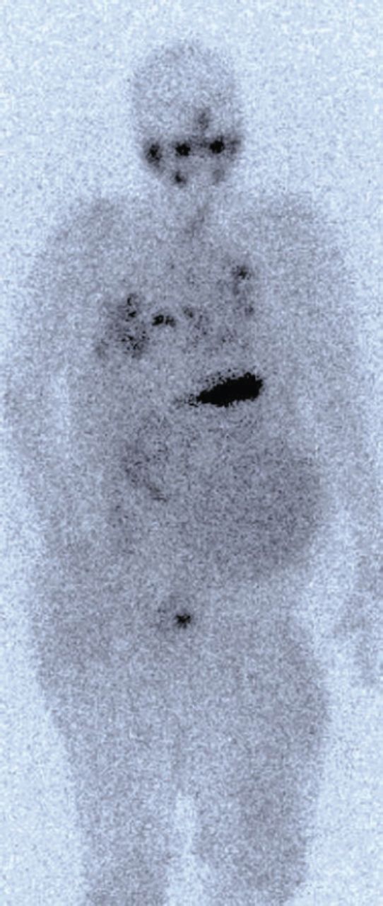
Adjuvant RAI was given to most of cases (39 patients, 92.8%). Median delay between surgery and adjuvant RAI was 8.5 weeks (range: 6-13). Doses were, 30 millicurie (mCi) in 5 patients (11.8%) for patients with primary size ≤2 cm without other risk factors; 100 mCi in 9 patients (21.4%) with positive margins, ETE and LVSI; 150 mCi in 23 patients (54.7%) with positive pathological lymph nodes and 200 mCi in 2 patients (4.8%) with distant metastasis. Postoperative radiation therapy (PORT) to thyroid bed and regional lymph nodes was given to 4 patients (3 patients with skeletal muscle invasion, one patient with tracheal invasion). Mean dose for PORT was 60 Gy (range: 60-66 Gy), given as daily 2 Gy/fraction, 5 days/week over 6-6.5 weeks (30-33 fractions). Median follow-up was 37.4 months (range: 6-60). Local recurrence rate (LRRs) were seen in 6/42 (14.2%) patients (Table 2). The LRRs were salvaged with neck resection (3 patients) followed by RAI (2 patients). Distant metastasis was seen in 8/42 (19%) patients. Among whom, 5 patients were treated with RAI, one patients with brain metastasis was managed with resection and radiation therapy and one patient with bone lesion was treated with palliative radiation therapy (Figure 1).

View this table:
  • View inline
  • View popup
  • Download powerpoint
Table 2

Treatment outcomes in our tall- cell variant papillary thyroid carcinoma.

Figure 1
  • Download figure
  • Open in new tab
  • Download powerpoint
Figure 1

Whole body scintigraphy showing iodine avid bilateral lungs, neck, and right femur uptake.

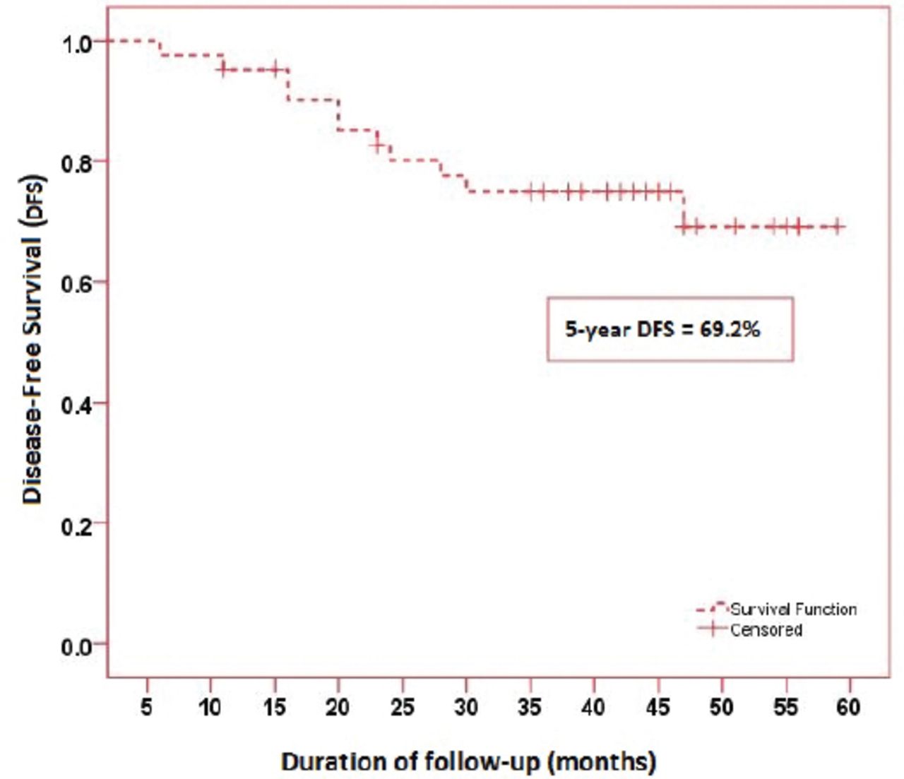
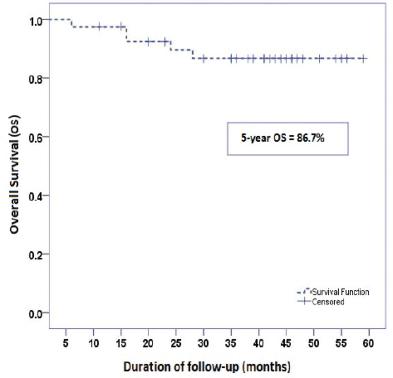
At the time of analysis, we found 5 year 82.3% LRC, 77.8% DMC, 69.2% DFS, and 86.7% OS (Figure 2 & 3). Further, univariate and multivariate analyses of different prognostic components for DFS and OS showed PTC-TVC as an important independent prognosticator (Odds ratio: 4.2; 95% confidence interval: 1.79-7.3; p=0.03).

Figure 2
  • Download figure
  • Open in new tab
  • Download powerpoint
Figure 2

Disease free survival of 42 cohort patients.

Figure 3
  • Download figure
  • Open in new tab
  • Download powerpoint
Figure 3

Overall survival of cohort 42 cohort patients.

Discussion

In the present study, PTC-TCV constituted 5.4% of all PTC cases, which is in agreement with the reported prevalence of 1.3-13% among of all PTC.1,8 The reason for this variation in reported prevalence can be explained by difficulty in distinguishing PTC-TCV from other histopathological variants, thus PTC-TCV is believed to be underdiagnosed in most of cases.9 In our cohort, women accounted for (73.8%) with the female to male ratio 3:1 is also in agreement with reported literature.10 Similarly, mean age of 52.3 years in our cohort study, is also in agreement with reported data.11 Further more, the multifocality (42.9%), pathological positive lymph nodes (66.7%) and distant metastasis (7.2%) at the time of diagnosis in our cohort were found to be similar to those reported in various studies.12 However, in disagreement with other studies, the percentage of ETE was comparatively low in our cohort (45.3% versus reported 63.9%). Mean primary tumor size was comparatively larger in our cohort (3.6 cm versus reported approximately 2.5-3 cm).1,5 Michels JJ et al13 also reported larger primary tumor size in their study of 56 patients with TCV. Due to these aggressive biological behavior, all patients in our cohort underwent total thyroidectomy. Although neck dissection (central and lateral) was performed in most of patients (78.6%), we recommend routine prophylactic central lymph node dissection at the time of thyroidectomy in PTC-TVC patients.1,13 Low occurrence of LRRs and distant metastasis in group, which received adjuvant RAI as part of management in our cohort, supports the notion that adjuvant RAI should be considered for all PTC-TVC patients, regardless of tumor size and lymph node status.6,14 In disagreement with similar reported data, PTC-TCV in our cohort had lower 5-year DFS rate (69.2%) after extensive treatment, which can be explained by our high risk cohort itself.15 In agreement with other related data, we found PTC-TVC as an important negative prognosticator.16

There were limitations in our study; A) small sample size, B) BRAF mutations were not analysed in our cohort, as current evidence suggests that PTC-TVC is associated with 80-100% BRAF mutations as compared with 63-83% in classic PTC,17 and C) rearranged during transcription/PTC (RET/PTC) gene rearrangements were also not examined. Among 13 different RET/PTC oncogenes, RET/PTC1 and RET/PTC3 represent the most common types.18 Recent data19 has shown a striking correlation between the RET/PTC3 rearrangement and the PTC-TCV phenotype (35.8% positive), which suggests that molecular pathogenesis in PTC-TCV may be different, which needs further investigations.

In conclusion, PTC-TVC is associated with aggressive biological behavior. Our results were found in agreement with previous studies. However, more molecular studies are warranted to address the role of BRAF mutations, and Rearranged during Transcription/PTC (RET/PTC) gene rearrangements in PTC-TVC.

Copyright

Whenever a manuscript contains material (tables, figures, etc.) which is protected by copyright (previously published), it is the obligation of the author to obtain written permission from the holder of the copyright (usually the publisher) to reproduce the material in Saudi Medical Journal. This also applies if the material is the authors own work. Please submit copies of the material from the source in which it was first published.

Footnotes

  • Disclosure. Authors have no conflict of interest, and the work was not supported or funded by any drug company.

  • Received June 6, 2016.
  • Accepted August 3, 2016.
  • Copyright: © Saudi Medical Journal

This is an open-access article distributed under the terms of the Creative Commons Attribution-Noncommercial-Share Alike 3.0 Unported, which permits unrestricted use, distribution, and reproduction in any medium, provided the original work is properly cited.

References

  1. ↵
    1. Wang X,
    2. Cheng W,
    3. Liu C,
    4. Li J
    (2016) Tall cell variant of papillary thyroid carcinoma: current evidence on clinicopathologic features and molecular biology. Oncotarget.
  2. ↵
    1. Axelsson TA,
    2. Hrafnkelsson J,
    3. Olafsdottir EJ,
    4. Jonasson JG
    (2015) Tall cell variant of papillary thyroid carcinoma: a population-based study in Icel and Thyroid, 25:216–220.
  3. ↵
    1. Solomon A,
    2. Gupta PK,
    3. LiVolsi VA,
    4. Baloch ZW
    (2002) Distinguishing tall cell variant of papillary thyroid carcinoma from usual variant of papillary thyroid carcinoma in cytologic specimens. Diagn Cytopathol 27:143–148.
    OpenUrlCrossRefPubMed
  4. ↵
    1. DeLellis RA LR,
    2. Heitz PU,
    3. Eng C
    (2004) World Health Organization Classification of Tumours: Pathology and Genetics of Tumours of Endocrine Organs (IARC Press, Lyon), pp 57–66.
  5. ↵
    1. Lam AK,
    2. Lo CY,
    3. Lam KS
    (2005) Papillary carcinoma of thyroid: A 30-yr clinicopathological review of the histological variants. Endocr Pathol 16:323–330.
    OpenUrlCrossRefPubMed
  6. ↵
    1. Ghossein R,
    2. Livolsi VA
    (2008) Papillary thyroid carcinoma tall cell variant. Thyroid 18:1179–1181.
    OpenUrlCrossRefPubMed
  7. ↵
    1. Leung AK,
    2. Chow SM,
    3. Law SC
    (2008) Clinical features and outcome of the tall cell variant of papillary thyroid carcinoma. Laryngoscope 118:32–38.
    OpenUrlCrossRefPubMed
  8. ↵
    1. Guan H,
    2. Vandenbussche CJ,
    3. Erozan YS,
    4. Rosenthal DL,
    5. Tatsas AD,
    6. Olson MT,
    7. et al.
    (2013) Can the tall cell variant of papillary thyroid carcinoma be distinguished from the conventional type in fine needle aspirates?A cytomorphologic study with assessment of diagnostic accuracy. Acta Cytol 57:534–542.
    OpenUrl
  9. ↵
    1. Okuyucu K,
    2. Alagoz E,
    3. Arslan N,
    4. Emer O,
    5. Ince S,
    6. Deveci S,
    7. et al.
    (2015) Clinicopathologic features and prognostic factors of tall cell variant of papillary thyroid carcinoma: comparison with classic variant of papillary thyroid carcinoma. Nucl Med Commun 36:1021–1025.
    OpenUrl
  10. ↵
    1. LiVolsi VA
    (2010) Papillary carcinoma tall cell variant (TCV): a review. Endocrine pathology 21:12–15.
    OpenUrlCrossRefPubMed
  11. ↵
    1. Kazaure HS,
    2. Roman SA,
    3. Sosa JA
    (2012) Aggressive variants of papillary thyroid cancer: incidence, characteristics and predictors of survival among 43,738 patients. Ann Surg Oncol 19:1874–1880.
    OpenUrlCrossRefPubMed
  12. ↵
    1. Regalbuto C,
    2. Malandrino P,
    3. Frasca F,
    4. Pellegriti G,
    5. Le Moli R,
    6. Vigneri R,
    7. et al.
    (2013) The tall cell variant of papillary thyroid carcinoma: clinical and pathological features and outcomes. J Endocrinol Invest 36:249–254.
    OpenUrl
  13. ↵
    1. Michels JJ,
    2. Jacques M,
    3. Henry-Amar M,
    4. Bardet S
    (2007) Prevalence and prognostic significance of tall cell variant of papillary thyroid carcinoma. Hum Pathol 38:212–219.
    OpenUrlCrossRefPubMed
  14. ↵
    1. Roman S,
    2. Sosa JA
    (2013) Aggressive variants of papillary thyroid cancer. Curr Opin Oncol 25:33–38.
    OpenUrlPubMed
  15. ↵
    1. Barbu CG,
    2. Florin A,
    3. Neamtu MC,
    4. Avramescu ET,
    5. Terzea D,
    6. Miron A,
    7. et al.
    (2015) Papillary thyroid carcinoma with anaplastic dedifferentiation in the lymph node metastasis - a rare form of presentation even for a tall cell variant. Rom J Morphol Embryol 56:527–531.
    OpenUrl
  16. ↵
    1. Jalisi S,
    2. Ainsworth T,
    3. Lavalley M
    (2010) Prognostic outcomes of tall cell variant papillary thyroid cancer: a meta-analysis. J Thyroid Res 2010:325602.
    OpenUrlCrossRefPubMed
  17. ↵
    1. Basolo F,
    2. Torregrossa L,
    3. Giannini R,
    4. Miccoli M,
    5. Lupi C,
    6. Sensi E,
    7. et al.
    (2010) Correlation between the BRAF V600E mutation and tumor invasiveness in papillary thyroid carcinomas smaller than 20 millimeters: analysis of 1060 cases. J Clin Endocrinol Metab 95:4197–4205.
    OpenUrlCrossRefPubMed
  18. ↵
    1. Gherardi E,
    2. Birchmeier W,
    3. Birchmeier C,
    4. Vande Woude G
    (2012) Targeting MET in cancer: rationale and progress. Nat Rev Cancer 12:89–103.
    OpenUrlCrossRefPubMed
  19. ↵
    1. Nikiforov YE
    (2002) RET/PTC rearrangement in thyroid tumors. Endocr Pathol 13:3–16.
    OpenUrlCrossRefPubMed
View Abstract
PreviousNext
Back to top

In this issue

Saudi Medical Journal: 37 (11)
Saudi Medical Journal
Vol. 37, Issue 11
1 Nov 2016
  • Table of Contents
  • Cover (PDF)
  • Index by author
Print
Download PDF
Email Article

Thank you for your interest in spreading the word on Saudi Medical Journal.

NOTE: We only request your email address so that the person you are recommending the page to knows that you wanted them to see it, and that it is not junk mail. We do not capture any email address.

Enter multiple addresses on separate lines or separate them with commas.
Tall cell variant papillary thyroid carcinoma in Saudi patients
(Your Name) has sent you a message from Saudi Medical Journal
(Your Name) thought you would like to see the Saudi Medical Journal web site.
Citation Tools
Tall cell variant papillary thyroid carcinoma in Saudi patients
Khalid H. Al-Qahtani, Mutahir A. Tunio, Mushabbab Al Asiri, Yasser Bayoumi, Walaa A. Alshehri, Naji J. Aljohani, Ahmed Amir Ali, Hanadi Fatani
Saudi Medical Journal Nov 2016, 37 (11) 1220-1224; DOI: 10.15537/smj.2016.11.15480

Citation Manager Formats

  • BibTeX
  • Bookends
  • EasyBib
  • EndNote (tagged)
  • EndNote 8 (xml)
  • Medlars
  • Mendeley
  • Papers
  • RefWorks Tagged
  • Ref Manager
  • RIS
  • Zotero
Share
Tall cell variant papillary thyroid carcinoma in Saudi patients
Khalid H. Al-Qahtani, Mutahir A. Tunio, Mushabbab Al Asiri, Yasser Bayoumi, Walaa A. Alshehri, Naji J. Aljohani, Ahmed Amir Ali, Hanadi Fatani
Saudi Medical Journal Nov 2016, 37 (11) 1220-1224; DOI: 10.15537/smj.2016.11.15480
Twitter logo Facebook logo Mendeley logo
  • Tweet Widget
  • Facebook Like
  • Google Plus One
Bookmark this article

Jump to section

  • Article
    • Abstract
    • Methods
    • Results
    • Discussion
    • Footnotes
    • References
  • Figures & Data
  • eLetters
  • References
  • Info & Metrics
  • PDF

Related Articles

  • No related articles found.
  • PubMed
  • Google Scholar

Cited By...

  • No citing articles found.
  • Google Scholar

More in this TOC Section

  • Hematological parameters in recent and past dengue infections in Jazan Province, Saudi Arabia
  • Longitudinal analysis of foodborne disease outbreaks in Saudi Arabia
  • Psychological stress and its association with bronchial asthma in Saudi Arabia
Show more Original Article

Similar Articles

CONTENT

  • home

JOURNAL

  • home

AUTHORS

  • home
Saudi Medical Journal

© 2025 Saudi Medical Journal Saudi Medical Journal is copyright under the Berne Convention and the International Copyright Convention.  Saudi Medical Journal is an Open Access journal and articles published are distributed under the terms of the Creative Commons Attribution-NonCommercial License (CC BY-NC). Readers may copy, distribute, and display the work for non-commercial purposes with the proper citation of the original work. Electronic ISSN 1658-3175. Print ISSN 0379-5284.

Powered by HighWire