
Main menu

  • Home
  • Content
    • Latest
    • Archive
    • home
  • Info for
    • Authors
    • Reviewers
    • Subscribers
    • Institutions
    • Advertisers
    • Join SMJ
  • About Us
    • About Us
    • Editorial Office
    • Editorial Board
  • More
    • Advertising
    • Alerts
    • Feedback
    • Folders
    • Help
  • Other Publications
    • NeuroSciences Journal

User menu

  • My alerts
  • Log in

Search

  • Advanced search
Saudi Medical Journal
  • Other Publications
    • NeuroSciences Journal
  • My alerts
  • Log in
Saudi Medical Journal

Advanced Search

  • Home
  • Content
    • Latest
    • Archive
    • home
  • Info for
    • Authors
    • Reviewers
    • Subscribers
    • Institutions
    • Advertisers
    • Join SMJ
  • About Us
    • About Us
    • Editorial Office
    • Editorial Board
  • More
    • Advertising
    • Alerts
    • Feedback
    • Folders
    • Help
  • Follow psmmc on Twitter
  • Visit psmmc on Facebook
  • RSS
Case ReportCase Report
Open Access

Urogenital myiasis caused by Psychoda albipennis

Mehmet G. Culha, Kamuran Turker, Sule Ozsoy and Ege C. Serefoglu
Saudi Medical Journal December 2016, 37 (12) 1401-1403; DOI: https://doi.org/10.15537/smj.2016.12.16312
Mehmet G. Culha
From the Department of Urology (Culha), Istanbul Training & Research Hospital, tbe Department of Infectious Diseases (Turker), the Department of Pathology (Ozsoy), and the Department of Urology (Serefoglu), Bagcilar Training & Research Hospital, Istanbul, Turkey
MD
  • Find this author on Google Scholar
  • Find this author on PubMed
  • Search for this author on this site
Kamuran Turker
From the Department of Urology (Culha), Istanbul Training & Research Hospital, tbe Department of Infectious Diseases (Turker), the Department of Pathology (Ozsoy), and the Department of Urology (Serefoglu), Bagcilar Training & Research Hospital, Istanbul, Turkey
MD
  • Find this author on Google Scholar
  • Find this author on PubMed
  • Search for this author on this site
Sule Ozsoy
From the Department of Urology (Culha), Istanbul Training & Research Hospital, tbe Department of Infectious Diseases (Turker), the Department of Pathology (Ozsoy), and the Department of Urology (Serefoglu), Bagcilar Training & Research Hospital, Istanbul, Turkey
MD
  • Find this author on Google Scholar
  • Find this author on PubMed
  • Search for this author on this site
Ege C. Serefoglu
From the Department of Urology (Culha), Istanbul Training & Research Hospital, tbe Department of Infectious Diseases (Turker), the Department of Pathology (Ozsoy), and the Department of Urology (Serefoglu), Bagcilar Training & Research Hospital, Istanbul, Turkey
MD, FECSM
  • Find this author on Google Scholar
  • Find this author on PubMed
  • Search for this author on this site
  • For correspondence: [email protected]
  • Article
  • Figures & Data
  • eLetters
  • Info & Metrics
  • References
  • PDF
Loading

Abstract

Myiasis is a parasitic infestation of vertebrate animals caused by the eggs and larvae of flies within the Diptera species. Psychoda albipennis is a rare cause of urogenital myiasis in humans. We present the case of a 42-year-old male diagnosed with urogenital myiasis caused by Psychoda albipennis.

Myiasis is derived from the Greek word ‘myia’, which means fly. It is an infestation of the skin, tissues or organs of humans or other vertebrate animals caused by the eggs and larvae of flies within the Diptera species.1 Myiasis can manifest in various body parts, such as the eye, nose, ear, skin, gastrointestinal system and urogenital system.2 Psychoda albipennis (P. albipennis) can thrive in the environment of a moist bathroom and may cause urogenital myiasis in humans.3 This insect typically lives in tropical or subtropical regions and usually appears during the summer. We report the case of a 42-year-old male who was diagnosed with urogenital myiasis caused by P. albipennis.

Case Report

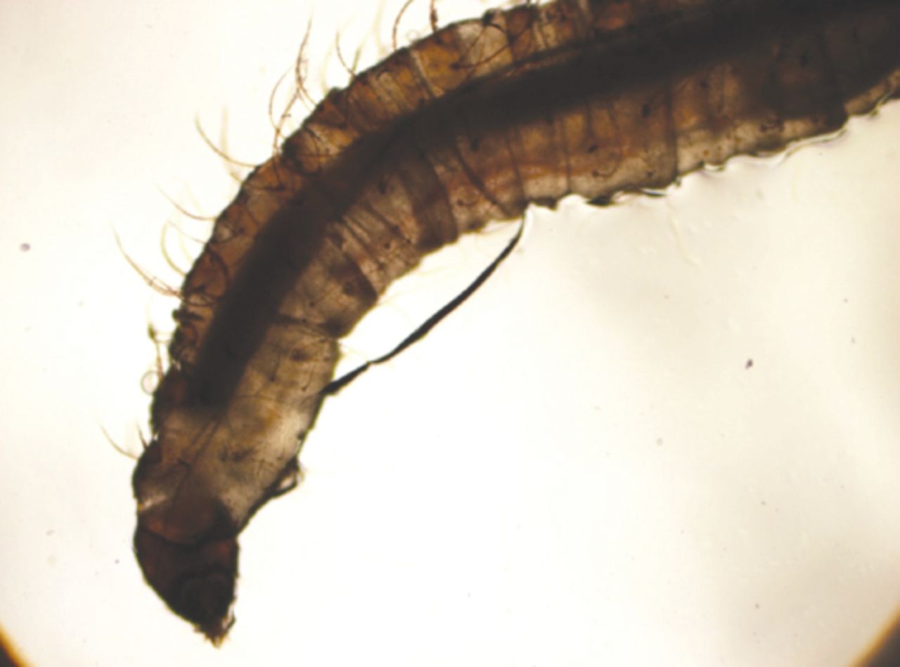
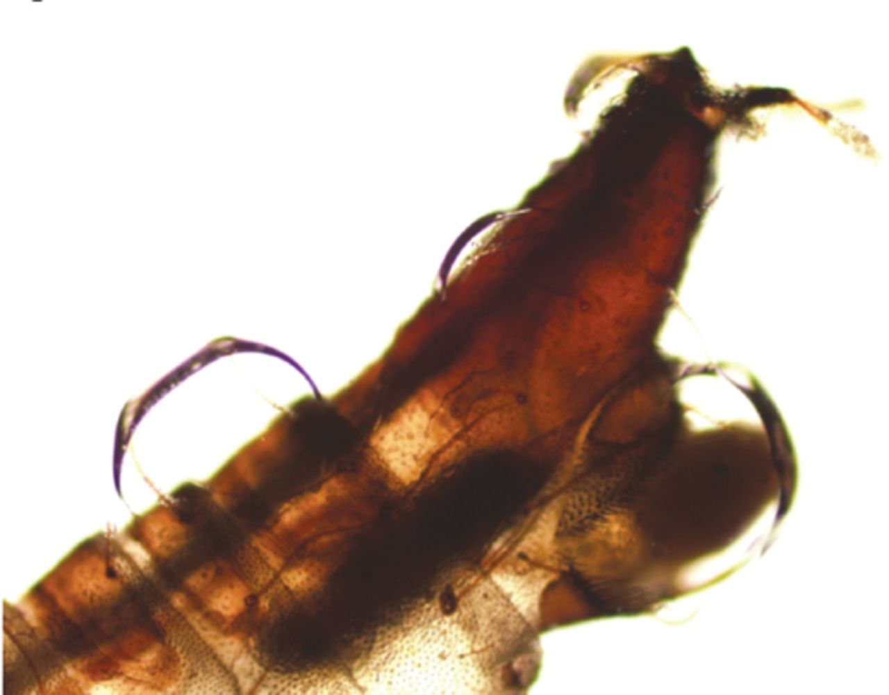
A 42-year-old male patient presented to the urology outpatient clinic with a complaint of moving, worm-like objects in his urine for the past 3 weeks. No history of travelling abroad, and his medical history was not significant. Physical examination was unremarkable other than mild obesity (body mass index: 32.28). During macroscopic examination of his urine, 2 0.8-cm-long worms were detected. The worms were examined under the microscope and they were diagnosed as being in the fourth larval stage of P. albipennis (Figures 1 & 2). Urinalysis results were remarkable, with 7 erythrocytes per high power field. The complete blood count and serum chemistry were within normal limits. Urinary ultrasonography did not reveal any pathology. The urine culture was negative for infection. He was scheduled for cystoscopic evaluation. He was also instructed to drink 3 liters of water per day and was prescribed oral nitrofurantoin 50 mg 3 times a day. Cystoscopic evaluation was performed one week after. Edema in the prostatic urethra was noted, with debris and sediment accumulation at the base of the bladder. He was discharged on the same day and he reported resolution of his symptoms at a follow-up visit one week postoperatively.

Figure 1
  • Download figure
  • Open in new tab
  • Download powerpoint
Figure 1

The fourth stage larvae of Psychoda albipennis.

Figure 2
  • Download figure
  • Open in new tab
  • Download powerpoint
Figure 2

Syphon of the Psychoda albipennis.

Discussion

Urogenital myiasis is a very rare clinical entity caused by Sarcophaga spp., Megaselia scalaris, Thyrsocnema incisilobata, Lucilia sericata, Eristalis tenax, Dermatobia hominis, Chrysomya bezziana, Fannia canilicularis, and P. albipennis.2,4-9 Psychoda albipennis is known to exist in temperate regions of Europe and China.10 It is common in various provinces of Turkey including Ankara, Edirne, Tekirdag, Istanbul, and Bursa.6 The larvae of P. albipennis thrive in damp and dirty areas, rotting fruit and vegetables, garbage piles and irrigation canals. The fourth stage larvae infest the urine of humans. Its gross appearance is of whitish-grey, slightly flat worms, approximately 3-5 mm in diameter. The larva has a double knob covered with long hair at the end as well as a syphon that narrows from the base to the tip.2,10

To our knowledge, this is the seventh case of urogenital myiasis caused by P. albipennis, and all previous reported cases were also from Turkey.2,3,5,6,11,12 Predisposing factors for urogenital myiasis are poor hygiene, limitation of mobility, urinary obstruction, sleeping without a blanket in the summer months and low socioeconomic levels.13 However, a significant portion of urogenital myiasis cases have been reported from developed countries and in patients with good hygiene.14 Our patient was living in an underdeveloped part of Istanbul; however, his personal hygiene appeared good. No other predisposing factors were identified. In patients with urogenital myiasis, symptoms often manifest as dysuria, polyuria, hematuria, flank pain, or vomiting.2,15,16 Urogenital myiasis can be misdiagnosed as an obstructing ureteral stone.6 Our patient did not present with any of these symptoms.

Treatment of urogenital myiasis varies according to localization of the infestation and severity of the symptoms. Removal of larvae, use of urinary tract antiseptics and antibiotics are all advocated, depending on the degree of symptoms.2,3,11,12 As with previously reported cases of urogenital myiasis caused by P. albipennis,2,6,11 we instructed our patient to drink 3 litres of water per day and prescribed a urinary tract antiseptic. We performed a cystoscopic examination to detect and remove the remaining larvae, none were observed at that time, most likely due to the one week delay between initial evaluation of the patient and time of the cystoscopy. However, signs of inflammation (edema and debris) were observed during the cystoscopy.

In conclusion, urogenital myiasis caused by P. albipennis is an extremely rare clinical entity. It can be managed with hydration and antiseptics. Health authorities must take appropriate precautions to maintain hygienic conditions, and urologists must be aware of these parasites for proper diagnosis and management of this infestation.

Footnotes

  • Disclosure. Authors have no conflict of interests, and the work was not supported or funded by any drug company.

  • Received July 21, 2016.
  • Accepted August 11, 2016.
  • Copyright: © Saudi Medical Journal

This is an open-access article distributed under the terms of the Creative Commons Attribution-Noncommercial-Share Alike 3.0 Unported, which permits unrestricted use, distribution, and reproduction in any medium, provided the original work is properly cited.

References

  1. ↵
    1. Cestari TF,
    2. Pessato S,
    3. Ramos-e-Silva M
    (2007) Tungiasis and myiasis. Clin Dermatol 25:158–164.
    OpenUrlCrossRefPubMed
  2. ↵
    1. Guven E,
    2. Kar S,
    3. Dogan N,
    4. Karaer Z
    (2008) [Urogenital myiasis caused by Psychoda albipennis in a woman]. Turkiye Parazitol Derg 32:174–176, Turkish.
    OpenUrlPubMed
  3. ↵
    1. Cicek M,
    2. Diker AI,
    3. Ipek DN,
    4. Tekin A,
    5. Dal T
    (2012) [Urogenital myiasis caused by Psychoda albipennis]. Turkiye Parazitol Derg 36:51–53, Turkish.
    OpenUrlPubMed
  4. ↵
    1. Raposo AA,
    2. Schettini AP,
    3. Massone C
    (2012) Concurrent primary and secondary myiasis on basal cell carcinoma. An Bras Dermatol 87:292–295.
    OpenUrlPubMed
  5. ↵
    1. Taylan-Ozkan A,
    2. Babur C,
    3. Kilic S,
    4. Nalbantoglu S,
    5. Dalkilic I,
    6. Mumcuoglu KY
    (2004) Urogenital myiasis caused by Psychoda albipennis (Diptera: Nematocera) in Turkey. Int J Dermatol 43:904–905.
    OpenUrlCrossRefPubMed
  6. ↵
    1. Demir AD,
    2. Iraz M,
    3. Ipek DN
    (2015) Urogenital myiasis caused by Psychoda albipennis in a child. Turk Pediatri Ars 50:65–68.
    OpenUrl
    1. Perez-Eid C,
    2. Mouffok N
    (1999) [Human urinary myiasis caused by Fannia canicularis (Diptera, Muscidae) larvae in Algeria]. Presse Med 28:580–581.
    OpenUrlPubMed
    1. Mumcuoglu I,
    2. Akarsu GA,
    3. Balaban N,
    4. Keles I
    (2005) Eristalis tenax as a cause of urinary myiasis. Scand J Infect Dis 37:942–943.
    OpenUrlCrossRefPubMed
  7. ↵
    1. Cilla G,
    2. Pico F,
    3. Peris A,
    4. Idigoras P,
    5. Urbieta M,
    6. Perez Trallero E
    (1992) [Human genital myiasis due to Sarcophaga]. Rev Clin Esp 190:189–190.
    OpenUrlPubMed
  8. ↵
    1. Zumpt F
    (1965) Myiasis in Man and Animals in the Old World (Butterwoths & Co. Ltd, London (UK)), p 267.
  9. ↵
    1. Kaya S,
    2. Arslan M,
    3. Karaer Z,
    4. Koksal I
    (2011) [Urogenital myiasis caused by Psychoda albipennis]. Turkish Parasitology Association 35:172–174.
    OpenUrl
  10. ↵
    1. Yenice MG,
    2. Demir T,
    3. Babur C,
    4. Nalbantoglu S,
    5. Kilic S
    (2011) [A case of urogenital myiasis caused by Psychoda albipennis (Diptera: Nematocera)]. Mikrobiyol Bul 45:558–564, Turkish.
    OpenUrlPubMed
  11. ↵
    1. Ramalingam S,
    2. Nurulhuda A,
    3. Bee LH
    (1980) Urogenital myiasis caused by Chrysomya bezziana (Diptera: Calliphoridae) in peninsular Malaysia. Southeast Asian J Trop Med Public Health 11:405–407.
    OpenUrlPubMed
  12. ↵
    1. Blejter J
    (2012) Tracheostomy wound myiasis in a child: case report and review of the literature. Case Rep Pediatr 2012:317862.
    OpenUrlPubMed
  13. ↵
    1. Passos MR1,
    2. Carvalho AV,
    3. Dutra AL,
    4. Goulart Filho RA,
    5. Barreto NA,
    6. Salles RS,
    7. et al.
    (1998) Vulvar myiasis. Infect Dis Obstet Gynecol 6:69–71.
    OpenUrlCrossRefPubMed
  14. ↵
    1. Wadhwa V,
    2. Kharbanda P,
    3. Rai S,
    4. Uppal B
    (2006) Urogenital myiasis due to Chrysomyia bezziana. Indian J Med Microbiol 24:70–71.
    OpenUrlCrossRefPubMed
PreviousNext
Back to top

In this issue

Saudi Medical Journal: 37 (12)
Saudi Medical Journal
Vol. 37, Issue 12
1 Dec 2016
  • Table of Contents
  • Cover (PDF)
  • Index by author
Print
Download PDF
Email Article

Thank you for your interest in spreading the word on Saudi Medical Journal.

NOTE: We only request your email address so that the person you are recommending the page to knows that you wanted them to see it, and that it is not junk mail. We do not capture any email address.

Enter multiple addresses on separate lines or separate them with commas.
Urogenital myiasis caused by Psychoda albipennis
(Your Name) has sent you a message from Saudi Medical Journal
(Your Name) thought you would like to see the Saudi Medical Journal web site.
Citation Tools
Urogenital myiasis caused by Psychoda albipennis
Mehmet G. Culha, Kamuran Turker, Sule Ozsoy, Ege C. Serefoglu
Saudi Medical Journal Dec 2016, 37 (12) 1401-1403; DOI: 10.15537/smj.2016.12.16312

Citation Manager Formats

  • BibTeX
  • Bookends
  • EasyBib
  • EndNote (tagged)
  • EndNote 8 (xml)
  • Medlars
  • Mendeley
  • Papers
  • RefWorks Tagged
  • Ref Manager
  • RIS
  • Zotero
Share
Urogenital myiasis caused by Psychoda albipennis
Mehmet G. Culha, Kamuran Turker, Sule Ozsoy, Ege C. Serefoglu
Saudi Medical Journal Dec 2016, 37 (12) 1401-1403; DOI: 10.15537/smj.2016.12.16312
Twitter logo Facebook logo Mendeley logo
  • Tweet Widget
  • Facebook Like
  • Google Plus One
Bookmark this article

Jump to section

  • Article
    • Abstract
    • Case Report
    • Discussion
    • Footnotes
    • References
  • Figures & Data
  • eLetters
  • References
  • Info & Metrics
  • PDF

Related Articles

  • No related articles found.
  • PubMed
  • Google Scholar

Cited By...

  • No citing articles found.
  • Google Scholar

More in this TOC Section

  • Cutaneous metastasis of signet ring cell adenocarcinoma of the colon
  • Giant left gastric artery aneurysm with intrathoracic extension
  • A rare case of a horseshoe kidney with a single left-sided ureter presented with recurrent urinary tract infection
Show more Case Report

Similar Articles

CONTENT

  • home

JOURNAL

  • home

AUTHORS

  • home
Saudi Medical Journal

© 2025 Saudi Medical Journal Saudi Medical Journal is copyright under the Berne Convention and the International Copyright Convention.  Saudi Medical Journal is an Open Access journal and articles published are distributed under the terms of the Creative Commons Attribution-NonCommercial License (CC BY-NC). Readers may copy, distribute, and display the work for non-commercial purposes with the proper citation of the original work. Electronic ISSN 1658-3175. Print ISSN 0379-5284.

Powered by HighWire