
Main menu

  • Home
  • Content
    • Latest
    • Archive
    • home
  • Info for
    • Authors
    • Reviewers
    • Subscribers
    • Institutions
    • Advertisers
    • Join SMJ
  • About Us
    • About Us
    • Editorial Office
    • Editorial Board
  • More
    • Advertising
    • Alerts
    • Feedback
    • Folders
    • Help
  • Other Publications
    • NeuroSciences Journal

User menu

  • My alerts
  • Log in

Search

  • Advanced search
Saudi Medical Journal
  • Other Publications
    • NeuroSciences Journal
  • My alerts
  • Log in
Saudi Medical Journal

Advanced Search

  • Home
  • Content
    • Latest
    • Archive
    • home
  • Info for
    • Authors
    • Reviewers
    • Subscribers
    • Institutions
    • Advertisers
    • Join SMJ
  • About Us
    • About Us
    • Editorial Office
    • Editorial Board
  • More
    • Advertising
    • Alerts
    • Feedback
    • Folders
    • Help
  • Follow psmmc on Twitter
  • Visit psmmc on Facebook
  • RSS
Research ArticleOriginal Article
Open Access

The efficacy of a percutaneous expandable titanium device in anatomical reduction of vertebral compression fractures of the thoracolumbar spine

Saleh S. Baeesa, Antonio Krueger, Francisco A. Aragón and David C. Noriega
Saudi Medical Journal January 2015, 36 (1) 52-60; DOI: https://doi.org/10.15537/smj.2015.1.9463
Saleh S. Baeesa
From the Division of Neurosurgery (Baeesa), Faculty of Medicine, King Abdulaziz University, Jeddah, Kingdom of Saudi Arabia, and the Department of Trauma, Hand, and Reconstructive Surgery (Krueger), Philipps University, Marburg, Germany, and the Spine Unit (Aragón, Noriega), Valladolid University Hospital, Royal Academy of Medicine and Surgery, Valladolid, Spain.
MBChB,FRCSC
  • Find this author on Google Scholar
  • Find this author on PubMed
  • Search for this author on this site
  • For correspondence: [email protected]
Antonio Krueger
From the Division of Neurosurgery (Baeesa), Faculty of Medicine, King Abdulaziz University, Jeddah, Kingdom of Saudi Arabia, and the Department of Trauma, Hand, and Reconstructive Surgery (Krueger), Philipps University, Marburg, Germany, and the Spine Unit (Aragón, Noriega), Valladolid University Hospital, Royal Academy of Medicine and Surgery, Valladolid, Spain.
MD
  • Find this author on Google Scholar
  • Find this author on PubMed
  • Search for this author on this site
Francisco A. Aragón
From the Division of Neurosurgery (Baeesa), Faculty of Medicine, King Abdulaziz University, Jeddah, Kingdom of Saudi Arabia, and the Department of Trauma, Hand, and Reconstructive Surgery (Krueger), Philipps University, Marburg, Germany, and the Spine Unit (Aragón, Noriega), Valladolid University Hospital, Royal Academy of Medicine and Surgery, Valladolid, Spain.
MD
  • Find this author on Google Scholar
  • Find this author on PubMed
  • Search for this author on this site
David C. Noriega
From the Division of Neurosurgery (Baeesa), Faculty of Medicine, King Abdulaziz University, Jeddah, Kingdom of Saudi Arabia, and the Department of Trauma, Hand, and Reconstructive Surgery (Krueger), Philipps University, Marburg, Germany, and the Spine Unit (Aragón, Noriega), Valladolid University Hospital, Royal Academy of Medicine and Surgery, Valladolid, Spain.
MD,PhD
  • Find this author on Google Scholar
  • Find this author on PubMed
  • Search for this author on this site
  • Article
  • Figures & Data
  • eLetters
  • Info & Metrics
  • References
  • PDF
Loading

Article Figures & Data

Figures

  • Tables
  • Figure 1
    • Download figure
    • Open in new tab
    • Download powerpoint
    Figure 1

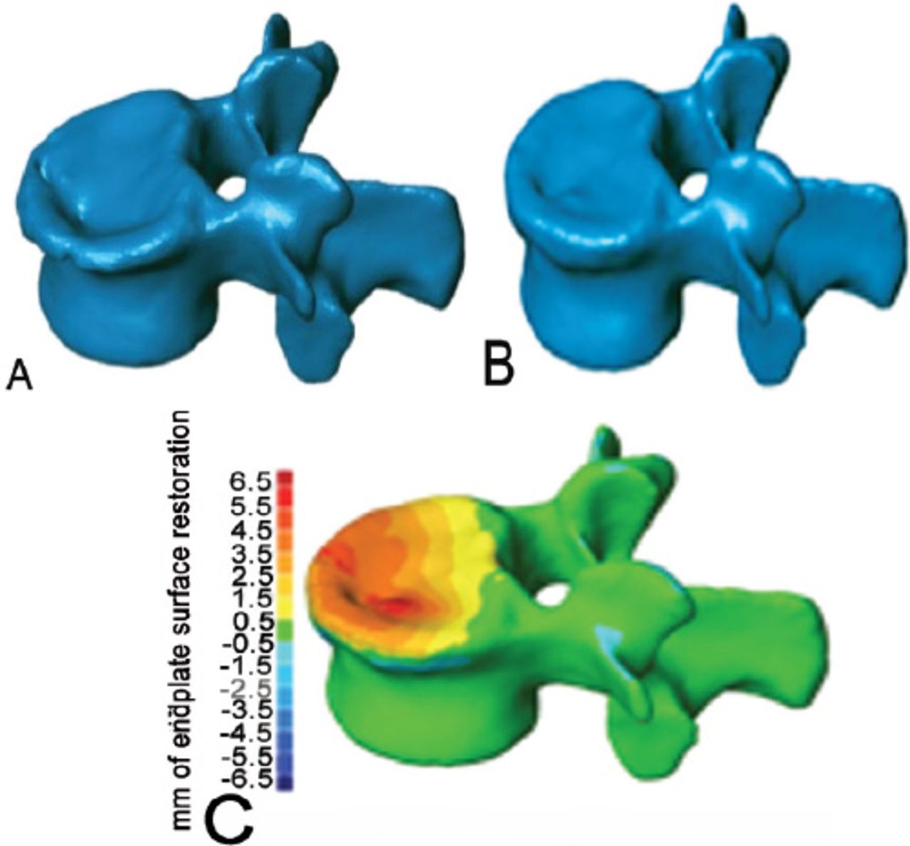
    An image showing: 3D computerized CT reconstruction of vertebral compression fractures before (A),and after (B) kyphoplasty with good restoration, and 3D topography reconstruction (C) demonstrates maximum restoration of height (red).

  • Figure 2
    • Download figure
    • Open in new tab
    • Download powerpoint
    Figure 2

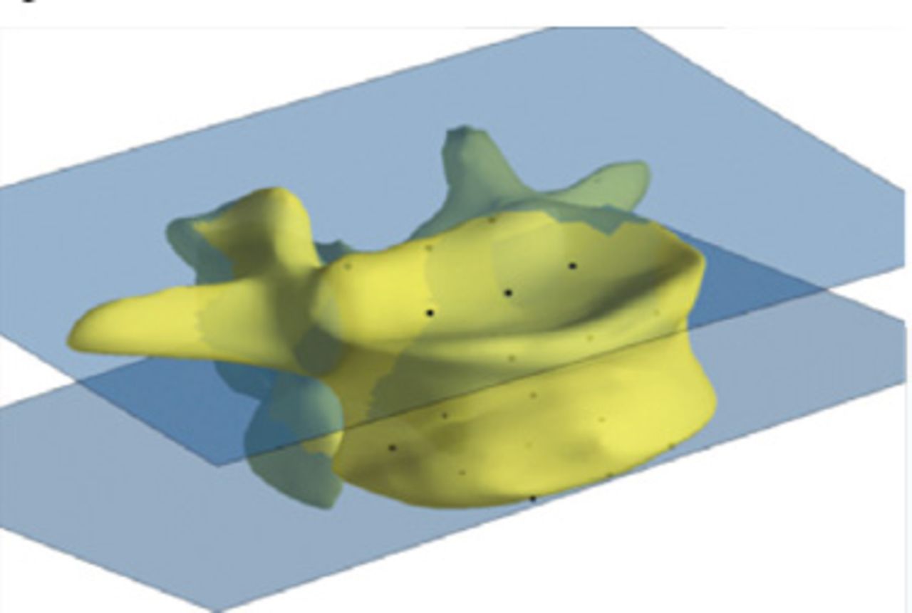
    A 3D computerized calculation of the kyphotic angle. The least-squares plane through these points is used to compute the angular orientation of the endplates. The angle measured is the difference between upper and lower vertebral endplates.

  • Figure 3
    • Download figure
    • Open in new tab
    • Download powerpoint
    Figure 3

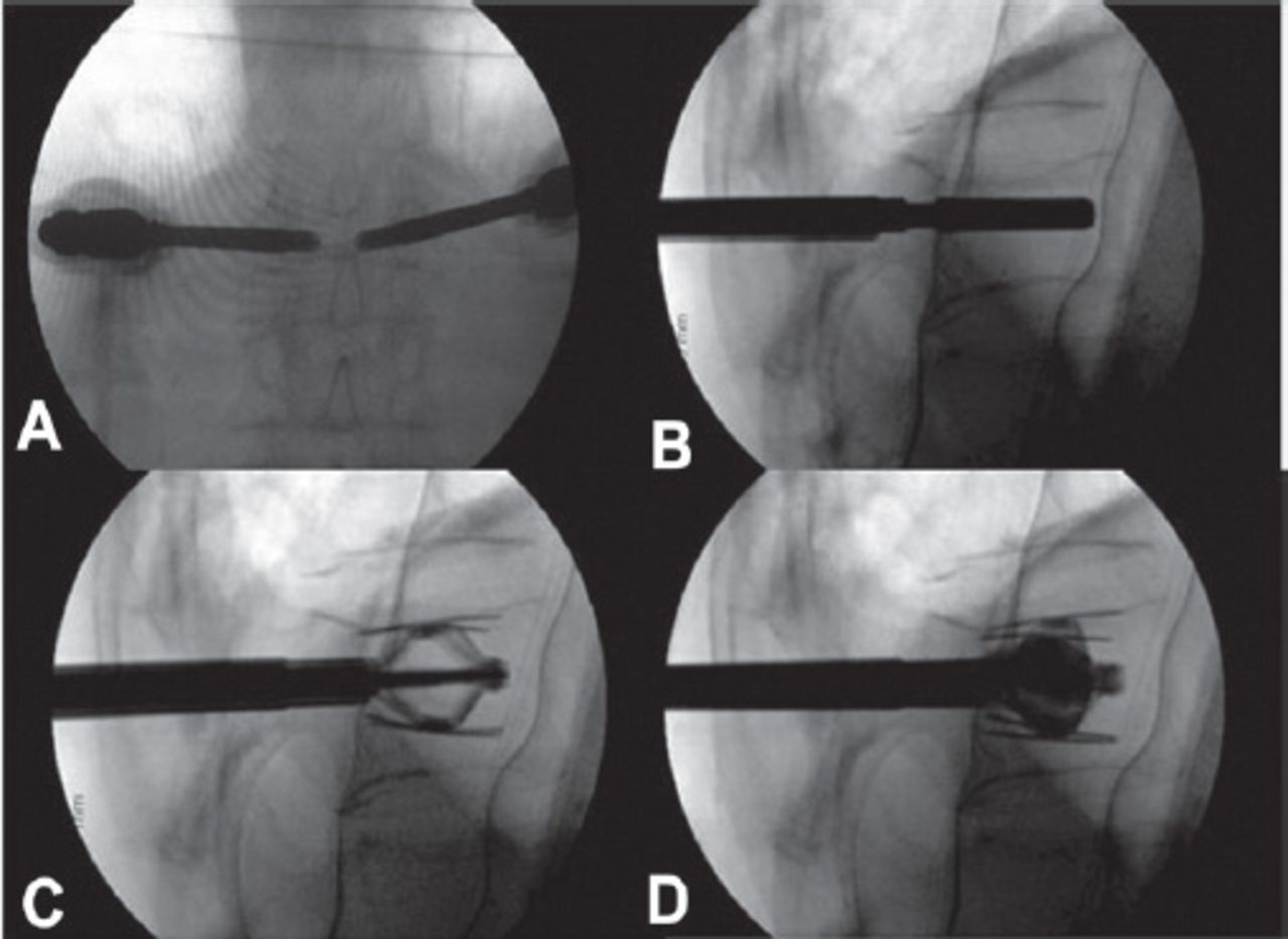
    Intraoperative radiographs demonstrating the main steps of the technique. The anterior-posterior (A) and lateral (B) view demonstrate bilateral percutaneous transpedicular placement of the implant. The implant is expanded (C) and after adequate restoration, the cement is injected under fluoroscopic monitoring.

  • Figure 4
    • Download figure
    • Open in new tab
    • Download powerpoint
    Figure 4

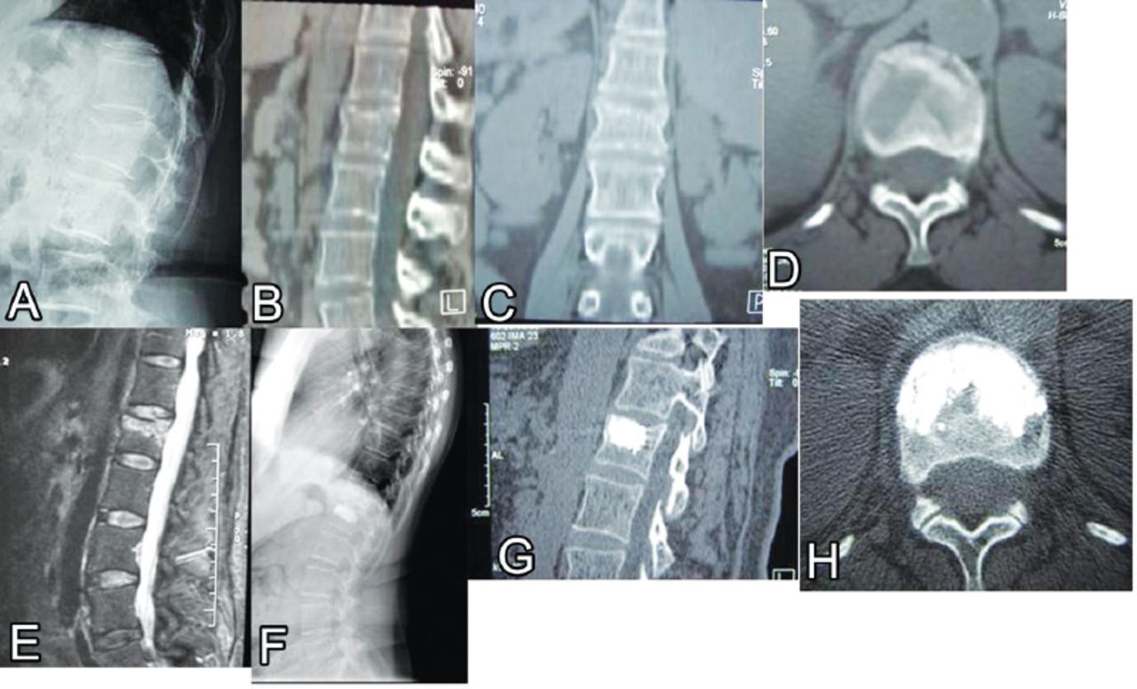
    A case illustration of a 42-year-old male presented after a trivial fall with severe lower back pain and normal neurological exam (visual analogue scale 9). Lateral radiograph (A) demonstrated vertebral compression fractures (VCF) (type A1.3). Sagittal B), coronal C), and axial D) CT scans demonstrate fracture of the upper endplate and kyphotic deformity. A T2-WI sagittal MRI scan E) demonstrated high signal intensity (edema) at the fracture site. One-year follow up lateral radiograph F) showing adequate fusion of the VCF was demonstrated on sagittal G) and axial H) CT scans.

  • Figure 5
    • Download figure
    • Open in new tab
    • Download powerpoint
    Figure 5

    Data distribution according to the location of vertebral compression fractures in the thoracolumbar spine.

Tables

  • Figures
  • Table 1
  • Table 2
  • Table 3
  • Table 4
  • Table 5
  • Table 6
PreviousNext
Back to top

In this issue

Saudi Medical Journal: 36 (1)
Saudi Medical Journal
Vol. 36, Issue 1
1 Jan 2015
  • Table of Contents
  • Cover (PDF)
  • Index by author
Print
Download PDF
Email Article

Thank you for your interest in spreading the word on Saudi Medical Journal.

NOTE: We only request your email address so that the person you are recommending the page to knows that you wanted them to see it, and that it is not junk mail. We do not capture any email address.

Enter multiple addresses on separate lines or separate them with commas.
The efficacy of a percutaneous expandable titanium device in anatomical reduction of vertebral compression fractures of the thoracolumbar spine
(Your Name) has sent you a message from Saudi Medical Journal
(Your Name) thought you would like to see the Saudi Medical Journal web site.
Citation Tools
The efficacy of a percutaneous expandable titanium device in anatomical reduction of vertebral compression fractures of the thoracolumbar spine
Saleh S. Baeesa, Antonio Krueger, Francisco A. Aragón, David C. Noriega
Saudi Medical Journal Jan 2015, 36 (1) 52-60; DOI: 10.15537/smj.2015.1.9463

Citation Manager Formats

  • BibTeX
  • Bookends
  • EasyBib
  • EndNote (tagged)
  • EndNote 8 (xml)
  • Medlars
  • Mendeley
  • Papers
  • RefWorks Tagged
  • Ref Manager
  • RIS
  • Zotero
Share
The efficacy of a percutaneous expandable titanium device in anatomical reduction of vertebral compression fractures of the thoracolumbar spine
Saleh S. Baeesa, Antonio Krueger, Francisco A. Aragón, David C. Noriega
Saudi Medical Journal Jan 2015, 36 (1) 52-60; DOI: 10.15537/smj.2015.1.9463
Twitter logo Facebook logo Mendeley logo
  • Tweet Widget
  • Facebook Like
  • Google Plus One
Bookmark this article

Jump to section

  • Article
    • Abstract
    • Methods
    • Results
    • Discussion
    • References
  • Figures & Data
  • eLetters
  • References
  • Info & Metrics
  • PDF

Related Articles

  • No related articles found.
  • PubMed
  • Google Scholar

Cited By...

  • No citing articles found.
  • Google Scholar

More in this TOC Section

  • The risk factors for cardiovascular disease and chronic kidney disease in patients with nonalcoholic fatty liver disease in Saudi Arabia
  • Prolonged flight exposure and its effects on sinonasal health among aircrew members
  • Identifying individuals at risk of post-stroke depression
Show more Original Article

Similar Articles

CONTENT

  • home

JOURNAL

  • home

AUTHORS

  • home
Saudi Medical Journal

© 2025 Saudi Medical Journal Saudi Medical Journal is copyright under the Berne Convention and the International Copyright Convention.  Saudi Medical Journal is an Open Access journal and articles published are distributed under the terms of the Creative Commons Attribution-NonCommercial License (CC BY-NC). Readers may copy, distribute, and display the work for non-commercial purposes with the proper citation of the original work. Electronic ISSN 1658-3175. Print ISSN 0379-5284.

Powered by HighWire