
Main menu

  • Home
  • Content
    • Latest
    • Archive
    • home
  • Info for
    • Authors
    • Reviewers
    • Subscribers
    • Institutions
    • Advertisers
    • Join SMJ
  • About Us
    • About Us
    • Editorial Office
    • Editorial Board
  • More
    • Advertising
    • Alerts
    • Feedback
    • Folders
    • Help
  • Other Publications
    • NeuroSciences Journal

User menu

  • My alerts
  • Log in

Search

  • Advanced search
Saudi Medical Journal
  • Other Publications
    • NeuroSciences Journal
  • My alerts
  • Log in
Saudi Medical Journal

Advanced Search

  • Home
  • Content
    • Latest
    • Archive
    • home
  • Info for
    • Authors
    • Reviewers
    • Subscribers
    • Institutions
    • Advertisers
    • Join SMJ
  • About Us
    • About Us
    • Editorial Office
    • Editorial Board
  • More
    • Advertising
    • Alerts
    • Feedback
    • Folders
    • Help
  • Follow psmmc on Twitter
  • Visit psmmc on Facebook
  • RSS
Case ReportCase Report
Open Access

Temporal bone metastasis as a sign of relapsing chronic lymphocytic leukemia

Hadeel M. Aljafar, Sari S. Alsuhibani, Mohammad S. Alahmari and Musaed A. Alzahrani
Saudi Medical Journal October 2015, 36 (10) 1233-1235; DOI: https://doi.org/10.15537/smj.2015.10.12259
Hadeel M. Aljafar
From the Division of Otorhinolaryngology, Department of Surgery (Aljafar, Alahmari, Alzahrani), King Fahd Specialist Hospital, and the Division of Otorhinolaryngology, Department of Surgery (Aljafar), Department of Radiology (Alsuhibani), King Fahd Hospital of the University, University of Dammam, Dammam, Kingdom of Saudi Arabia
MD
  • Find this author on Google Scholar
  • Find this author on PubMed
  • Search for this author on this site
Sari S. Alsuhibani
From the Division of Otorhinolaryngology, Department of Surgery (Aljafar, Alahmari, Alzahrani), King Fahd Specialist Hospital, and the Division of Otorhinolaryngology, Department of Surgery (Aljafar), Department of Radiology (Alsuhibani), King Fahd Hospital of the University, University of Dammam, Dammam, Kingdom of Saudi Arabia
MD
  • Find this author on Google Scholar
  • Find this author on PubMed
  • Search for this author on this site
Mohammad S. Alahmari
From the Division of Otorhinolaryngology, Department of Surgery (Aljafar, Alahmari, Alzahrani), King Fahd Specialist Hospital, and the Division of Otorhinolaryngology, Department of Surgery (Aljafar), Department of Radiology (Alsuhibani), King Fahd Hospital of the University, University of Dammam, Dammam, Kingdom of Saudi Arabia
MD
  • Find this author on Google Scholar
  • Find this author on PubMed
  • Search for this author on this site
Musaed A. Alzahrani
From the Division of Otorhinolaryngology, Department of Surgery (Aljafar, Alahmari, Alzahrani), King Fahd Specialist Hospital, and the Division of Otorhinolaryngology, Department of Surgery (Aljafar), Department of Radiology (Alsuhibani), King Fahd Hospital of the University, University of Dammam, Dammam, Kingdom of Saudi Arabia
MD
  • Find this author on Google Scholar
  • Find this author on PubMed
  • Search for this author on this site
  • For correspondence: [email protected]
  • Article
  • Figures & Data
  • eLetters
  • Info & Metrics
  • References
  • PDF
Loading

Abstract

Otologic manifestations in chronic lymphocytic leukemia (CLL) are common presentations. However, temporal bone metastasis is rarely described as a sign of relapsing CLL. A 65-year-old male diabetic patient known to have CLL on remission presented to the outpatient otolaryngology clinic with a one month history of progressive bilateral otalgia and right otorrhea, despite multiple courses of antibiotics. He was admitted with suspicion of malignant otitis externa. Left ear showed large hemorrhagic bullae on the posterior segment of tympanic membrane. Left sided facial paralysis developed on the third day of admission. Full recovery of facial paralysis is achieved by 10 days course of corticotherapy. Histological examination of middle ear tissue biopsy showed infiltration by monotonous small lymphoid cells, showing round nuclei, condensed chromatin suggestive of CLL. Although rare, unusual otologic manifestations should raise the suspicion of a temporal bone metastasis as a sign of relapsing CLL.

The incidence of otologic complains in leukemia patients ranges from 16.8-48%.1 Hearing loss, conductive, or sensorineural, is the most common symptom. In addition, patients may complain of tinnitus, vertigo, otalgia, otorrhea, and otorrhagia.2 Although, otologic manifestations are very common in those patients, leukemic infiltration of the temporal bone is very rare, and only few cases are reported in the literature. We report a case of relapsing chronic lymphocytic leukemia (CLL) presenting with otitis media and lower motor neuron facial paralysis. Our objective in presenting this particular case is to highlight the rare temporal bone complications and management in CLL.

Case Report

A 65-year-old diabetic patient was admitted to the hospital with suspicion of skull base osteomyelitis. He presented with bilateral persistent otitis media for one month not responding to antibiotic therapy with severe otalgia. Past medical history revealed CLL 15 years ago. He was in complete remission for 8 years before having a recurrence presented as nasopharyngeal mass with secretory otitis media. A second relapse was detected last year where histology of a nasopharyngeal mass showed evidence of Richter’s transformation to high-grade lymphoma. He was started on cyclophosphamide, Hydroxydaunorubicin, oncovin, and prednisone (CHOP) chemotherapy regimen for 4 months and then continued on Rituximab to complete a 6-month cycle. On admission, the right ear showed purulent otorrhea with edematous external auditory canal. Left ear showed large hemorrhagic bullae on the posterior segment of tympanic membrane that gives the impression of bullous myringitis.

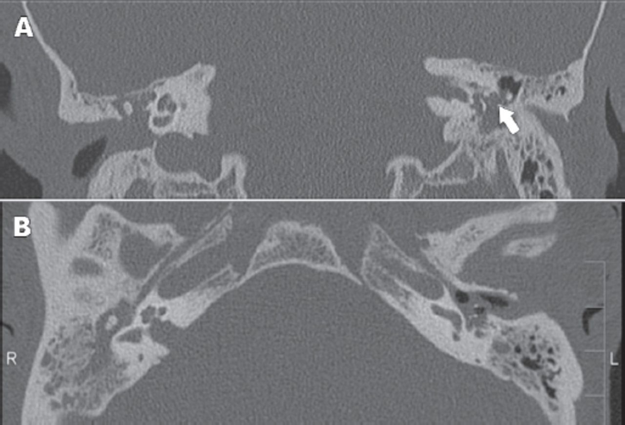
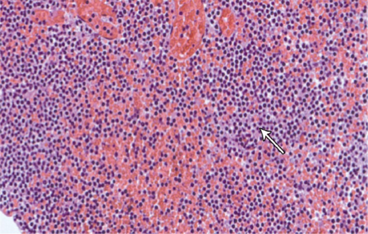
Nasopharyngoscopy revealed a non-obstructive central nasopharyngeal mass while the rest of the otorhinolaryngology examination was clear. Audiogram showed bilateral, moderate to severe mixed hearing loss. Temporal bone CT scan revealed bilateral opacification of the middle ears and mastoids cavities with dehiscence of the tympanic portion of the facial nerve bilaterally (Figure 1). No other bony erosions were noted. A complete blood count revealed hemoglobin - 10.8 g/dL (normal value [NV] - 13-17 g/dL), platelet - 494,000 mcL (NV - 150,000-450,000 mcL), and white blood cells - 108,000 mcL (NV - 4,500-10,000 mcL) with 85% lymphocytes. He was started on intravenous antibiotics, and on the third day of admission, he developed left sided lower motor neuron facial paralysis. He was sent to the surgical theater for myringotomy and ventilation tube insertions. His left tympanic membrane (TM) was perforated, and granulation tissue can be seen filling the mesotympanum and protruding through it. Biopsy of this granulation tissue was consistent with a lymphoproliferative malignancy (Figure 2). Bone marrow aspirate confirmed the diagnosis of a relapsing CLL. Cytogenetic chromosomal analysis revealed 71% trisomy 12 (which is the most common cytogenetic change in CLL, associated with an intermediate prognosis), and 13% monosomy p53. He was started on Ibrutinib, a long term daily oral chemotherapy that resulted in complete remission. Facial nerve paralysis was completely recovered with a residual TM perforation on the left ear. Granulation tissue of the middle ear disappeared completely at one month follow up after treatment.

Figure 1
  • Download figure
  • Open in new tab
  • Download powerpoint
Figure 1

Coronal (A) and Axial (B) CT scan cuts of the temporal bones showing homogenous opacity of the mastoid and middle ear cavity. Dehiscent fallopian canal of the facial nerve is visible on the left side (arrow).

Figure 2
  • Download figure
  • Open in new tab
  • Download powerpoint
Figure 2

Fragment of middle ear granulation tissue infiltrated by monotonous small lymphoid cells showing round nuclei, condensed chromatin, inconspicuous nucleoli, and scanty cytoplasm (arrow).

Discussion

An elderly diabetic patient presenting with otalgia and facial nerve paralysis has most probably skull base osteomyelitis. However, this present study shows that those findings can be signs of relapsing CLL. Chronic lymphocytic leukemia is characterized by proliferation of (mono) clonal mature B-lymphocytes in peripheral blood, bone marrow, and lymphoid tissues with a characteristic immunophenotype.3 In a study of the frequency of genetic abnormalities in leukemic B-cells, 111 out of 180 CLL cases (62%) displayed one or more cytogenetic abnormalities. In the whole series, del(13q-) was the most frequently observed abnormality (35%) followed by trisomy 12 (23%), 11q- (9.4%), and 17p- (8%).4 Peripheral facial palsy (PFP) is most commonly caused by a benign self-limiting inflammatory neuropathy condition, known as Bell’s palsy.5 However, systemic diseases can present with PFP including Guillain-Barré syndrome, sarcoidosis, syphilis, leukemia, Lyme disease, meningitis, and acute HIV infection.6

Temporal bone metastasis from distant primary carcinoma can mimic any local inflammatory or infectious process. Metastasis can reach the temporal bone through any of these patterns: a) hematogenous spread; b) tumor cells gaining access to the cerebrospinal fluid and disseminating through the subarachnoid space into the internal auditory canal; c) direct extension; d) leptomeningeal extension from an intracranial primary tumor; and e) lymphomatous infiltration.7 Druss8 in 1945, was the first otologist to call attention to the fact that patients with leukemia can present with secondary otological complications.

Buyukavci et al9 reported that bilateral PFP is a warning sign of leukocyte infiltration. The CLL can present with otologic symptoms in up to half of the affected persons, such as acute otitis media and acute mastoiditis.10 However, it is important to keep in mind the possibility of temporal bone metastasis of CLL with unusual presentations, including facial paralysis and sensorineural hearing loss.

In conclusion, unusual otologic manifestations in patients with past history of CLL should alert us to the possibility of temporal bone metastasis. Full history and physical examination is mandatory in addition to proper imaging and histological exams.

Related Articles

Basakran NS. CD44 as a potential diagnostic tumor marker. Saudi Med J 2015; 36: 273-279.

Raja-Sabudin RZ, Othman A, Ahmed-Mohamed KA, Ithnin A, Alauddin H, Alias H, et al. Immature reticulocyte fraction is an early predictor of bone marrow recovery post chemotherapy in patients with acute leukemia. Saudi Med J 2014; 35: 346-349.

Hou S, Yang W. A meta-analysis on Rituximab combined CHOP chemotherapy for non-Hodgkin lymphoma in China. Saudi Med J 2011; 32: 675-678.

Acknowledgment

We thank our colleagues Dr. Heba N. Raslan, MD, Consultant Hematopathology, and Dr Ahmed Alsaghier, MD, Consultant Hemato-Oncology, King Fahd Specialist Hospital, Dammam, Kingdom of Saudi Arabia for their insight and expertise that greatly assisted this case report.

Footnotes

  • Disclosure. Authors have no conflict of interests, and the work was not supported or funded by any drug company.

  • Received May 10, 2015.
  • Accepted August 19, 2015.
  • Copyright: © Saudi Medical Journal

This is an open-access article distributed under the terms of the Creative Commons Attribution-Noncommercial-Share Alike 3.0 Unported, which permits unrestricted use, distribution, and reproduction in any medium, provided the original work is properly cited.

References

  1. ↵
    1. Paparella MM,
    2. Berlinger NT,
    3. Oda M,
    4. el-Fiky F
    (1973) Otological manifestations of leukemia. Laryngoscope 83:1510–1526.
    OpenUrlCrossRefPubMed
  2. ↵
    1. Terao K,
    2. Cureoglu S,
    3. Schachern PA,
    4. Paparella MM,
    5. Morita N,
    6. Nomiya S,
    7. et al.
    (2011) Pathologic correlations of otologic symptoms in acute lymphocytic leukemia. Am J Otolaryngol 32:13–18.
    OpenUrlPubMed
  3. ↵
    1. Chastain EC,
    2. Duncavage EJ
    (2015) Clinical prognostic biomarkers in chronic lymphocytic leukemia and diffuse large B-cell lymphoma. Arch Pathol Lab Med 139:602–607.
    OpenUrl
  4. ↵
    1. Quijano S,
    2. López A,
    3. Rasillo A,
    4. Sayagués JM,
    5. Barrena S,
    6. Sánchez ML,
    7. et al.
    (2008) Impact of trisomy 12, del(13q), del(17p), and del(11q) on the immunophenotype, DNA ploidy status, and proliferative rate of leukemic B-cells in chronic lymphocytic leukemia. Cytometry B Clin Cytom 74:139–149.
    OpenUrlPubMed
  5. ↵
    1. Chan EH,
    2. Tan HM,
    3. Tan TY
    (2005) Facial palsy from temporal bone lesions. Ann Acad Med Singapore 34:322–329.
    OpenUrlPubMed
  6. ↵
    1. Todd NW Jr.,
    2. Bowman CA
    (1984) Acute myelogenous leukemia presenting as atypical mastoiditis with facial paralysis. Int J Pediatr Otorhinolaryngol 7:173–177.
    OpenUrlCrossRefPubMed
  7. ↵
    1. Cureoglu S,
    2. Tulunay O,
    3. Ferlito A,
    4. Schachern PA,
    5. Paparella MM,
    6. Rinaldo A
    (2004) Otologic manifestations of metastatic tumors to the temporal bone. Acta Otolaryngol 124:1117–1123.
    OpenUrlPubMed
  8. ↵
    1. Druss JG
    (1945) Aural manifestations of leukemia. Arch Otolaryngol 42:267–274.
    OpenUrlCrossRefPubMed
  9. ↵
    1. Buyukavci M,
    2. Tan H,
    3. Akdag R
    (2002) An alarming sign for serious diseases in children: bilateral facial paralysis. Pediatr Neurol 27:312–313.
    OpenUrlPubMed
  10. ↵
    1. Leite da Silveira P,
    2. Gonçalves Silva V,
    3. Rizzato Paschoal J,
    4. Nizam Pfeilsticker L
    (2015) Bilateral peripheral facial palsy and mastoid infiltration as symptoms of relapsed acute myeloid leukemia. Eur Ann Otorhinolaryngol Head Neck Dis 132:41–43.
    OpenUrl
PreviousNext
Back to top

In this issue

Saudi Medical Journal: 36 (10)
Saudi Medical Journal
Vol. 36, Issue 10
1 Oct 2015
  • Table of Contents
  • Cover (PDF)
  • Index by author
Print
Download PDF
Email Article

Thank you for your interest in spreading the word on Saudi Medical Journal.

NOTE: We only request your email address so that the person you are recommending the page to knows that you wanted them to see it, and that it is not junk mail. We do not capture any email address.

Enter multiple addresses on separate lines or separate them with commas.
Temporal bone metastasis as a sign of relapsing chronic lymphocytic leukemia
(Your Name) has sent you a message from Saudi Medical Journal
(Your Name) thought you would like to see the Saudi Medical Journal web site.
Citation Tools
Temporal bone metastasis as a sign of relapsing chronic lymphocytic leukemia
Hadeel M. Aljafar, Sari S. Alsuhibani, Mohammad S. Alahmari, Musaed A. Alzahrani
Saudi Medical Journal Oct 2015, 36 (10) 1233-1235; DOI: 10.15537/smj.2015.10.12259

Citation Manager Formats

  • BibTeX
  • Bookends
  • EasyBib
  • EndNote (tagged)
  • EndNote 8 (xml)
  • Medlars
  • Mendeley
  • Papers
  • RefWorks Tagged
  • Ref Manager
  • RIS
  • Zotero
Share
Temporal bone metastasis as a sign of relapsing chronic lymphocytic leukemia
Hadeel M. Aljafar, Sari S. Alsuhibani, Mohammad S. Alahmari, Musaed A. Alzahrani
Saudi Medical Journal Oct 2015, 36 (10) 1233-1235; DOI: 10.15537/smj.2015.10.12259
Twitter logo Facebook logo Mendeley logo
  • Tweet Widget
  • Facebook Like
  • Google Plus One
Bookmark this article

Jump to section

  • Article
    • Abstract
    • Case Report
    • Discussion
    • Acknowledgment
    • Footnotes
    • References
  • Figures & Data
  • eLetters
  • References
  • Info & Metrics
  • PDF

Related Articles

  • No related articles found.
  • PubMed
  • Google Scholar

Cited By...

  • No citing articles found.
  • Google Scholar

More in this TOC Section

  • Cutaneous metastasis of signet ring cell adenocarcinoma of the colon
  • Giant left gastric artery aneurysm with intrathoracic extension
  • A rare case of a horseshoe kidney with a single left-sided ureter presented with recurrent urinary tract infection
Show more Case Report

Similar Articles

CONTENT

  • home

JOURNAL

  • home

AUTHORS

  • home
Saudi Medical Journal

© 2025 Saudi Medical Journal Saudi Medical Journal is copyright under the Berne Convention and the International Copyright Convention.  Saudi Medical Journal is an Open Access journal and articles published are distributed under the terms of the Creative Commons Attribution-NonCommercial License (CC BY-NC). Readers may copy, distribute, and display the work for non-commercial purposes with the proper citation of the original work. Electronic ISSN 1658-3175. Print ISSN 0379-5284.

Powered by HighWire