
Main menu

  • Home
  • Content
    • Latest
    • Archive
    • home
  • Info for
    • Authors
    • Reviewers
    • Subscribers
    • Institutions
    • Advertisers
    • Join SMJ
  • About Us
    • About Us
    • Editorial Office
    • Editorial Board
  • More
    • Advertising
    • Alerts
    • Feedback
    • Folders
    • Help
  • Other Publications
    • NeuroSciences Journal

User menu

  • My alerts
  • Log in

Search

  • Advanced search
Saudi Medical Journal
  • Other Publications
    • NeuroSciences Journal
  • My alerts
  • Log in
Saudi Medical Journal

Advanced Search

  • Home
  • Content
    • Latest
    • Archive
    • home
  • Info for
    • Authors
    • Reviewers
    • Subscribers
    • Institutions
    • Advertisers
    • Join SMJ
  • About Us
    • About Us
    • Editorial Office
    • Editorial Board
  • More
    • Advertising
    • Alerts
    • Feedback
    • Folders
    • Help
  • Follow psmmc on Twitter
  • Visit psmmc on Facebook
  • RSS
Case ReportCase Report
Open Access

A solitary fibrous tumor of the upper limb

Bisher N. Al-Shanawani, Mohammad M. Al-Qattan, Maha M. Arafah and Muhammed I. Al-Motairi
Saudi Medical Journal January 2015, 36 (2) 236-238; DOI: https://doi.org/10.15537/smj.2015.2.10524
Bisher N. Al-Shanawani
From the Division of Plastic Surgery (Al-Shanawani, Al-Qattan, Al-Motairi), and the Department of Pathology (Arafah), King Saud University, Riyadh, Kingdom of Saudi Arabia.
MBBS
  • Find this author on Google Scholar
  • Find this author on PubMed
  • Search for this author on this site
Mohammad M. Al-Qattan
From the Division of Plastic Surgery (Al-Shanawani, Al-Qattan, Al-Motairi), and the Department of Pathology (Arafah), King Saud University, Riyadh, Kingdom of Saudi Arabia.
MBBS
  • Find this author on Google Scholar
  • Find this author on PubMed
  • Search for this author on this site
  • For correspondence: [email protected]
Maha M. Arafah
From the Division of Plastic Surgery (Al-Shanawani, Al-Qattan, Al-Motairi), and the Department of Pathology (Arafah), King Saud University, Riyadh, Kingdom of Saudi Arabia.
MBBS
  • Find this author on Google Scholar
  • Find this author on PubMed
  • Search for this author on this site
Muhammed I. Al-Motairi
From the Division of Plastic Surgery (Al-Shanawani, Al-Qattan, Al-Motairi), and the Department of Pathology (Arafah), King Saud University, Riyadh, Kingdom of Saudi Arabia.
MBBS
  • Find this author on Google Scholar
  • Find this author on PubMed
  • Search for this author on this site
  • Article
  • Figures & Data
  • eLetters
  • Info & Metrics
  • References
  • PDF
Loading

Abstract

Solitary fibrous tumors (SFT) of the upper limb are extremely rare, and we report this tumor in the arm of a 30-year-old male. He is presented with a 22 cm painless mass. Complete surgical excision was performed. The histological diagnosis of SFT was based on the presence of ectatic blood vessels and positive staining for CD34 and vimentin. He remains disease-free at the 3-year follow-up interval. The report aims to increase the awareness of the criteria for the histological diagnosis of SFT, as well as the principles of their surgical excision and follow-up.

Solitary fibrous tumors (SFT) are rare spindle-cell mesenchymal tumors, which are also known as fibrous mesotheliomas. As the name implies, the tumor is mainly seen in the pleura, the mesothelial lining of the lungs. In 5% of cases, it is accompanied by hypoglycemia secondary to tumor production of insulin-like growth factor II; and this is most often seen in tumors located in the pelvis and retroperitoneum.1 The majority of the tumors are histologically benign; however, atypical features have been described in some, and malignant cases have been reported.2 Extra thoracic SFT are very rare, and those involving the upper limb are extremely rare. In 2003, Akisue et al3 reviewed the literature on SFT of the limbs and could only find 9 cases involving the upper limbs. Since 2003, few other upper limb tumors have been reported.2,4-7 Our objective in presenting this particular case is to increase awareness, and focus on the criteria for histological diagnosis of SFT, as well as the principles of their surgical excision and follow-up.

Case Report

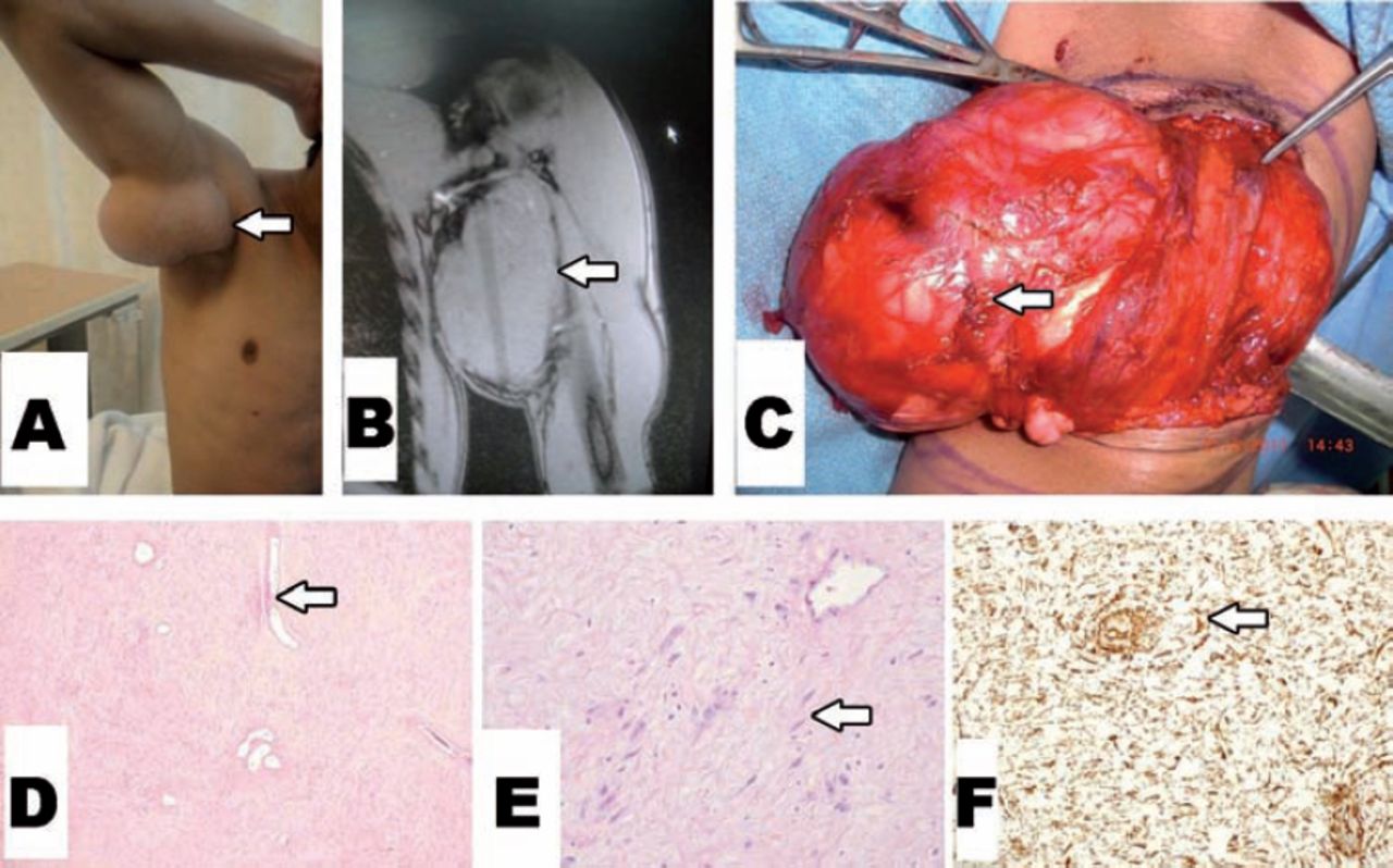
A 30-year-old man presented with a one-year history of a slowly growing mass of the left upper-medial arm (Figure 1A). There was no pain or neurovascular-related symptoms. On MRI (Figure 1B), a well circumscribed mass with intermediate signal intensity on T1 and T2 weighted images was seen. A metastatic work-up including CT scan of the chest and ultrasound of the abdomen was negative. Surgical excision was performed under general anesthesia. A well-circumscribed tumor was found with a firm whitish capsule (Figure 1C). The tumor mass along with the capsule was excised. The mass measured 22 × 10 cm and cut-sections showed a multi-nodular mass with a smooth surface feature. Histological examination showed a benign hypocellular fibrous tumor with ectatic blood vessels (Figure 1D); composed of fibroblasts separated by thin bands of collagen (Figure 1E). There were no areas of tumor cell necrosis, and mitotic figures were rare. There was no pleomorphism or atypia. All resection margins were free of tumor. Tumor cells stained positive for CD34 (Figure 1F), and vimentin; and negative for desmin, actin, cytokeratin, and S-100 protein. The final diagnosis was a benign solitary fibrous tumor. He did well after surgery and after 3-years is disease free.

Figure 1
  • Download figure
  • Open in new tab
  • Download powerpoint
Figure 1

Clinical photo showing: A) preoperative appearance, B) MRI showing an iso-intense mass, C) surgical excision of the mass along with its capsule, D) the tumor is hypocellular with ectatic blood vessels (hematoxylin and eosin stain x40), E) high power showing the fibroblasts surrounded by thin collagen (hematoxylin and eosin stain x200), and F) positive CD34 immunostain (x100).

Discussion

Solitary fibrous tumors of the upper limb are extremely rare, and less than 20 cases were reported in the literature.2-9 Historically, SFT were known as hemangiopericytomas because the presence of stag-horn vascular pattern was thought to be an indication of a pericytic origin. Later, this stag-horn vascular feature was found to be present in several other benign, and malignant tumors such as infantile myofibromatosis, and synovial sarcoma.10

The differential diagnosis of SFT is a long list of benign fibrous tumors that are known to affect the upper limb of children and adults. All benign fibrous tumors are mainly composed of fibroblasts (spindle cells) separated by collagen bands. The diagnosis of the type of tumor will be based on other histological features and on immunohistological analysis.11,12 An accurate histological diagnosis is essential because it will determine the behavior, risk of recurrence, and the adequacy of surgical resection. For example, nodular fasciitis has a very low risk of recurrence and may even regress spontaneously; while desmoid tumors are locally aggressive with a high risk of recurrence and hence require wide local excision. The diagnosis of nodular fasciitis is based on the presence of myxoid areas and positivity for actin staining, while desmoid tumors are positive for desmin staining. Therefore, an experienced histopathologist is an essential member of the team for managing fibrous tumors of the limb. The most characteristic histological feature of SFT is the presence of ectatic or stag-horn blood vessels; although other distinct histologic patterns have been described.6 Furthermore, SFT cells will stain positive for CD34 and vimentin; and will stain negative for actin, desmin, and other epithelial (such as cytokeratin), and neural (such as S-100 protein) markers. Finally, all benign-looking fibrous tumors should be carefully examined for any areas of atypia or polymorphism. These features suggests the presence of a malignant component, and Baldi et al13 have reported a distant metastases from tumors with such features. The tumor in our patient had no such features. However, it should be emphasized that histological features will not always predict biological behavior.

Surgically, the tumor mass should be removed along with its capsule since failure to do so is associated with increased risk of local recurrence.14 Other factors that are known to be associated with local recurrence include positive surgical margins, accidentally going through the capsule during surgery, high cellularity, and the presence of frequent mitotic figures.13 None of these features were present in our patient and there was no local recurrence through the 3-year follow-up visit. Long-term follow-up is recommended for benign SFT since they may have a very slow growth, and late recurrences have been reported.13 The histological features of the tumor of our patient are associated with a low risk of recurrence, and surgical excision was curative. Long-term follow-up is planned for our patient.

In conclusion, we report a rare case of SFT of the upper limb and emphasize the principles of excision, the diagnostic criteria, and follow-up of similar cases. Excision should include the capsule surrounding the tumor mass. The diagnosis should be confirmed by the presence of stag-horn vessels and positivity for CD 34 and vimentin. Long term follow up is required even for tumors with rare mitotic figures.

Case Reports

Case reports will only be considered for unusual topics that add something new to the literature. All Case Reports should include at least one figure. Written informed consent for publication must accompany any photograph in which the subject can be identified. Figures should be submitted with a 300 dpi resolution when submitting electronically or printed on high-contrast glossy paper when submitting print copies. The abstract should be unstructured, and the introductory section should always include the objective and reason why the author is presenting this particular case. References should be up to date, preferably not exceeding 15.

Footnotes

  • Disclosure. Authors have no conflict of interests, and the work was not supported or funded by any drug company. This work is supported and funded by the College of Medicine Research Center, Deanship of Scientific Research, King Saud University, Riyadh, Kingdom of Saudi Arabia.

  • Received October 19, 2014.
  • Accepted December 15, 2014.
  • Copyright: © Saudi Medical Journal

This is an open-access article distributed under the terms of the Creative Commons Attribution-Noncommercial-Share Alike 3.0 Unported, which permits unrestricted use, distribution, and reproduction in any medium, provided the original work is properly cited.

References

  1. ↵
    1. Fukasawa Y,
    2. Takada A,
    3. Tateno M,
    4. Sato H,
    5. Koizumi M,
    6. Tanaka A,
    7. et al.
    (1998) Solitary fibrous tumor of the pleura causing recurrent hypoglycemia by secretion of insulin-like growth factor II. Pathol Int 48:47–52.
    OpenUrlCrossRefPubMed
  2. ↵
    1. Lee JC,
    2. Fletcher CD
    (2011) Malignant fat-forming solitary fibrous tumor (so-called “lipomatous hemangiopericytoma”): clinicopathologic analysis of 14 cases. Am J Surg Pathol 35:1177–1185.
    OpenUrlCrossRefPubMed
  3. ↵
    1. Akisue T,
    2. Matsumoto K,
    3. Kizaki T,
    4. Fujita I,
    5. Yamamoto T,
    6. Yoshiya S,
    7. et al.
    (2003) Solitary fibrous tumor in the extremity: case report and review of the literature. Clin Orthop Relat Res 411:236–244.
    OpenUrlCrossRefPubMed
  4. ↵
    1. Rakheja D,
    2. Wilson KS,
    3. Meehan JJ,
    4. Schultz RA,
    5. Maale GE,
    6. Timmons CF
    (2004) Extrapleural benign solitary fibrous tumor in the shoulder of a 9-year-old girl: case report and review of the literature. Pediatr Dev Pathol 7:653–660.
    OpenUrlCrossRefPubMed
    1. Sookur PA,
    2. Saifuddin A
    (2011) Indeterminate soft-tissue tumors of the hand and wrist: a review based on a clinical series of 39 cases. Skeletal Radiol 40:977–989.
    OpenUrlCrossRefPubMed
  5. ↵
    1. Terada T
    (2011) Solitary fibrous tumor of the shoulder showing diverse distinct histologic patterns. Int J Dermatol 50:208–211.
    OpenUrlPubMed
  6. ↵
    1. Insabato L,
    2. Siano M,
    3. Somma A,
    4. Gentile R,
    5. Santangelo M,
    6. Pettinato G
    (2009) Extrapleural solitary fibrous tumor: a clinicopathologic study of 19 cases. Int J Surg Pathol 17:250–254.
    OpenUrlCrossRefPubMed
    1. Wilky BA,
    2. Montgomery EA,
    3. Guzzetta AA,
    4. Ahuja N,
    5. Meyer CF
    (2013) Extrathoracic location and “borderline” histology are associated with recurrence of solitary fibrous tumors after surgical resection. Ann Surg Oncol 20:4080–4089.
    OpenUrl
  7. ↵
    1. Demicco EG,
    2. Park MS,
    3. Araujo DM,
    4. Fox PS,
    5. Bassett RL,
    6. Pollock RE,
    7. et al.
    (2012) Solitary fibrous tumor: a clinicopathological study of 110 cases and proposed risk assessment model. Mod Pathol 25:1298–1306.
    OpenUrlCrossRefPubMed
  8. ↵
    1. Gengler C,
    2. Guillou L
    (2006) Solitary fibrous tumour and haemangiopericytoma: evolution of a concept. Histopathology 48:63–74.
    OpenUrlCrossRefPubMed
  9. ↵
    1. Langman G
    (2011) Solitary fibrous tumor: A pathological enigma and clinical dilemma. J Thorac Dis 3:86–87.
    OpenUrlPubMed
  10. ↵
    1. Chandanwale SS,
    2. Gore CR,
    3. Sammi AB,
    4. Shah KR,
    5. Kaur PR
    (2014) Recurrent solitary fibrous tumor in distal lower extremity: An extremely rare entity. Int J Appl Basic Med Res 4:134–136.
    OpenUrl
  11. ↵
    1. Baldi GG,
    2. Stacchiotti S,
    3. Mauro V,
    4. Dei Tos AP,
    5. Gronchi A,
    6. Pastorino U,
    7. et al.
    (2013) Solitary fibrous tumor of all sites: outcome of late recurrences in 14 patients. Clin Sarcoma Res 3:4.
    OpenUrlPubMed
  12. ↵
    1. Andres JO,
    2. Aurich M,
    3. Lang T,
    4. Wagner A
    (2006) Solitary fibrous tumor in the thigh: review of the literature. J Cancer Res Clin Onco 132:69–75.
    OpenUrl
PreviousNext
Back to top

In this issue

Saudi Medical Journal: 36 (2)
Saudi Medical Journal
Vol. 36, Issue 2
1 Jan 2015
  • Table of Contents
  • Cover (PDF)
  • Index by author
Print
Download PDF
Email Article

Thank you for your interest in spreading the word on Saudi Medical Journal.

NOTE: We only request your email address so that the person you are recommending the page to knows that you wanted them to see it, and that it is not junk mail. We do not capture any email address.

Enter multiple addresses on separate lines or separate them with commas.
A solitary fibrous tumor of the upper limb
(Your Name) has sent you a message from Saudi Medical Journal
(Your Name) thought you would like to see the Saudi Medical Journal web site.
Citation Tools
A solitary fibrous tumor of the upper limb
Bisher N. Al-Shanawani, Mohammad M. Al-Qattan, Maha M. Arafah, Muhammed I. Al-Motairi
Saudi Medical Journal Jan 2015, 36 (2) 236-238; DOI: 10.15537/smj.2015.2.10524

Citation Manager Formats

  • BibTeX
  • Bookends
  • EasyBib
  • EndNote (tagged)
  • EndNote 8 (xml)
  • Medlars
  • Mendeley
  • Papers
  • RefWorks Tagged
  • Ref Manager
  • RIS
  • Zotero
Share
A solitary fibrous tumor of the upper limb
Bisher N. Al-Shanawani, Mohammad M. Al-Qattan, Maha M. Arafah, Muhammed I. Al-Motairi
Saudi Medical Journal Jan 2015, 36 (2) 236-238; DOI: 10.15537/smj.2015.2.10524
Twitter logo Facebook logo Mendeley logo
  • Tweet Widget
  • Facebook Like
  • Google Plus One
Bookmark this article

Jump to section

  • Article
    • Abstract
    • Case Report
    • Discussion
    • Footnotes
    • References
  • Figures & Data
  • eLetters
  • References
  • Info & Metrics
  • PDF

Related Articles

  • No related articles found.
  • PubMed
  • Google Scholar

Cited By...

  • No citing articles found.
  • Google Scholar

More in this TOC Section

  • Cutaneous metastasis of signet ring cell adenocarcinoma of the colon
  • Giant left gastric artery aneurysm with intrathoracic extension
  • A rare case of a horseshoe kidney with a single left-sided ureter presented with recurrent urinary tract infection
Show more Case Report

Similar Articles

CONTENT

  • home

JOURNAL

  • home

AUTHORS

  • home
Saudi Medical Journal

© 2025 Saudi Medical Journal Saudi Medical Journal is copyright under the Berne Convention and the International Copyright Convention.  Saudi Medical Journal is an Open Access journal and articles published are distributed under the terms of the Creative Commons Attribution-NonCommercial License (CC BY-NC). Readers may copy, distribute, and display the work for non-commercial purposes with the proper citation of the original work. Electronic ISSN 1658-3175. Print ISSN 0379-5284.

Powered by HighWire