


Abstract
Objectives: To investigate the potential protective effect of resveratrol (RES) on aluminum chloride (AlCl3)-induced nephrotoxicity in rats.
Methods: This experimental study was conducted from April to June 2015 at the Medical College of King Khalid University, Abha, Kingdom of Saudi Arabia. The experiments were performed on 24 male Wistar rats. The rats were randomly allocated into 4 groups; 1) group A: control rats received only normal saline, 2) group B: received RES dissolved in normal saline, 3) group C: model group and received AlCl3 dissolved in normal saline and 4) group D: RES treated group and received concomitant doses of RES+AlCl3. All treatments were administered for consecutive 40 days. After 40 days of treatments, kidney function tests, oxidative stress parameters and histopathological assay were evaluated.
Results: all findings clearly showed significant deteriorations in kidney function and architectures after AlCl3 exposure. This was accompanied by increased renal oxidative stress and inflammation suggesting strong pro-oxidant activity of AlCl3 in spite of its non-redox status. Resveratrol co-treatment with AlCl3 to the rats showed significant improvement in all biochemical and histological parameters related to kidney function and structure.
Conclusion: The findings of the current study showed that RES pre-administration to rats ameliorates renal damage and improves renal function in AlCl3 intoxicated rats in a mechanism related to its antioxidant potential.
A major concern of aluminum (Al) toxicity in humans and animals has been raised during the last decades.1 In 2007, Al was included in the priority list of hazardous substances identified by The Agency for Toxic Substances and Disease Registry.2 Exposure to Al is very common during daily life due to the facts that it is widely distributed in the environment, and extensively used in daily life.3 Major sources of Al exposure include, but are not limited to foods especially corn, yellow cheese, grain products (flour), salt and spices, vegetables and tea leaves, cosmetics, cookware and containers.4 Also, Al could be found in the drinking water, which is usually added for purification purposes.4 Environmental pollution, especially waste water exposes people to a higher risk of Al toxicity.5 Aluminum gets access to the human body via the gastrointestinal and the respiratory tracts.3 In the medical field, Al is used as a major constituent of drugs such as antacids, phosphate binders, buffered aspirins, vaccines and injectable allergens.4 However, elimination of Al from the human body is very limited and the primary route of its elimination is through urine.6 Higher doses of Al could increase the risk of renal Al retention and hence, induced nephrotoxicity.6 It has been reported that Al can cause degeneration of the renal tubular cells through generation of reactive oxygen species (ROS) which cause oxidative damage to cellular lipids, proteins, and DNAs.7 Aluminum intoxication lowers the intracellular levels of reduced glutathione.8,9 Also, Al salts may inhibit enzymes like acid and alkaline phosphatases and phosphodiesterase.10 In recent years, there has been a general consensus concerning the positive effects of resveratrol (RES) in prevention and treatment of various disease conditions due to its potent anti-inflammatory and antioxidant potentials.11,12 Suggested possible targets for the action of RES included endothelial nitric oxide synthase (eNOS),13 the mitogen-activated protein kinase (MAPK),14 the hemeoxygenase-1 (HO-1) and the nuclear factor E2-related factor-2 (Nfr2) and nuclear factor-kappa B (NF-κB).15 In its role in protection against environmental hazards, in a novel study, Elewa et al,15 showed that RES might be a promising agent in the treatment of cadmium chloride induced testicular damage by enhancing endogenous antioxidant potential, reducing inflammation and its ability to act as antiapoptotic agent. Similarly, in our labs, a protective effect against RES against Al chloride induced testicular damage was reported.16 Based on these findings and possible mechanisms of actions of RES, It was hypothesized that RES could be a promising agent against Al chloride induced renal damage. Therefore, the aim of the present study is to investigate the potential protective effect of RES on aluminum chloride (AlCl3)-induced nephrotoxicity in rats.
Methods
Drugs
The active form of resveratrol (RES), (trans-Resveratrol), and the crystalline form of AlCl3 were purchased from Sigma-Aldrich (St. Louis, MO, USA) and freshly prepared by dissolving in a saline solution (0.9% NaCl) of 20% hydroxypropyl cyclodextrin (American Maize-Products Co., Hammond, IN, USA) to a final concentration of 20 mg/ml as previously published.15 This dose selected for RES was based on a previous study that showed antioxidant, anti-inflammatory, and anti-apoptotic effects of RES.15 The AlCl3 was dissolved in 0.9% saline at a final concentration of 20 mg/kg/bwt (1/20 LD50).16,17 These dose selected for AlCl3 is based on previous dose-response studies,16 and as previously reported18 that showed severe renal damage at a similar dose and time administration.16,18
Animals
This experimental study was conducted from April-June 2015 at King Khalid University, Abha, Kingdom of Saudi Arabia (KSA) on 24 male Wistar rats of 8 weeks old (190-200 g). Rats were housed in a 4 rat-cages. Rats in all treatment groups were preconditioned for one week prior to implementing the treatment protocol. During this time, rats received standard chow diet and water ad-libitum and were kept at room temperature of 22 ± 2 °C, relative humidity of 55 ± 10% and a light/dark cycle of 12 hours. All experimental procedures involving animals and their care in the current study were conducted in accordance with the guidelines of the Institutional Animal Care and Use Committee of the King Khalid University, which are in compliance with the national and international laws and policies (seventh edition). All efforts were made to minimize animal suffering including anesthesia, blood sampling, and surgical procedures.
Experimental procedure
After an adaptation period, the rats were randomly allocated into 4 groups (n=4): Group A: received 1 ml saline solution containing 20% hydroxypropyl cyclodextrin as vehicle; Group B: received 1 ml RES solution; Group C: received 1 ml AlCl3 solution with a concomitant dose of 1 ml saline solution containing 20% hydroxypropyl cyclodextrin and Group D: received 1 ml RES solution and a concomitant dose of 1 ml AlCl3 solution. However, among these groups we also ran other 2 groups of control rats and AlCl3 intoxicated rats, which were administered normal saline only, and we found no significant diffenerences when compared with the corresponding groups that normal saline containing 20% hydroxypropyl cyclodextrin in all the below mentioned parameters, thus these groups were not included in the current study and analysis.
Resveratrol and AlCl3 were administered in a final concentration of 20 mg/kg/bwt and all treatments were administered for a total period of 40 days on a daily basis. Treatments were given to all groups orally namely, oral gavage. During this period, the rats were adapted weekly to metabolic cages (these cages offer larg space for rats to move freely without any restrained stress). At the end of day 40 and after a 12 hours fasting, rats were placed in their metabolic cages and the urine samples were collected into tubes containing 20 µL of 2.5 mol/L HCl over 24 hours. Then, the volume of urine were measured individually and filtered with 0.2 µm Millipore filters and stored at -78°C to measure the levels of urinary creatinine (Cr) levels. After collection of urine, all rats were anesthetized with light diethyl ether and 2 ml blood were collected directly by cardiac puncture, placed into plain tubes, which were allowed to clot and then centrifuged at 5000 rpm for 10 minutes at room temperature to collect serum. Serum samples were stored at -80°C for further analysis of urea and creatinine levels using commercial available kits according to manufacturer’s instruction.
Creatinine clearance (Ccr)
Assay colorimetric kit for determination of serum and urinary Cr concentration (Cat., no. 700460 & Cat., no. 500701) were purchased from Cayman Company (Ann Arbor, MI, USA). All analyses were performed in accordance with the manuals provided by the manufacturers.
The Ccr was calculated using the following equation19 Ccr (mL/min/kg) = [urinary Cr (mg/dL) × urinary volume (mL) / serum Cr (mg/dL)]×[1000/body weight (g)]×[1/1440 (min)].
Tissue collection
Twelve hours after blood collection, all animals were anesthetized by diethyl ether and killed by decapitation. Both kidneys from each rat of each group were quickly collected, weighed, and washed with phosphate buffered saline, pH 7.4, containing 0.16 mg/mL heparin to remove any red blood cells or clots. Then one kidney from each rat was homogenized in the appropriate buffer as previously described.15,16 The resultant supernatants were kept in separate tubes and stored at -20 °C. However, the other kidney obtained from each rat was placed in 10% formalin solution for histopathological evaluation.
Biochemical analysis
Total protein concentration was measured in the tissue homogenates by Bradford assay. Levels of lipid peroxidation in the kidney homogenates were measured by the thiobarbituric acid reaction using the commercially available kits (MDA, Cat No. NWK-MDA01) as per Northwest life science specialties (LCC, Vancouver, WA 98662, Canada) instructions and MDA levels were expressed as nmol/mg protein. Assay activities of superoxide dismutase (SOD, Cat. NO.706002), glutathione peroxidase (GPx, Cat. NO.703102), and levels of reduced glutathione (GSH, Cat NO.703002) were purchased from Cayman Chemical, MI, USA. The calculated SOD and GPx activities were expressed as U/mg protein and as nmol/min/g protein, respectively. Calculated GSH levels were expressed as µmol/mg protein.
Inflammatory mediators assay
Levels of tumor necrosis factor-alpha (TNF-α) and interleukin-6 (IL-6) in kidney homogenates were determined by enzyme-linked immunosorbent assay (ELISA) (Cat no. ab46070, Abcam, Cambridge, MA, USA and Cat No. ELR-IL6-001, RayBio, MO, USA). Renal levels of TNF-α and IL-6 levels were expressed as pg/mg protein.
Histopathological evaluation
Kidney specimens from all groups of rats were processed routinely in 10% formalin solution and embedded in paraffin. Tissue sections of 5 µm were obtained and stained with Hematoxylin and Eosin. All histopathological examinations were performed under a light microscope (NIKON, Japan) by different histologists at the College of Medicine at King Khalid University who were blinded to all tissue specimens regarding their group. A minimum of 10 fields for each kidney were examined.
Statistical analysis
Statistical analyses were performed by using Graphpad prism statistical software package (version 6). The data are represented as mean ± SD. All comparisons were analyzed by one-way ANOVA followed by post hoc tukeys t-test and accepted as significant at p<0.05.
Results
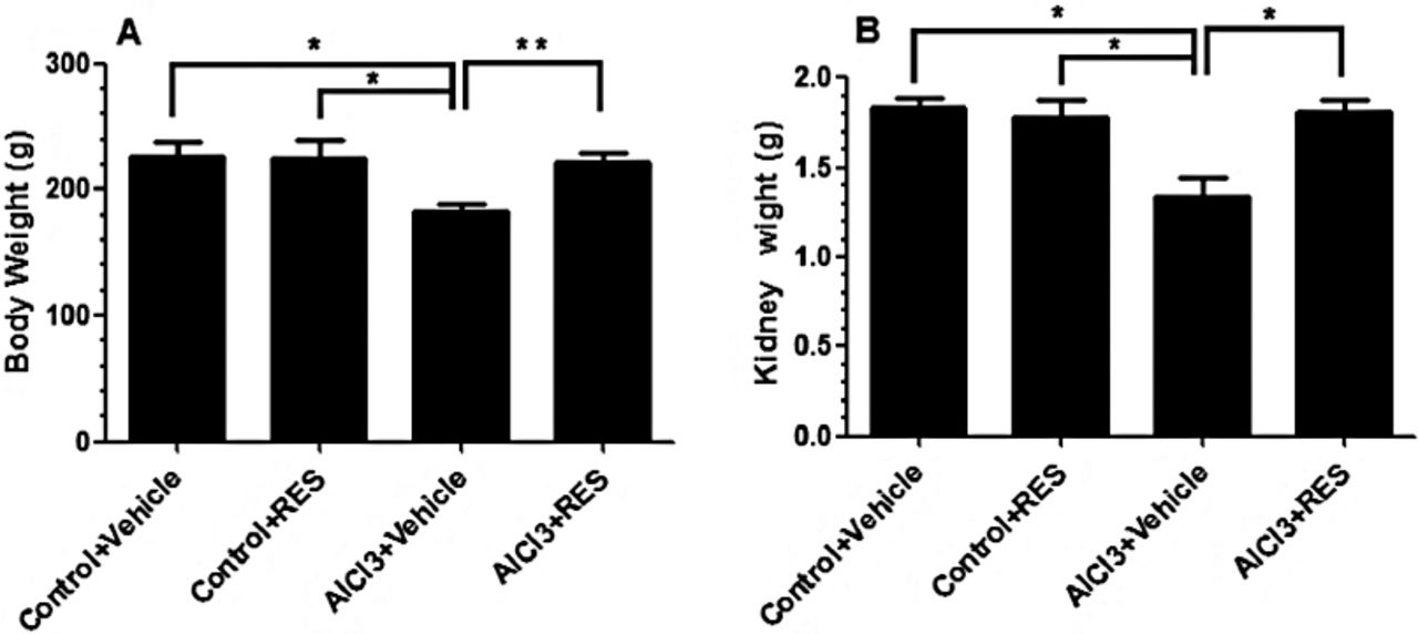
Changes in body weight and relative kidneys weight changes in the final body and kidneys weights are shown in Figures 1A & 1B. In all experimental groups under various treatments, no mortality was observed during the period of the study. In this study, while there were no significant changes in final body and kidney weights between control rats that received the vehicle or normal saline, significant decreases in both parameters (p<0.01) (20.6% and 23.2%) were observed in AlCl3 intoxicated animals when compared with control rats receiving vehicle. In contrast, the AlCl3 group of rats that received RES showed significantly higher body weights and relative kidney weights (p=0.011, p=0.008) than the corresponding levels seen in the AlCl3 intoxicated rats. No significant differences were seen in the levels of these parameters when AlCl3 + RES group was compared with the control group that received the vehicle.

Changes in the total body and individual kidney weights in all groups. Values are expressed as mean ± standard deviation for 6 rats/group. Values were considered significantly different at p<0.05. * p<0.01, ** p<0.001. Res - resveratrol, AlCl3 - aluminum chloride
Markers of kidney function
Indices of kidney function are shown in Figures 2A-2D. There were no significant changes in the levels of urea and Cr and urinary levels of Cr or urine volume when control rats that received the vehicle were compared with control rats that received the vehicle. However, the levels of serum urea and Cr significantly (p<0.0001) increased (158.4.% & 258.5) (Figures 2A & 2B) where urine volume and urine creatinine levels were significantly (p<0.0001) decreased (65.7% & 53.7%) (Figures 2C & 2D) in AlCl3 intoxicated rats in comparison with the control group that received the vehicle. Also, AlCl3 intoxication caused significant reductions (p<0.0001) in Cr clearance in both AlCl3 intoxicated rats received vehicle (94.9%,) (Figure 3). Interestingly, the levels of all these parameters showed significant improvement toward their normal levels seen in the control rats received the vehicle when RES was concomitantly administered with AlCl3.

Kidney function parameters in the serum and urine of all groups. Values are expressed as mean ± standard deviation for 6 rats/group. Values were considered significantly different at p<0.05. ***p<0.0001. Res - resveratrol, AlCl3 - aluminum chloride

Kidney creatinine clearance values in all groups. Values are expressed as mean ± standard deviation for 6 rats/group. Values were considered significantly different at p<0.05. **p<0.00, ***p<0.0001. Res - resveratrol, AlCl3 - aluminum chloride
Oxidative stress parameters in the renal homogenates
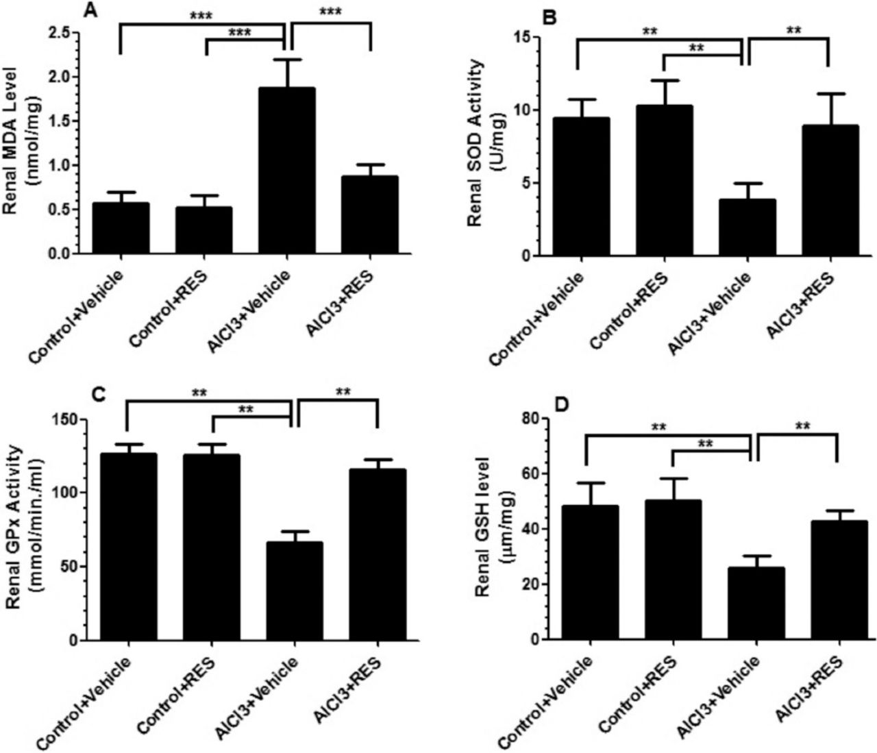
Figure 4 showed no significant change in the levels of malondialdehyde (MDA) (Figure 4A), and reduced glutathione (GSH) (Figure 4D), or in the activities of total superoxide dismutase (SOD) (Figure 4B), or glutathione peroxidase (GPx) (Figure 4C) in the groups of control rats received vehicle or RES. Significant depression (p<0.001) in the activities of SOD (59.1%), GPx (47.2%), as well as levels of GSH (45.8%) with a concurrent significant enhancement (p<0.0001) in levels of MDA (229.3%) were reported in the renal homogenates of AlCl3 intoxicated animals received vehicle. Meanwhile, concomitant administration of RES with AlCl3 to rats resulted in significant increases (p<0.001) in the activities of SOD and GPx and in GSH levels and resulted in a significant decrease (p<0.0001) in MDA levels in their renal homogenates as compared with AlCl3 intoxicated rats and returned to their normal levels seen in control rats that received the vehicle.

Oxidative stress related parameter evaluation in the renal homogenates of all groups. Values are expressed as mean ± standard deviation for 6 rats/group. Values were considered significantly different at p<0.05. **P<0.00, ***P<0.0001. Res - resveratrol, AlCl3 - aluminum chloride
Inflammatory cytokines
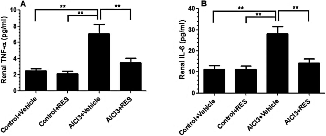
Figure 5 shows significant elevations (p<0.001) of 3 fold increases in TNF-α and IL-6 in AlCl3 intoxicated animals as compared with control rats that received the vehicle. However, concomitant administration of RES with AlCl3 resulted in significant decreases (p<0.001) in the levels of both cytokines as compared with AlCl3 intoxicated animals. However, ANOVA analysis revealed no significant changes in the levels of these mediators when compared between AlCl3 intoxicated rats that received RES, and those corresponding levels measured in the control group that receive the vehicle.

Levels of tumor necrosis factor-a (TNF-α) and interleukin 6 (IL-6) in the renal homogenates of in all groups. Values are expressed as mean ± standard deviation for 6 rats/group. Values were considered significantly different at p<0.05. **p<0.001, Res - resveratrol, AlCl3 - aluminum chloride
Histology findings of liver and kidney
Histology of the kidney in the control rats that received or RES treated rats showed normal structure, Figures 6A & 6B. kidney sections of the AlCl3 intoxicated group (Figures 6C & 6D) showed mild thickening of the basement membrane along with mild changes in the density of mesenchyme, atrophy, and degeneration of glomerular capillaries with increased Bowman’s space (urinary space) and mild tubular necrosis. Macrophage infiltration was also dominant (Figure 6D). However, rats administered AlCl3 and RES (Figures 6E & 6F) showed improvement in their histological architectures including normal glomerulus, normal basement membrane and capillaries. Moreover, the kidney of these rats showed normal urinary space with less degeneration in the tubules. Macrophage infiltration was rarely seen.

Photomicrographs of kidneys obtained from all groups of rats. A and B) were taken from control + vehicle and control + RES treated rats. These groups show normal architecture of kidney with prominent Bowman’s capsule, epithelial cells and normal tubules. C and D) were taken from AlCl3 intoxicated rats. The kidney sections of these rats shows mild thickening of the basement membrane along with changes in the density of mesenchyme, atrophy and degeneration of glomerular capillaries with increased Bowman’s space (urinary space) and tubular necrosis. Glomerular capillaries showed severe shrinkage and damage. Also, severe infiltration was seen (D). E and F) were taken from treated rats and shows normal architecture of glomerular capillaries, intact epithelial cells with the presence of some degeneration in the tubules and slight shrinkage of the glomerular capillaries.
Discussion
The present study was undertaken to determine whether RES can prevent and/or reduce AlCl3 induced renal damage by examining different biochemical and histological parameters related to kidney function of intoxicated and treated rats. Findings clearly showed significant alterations in kidney function in histopathological status after AlCl3 exposure associated with increased renal oxidative stress and inflammation suggesting the strong pro-oxidant activity of AlCl3 in spite of its non-redox status.20 However, RES co-treatment with AlCl3 showed significant improvement in all biochemical and histological parameters related to kidney function and structure. This data shows that RES is able to ameliorate AlCl3 induced nephrotoxicity by improving levels of endogenous antioxidants and reduction of inflammatory biomarkers.
Aluminum accumulates in all tissues of mammals, such as the kidneys, liver, heart, blood, bones and brain,21 and it was found that one of the main organs targeted by Al exposure is the kidney, which plays a major role in preventing accumulation of Al by excreting it out through urine.6 Different mechanisms of renal excretion of Al have been suggested. These include glomerular filtration,22 tubular reabsorption of filtered Al and secretion in distal nephron,23 and excretion in the distal tubules.24 Hence, Al accumulates in the kidneys and induces renal toxicity.20
Indeed, Al accumulation in the kidney has been related to worsening renal function.20,24 It has been reported that the kidney may be exposed to high concentrations of Al during the normal process of renal excretion making the kidney vulnerable to Al-mediated toxicity, which is dependent on the route of exposure.1 In this regard, most of the published studies have investigated the toxic effects of AlCl3 in animals after intraperitoneal or parenteral administration, which does not exhibit the main route of human exposure.1,25
It is well reported that Al enters the body via 2 major routes, pulmonary and oral. Although only a small portion of Al is absorbed through the gastrointestinal tract, oral intake is associated with the greatest toxicological implications.1,25 For this reason, in this study, rats were treated with AlCl3 for 40 days through an intra-gastric tube to mimic chronic toxicity of Al as may occur in humans. The high dose of orally administered AlCl3 was chosen since the intestine plays the role of a protective barrier against Al toxicity since only a small fraction (0.1-0.5%) of ingested Al is absorbed.26
This study showed that AlCl3 induced elevations in serum urea and creatinine levels with the concomitant decrease in creatinine clearance. These findings accord to those obtained by Kowalczyk et al27 who reported that Al has a significant role in the pathogenesis of renal dysfunction and in many clinical disorders. Chronic exposure of AlCl3 in rats has shown nephrotoxicity and glomerular tuft, and renal tubules were reported to be the primary sites of renal damage.28 Aluminum chloride is excreted mainly by the kidneys, and it causes marked degeneration of tubules. Increased serum urea and creatinine concentration can be a consequence of critical accumulation of this metal in the kidneys, eventually resulting in renal failure.29
The kidneys are involved in the excretion of various xenobiotics, pollutants, and toxins hence, they are prone to liberate high quantities of free radicals, which contribute to high oxidative stress that is involved in the pathogenesis of kidney damage.30 Aluminum chloride toxicity appears to be mediated, in part, by free-radical generation. To date, evidence has shown that the toxic effects associated with AlCl3 are due to the generation of ROS, which in turns results in the oxidative deterioration of cellular lipids, proteins, and DNA.31 It was demonstrated that Al may alter the activity and levels of a number of components of the tissue antioxidant defense system, such as GSH, SOD leading to enhance production of free radicals especially ROS and development of lipid peroxidation.32
Lipid peroxidation of biological membranes leads to a loss of membrane fluidity, changes in membrane potential, an increase in membrane permeability and alterations in receptor functions.33 In the same line, our data support this evidence; thiobarbituric acid reactive substances (TBARS) levels as a marker of lipid peroxidation were significantly increased with a concomitant decrease in the levels of GSH and activities of SOD and GPx in the kidney homogenates of intoxicated rats. Although Al is not a transition metal, and therefore, cannot initiate peroxidation, many studies have searched for mechanisms between aluminum Al and oxidative damage in tissues.33
A previous study34 reported that exposure to Al could promote disruptions in the mineral balance, resulting in Al ions replacing iron and magnesium, which would then lead to a reduction in Fe2+ binding to ferritin. Free iron ions released from biological complexes by Al can catalyze hydroperoxides decomposition to hydroxyl radicals via Fenton’s reaction. This high hydroxyl radical reactivity could initiate the peroxidation of membrane lipids, causing membrane damage.34
Also, AlCl3 resulted in significant elevations in the levels of pro-inflammatory cytokines; including TNF-α and IL-6, which fits with many previous reports.35 Kidney sections of the AlCl3 treated group showed mild thickening of the basement membrane along with mild changes in the density of mesenchyme, atrophy and degeneration of glomerular capillaries with increased Bowman’s space (urinary space) and mild tubular necrosis with increased and dominant macrophage infiltration. Aluminum chloride affects the Bowman’s capsule by increased thickening of its basement membrane, which is the first step in the filtration of blood to form urine. Aluminum chloride also resulted in atrophy of glomerulus capillaries and damaging of both proximal and distal tubules. These findings agree with previous findings of in vivo and in vitro studies.36,37
In this study, administration of RES to AlCl3 treated rats effectively improved renal function, as concluded from 1) decreased serum urea and creatinine concentrations and enhancing of Cr clearance, 2) ameliorated oxidative stress and inflammatory cytokines, and 3) attenuated histological changes characteristic of AlCl3 nephrotoxicity. Previously, it was reported that treatment with RES (5 mg or 10 mg/kg orally) for 2 weeks improved urinary protein excretion, renal dysfunction, and renal oxidative stress in streptozotocin- (STZ-) induced diabetic kidneys.38 Furthermore, pretreatment with RES (25 mg/kg, intraperitoneal injection) attenuated signs of cisplatin-induced renal injury.39
This study also suggested that the main mechanism by which RES acts in ameliorating AlCl3 induced renal damage is due to its potent and anti-inflammatory effect. Most of the previous studies accord with our findings as they have shown that resveratrol can directly scavenge ROS, such as superoxide and toxic hydroxyl radicals.40 In addition to scavenging ROS, administered RES modulated the expression and activity of antioxidant enzymes, such as SOD, GPx, and catalase, through transcriptional regulation via nuclear factor E2-related factor 2 (Nrf2), activator proteins (AP)-1, forkhead box O (FOXO), and SP-1 or through enzymatic modification.41 Also, Kitada et al42 reported that RES treatment (400 mg/kg, orally, administered at concentration of 0.3% resveratrol) alleviated albuminuria and histological mesangial expansion and reduced the increased levels of renal oxidative stress and inflammation in the kidneys of db/db mice through the scavenging of ROS and normalizing manganese (Mn)-SOD function by decreasing its levels of nitrosative modification.
Additionally, Chen et al43 reported that RES treatment improved diabetes-induced glomerular hypertrophy and urinary albumin excretion; reduced the expression of glomerular fibronectin, collagen IV, and transforming growth factor; reduced the thickness of the glomerular basement membrane; and reduced nephrin expression in the kidneys of STZ-induced diabetic rats. Furthermore, Wu et al44 demonstrated that RES has protective effects on diabetic kidneys by modulating the SIRT1/FOXO1 pathway. Such pathways may be considered for further research in RES protection in our animal model.
Despite of these finding and the clear renal nephrotoxic effect of AlCl3 in our model, this study had some limitation. As previously discussed, very low fraction of AlCl3 is expected to be absorbed using oral administration and unfortunately, we were not able to determine the levels of accumulated AlCl3 in the kidney tissues that reflects the damaging effect of our dose used in this study. Also, further experiments at the molecular levels are required to investigate the mechanism by which RES reverses AlCl3 induced nephrotoxicity.
In conclusion, this study shows a protective effect of RES against AlCl3 induced renal damage. Reduction of oxidative stress and inflammation may be considered as the main pathways of action.
Illustrations, Figures, Photographs
All figures or photographs should be submitted in a high resolution (minimum 300 DPI) electronic version saved in jpeg or tiff format. Original hard copies of all figures may be requested when necessary. Photographs will be accepted at the discretion of the Editorial Board. All lettering, arrows, or other artwork must be done by an artist or draftsman. If arrows are used please ensure they appear in a different color to the background color, preferably black with a white border, or white with a black border. If arrows distinguish different items on the figure then different arrow styles should be used ie. long, short, wide, narrow. Written informed consent for publication must accompany any photograph in which the subject can be identified. Written copyright permission, from the publishers, must accompany any illustration that has been previously published.
Acknowledgment
The author would like to thank Prof. Mohammed Hairdah, Professor of Physiology and Ethical Committee member at King Khalid University for his valuable collaboration and support to accomplish this work. Also, author would like to thank Mr. Mohmoud Al Khateeb, Lecturer of Physiology at College of Medicine, King Khalid University for all technical and experimental support.
Footnotes
Disclosure. Author has no conflict of interests, and the work was not supported or funded by any drug company.
- Received October 27, 2015.
- Accepted February 11, 2016.
- Copyright: © Saudi Medical Journal
This is an open-access article distributed under the terms of the Creative Commons Attribution-Noncommercial-Share Alike 3.0 Unported, which permits unrestricted use, distribution, and reproduction in any medium, provided the original work is properly cited.
References
In this issue


Jump to section
Related Articles
Cited By...
- No citing articles found.