


Abstract
Objectives: To gain preliminary insight by exploring ulnar variance changes in a Saudi-based sample.
Methods: This 6-month (December 2013 to June 2014) cross-sectional study was conducted on a randomly selected healthy adult volunteers with a sample size of 104, at King Abdulaziz University Hospital, Jeddah, Saudi Arabia. Posteroanterior (PA), anteroposterior (AP), and PA grip views are taken. The variables of interest were the PA, AP, and PA fist measurements of both right and left wrists. An independent t-test was used to compare means between groups.
Results: A total of 104 volunteers were recruited. Among 17 participants who had a negative ulnar variance on right PA views, a significantly high proportion (n=9; 56.2%) maintained a negative value on fist views; 7 participants (43.8%) had a neutral ulnar variance while none (0%) had a positive value (p<0.001). Similarly, a significant proportion of participants who had neutral, or positive values on right PA views maintained the same values on right fist views (p<0.001). On radiographs of the right wrist, the ulnar variance decreased with a change in wrist position, with an absolute difference in magnitude of 2.13 (p<0.001) between PA and AP views. Similarly, the ulnar variance on the left side decreased significantly between PA and AP views (absolute difference in magnitude, 1.68; p<0.001).
Conclusions: Ulnar variance changes in our sample are similar to what is reported in the literature.
Previous clinical studies have measured ulnar variance (UV), which is the length of the ulna compared to the radius.1,2 The ulnar variance may vary among different individuals, and in the lifespan of an individual, the length of the ulna to the radius may change.1 A standard radiograph is required to determine ulnar variance.3 A posteroanterior (PA) view taken with the wrist in pronation position, the elbow flexed to 90 degrees, and the shoulder abducted to 90 degrees is, in general, the standard view.2 Negative ulnar variance is an associate with Kienbock’s disease, avascular necrosis of scaphoid and scapholunate dissociations.3 A positive ulnar variance is harmful to the ulnar compartment of the wrist as it causes degradation and perforation of the triangular fibrocartilage complex and carpal bones cartilaginous wear (ulnar impaction syndrome).3 Mann et al4 reported that the association between UV and load attributes to the biomechanics and anatomical differences within individuals with different UV. The burden that is borne by the radius is 80% and 20% by the ulna with neutral ulnar variance; a positive ulnar variance increases the load carried by the ulnocarpal joint from 18% to 42%.5-7 To cover, or to solve this medical need, the current study was designed to gain preliminary insight by exploring ulnar variance changes in a Saudi-based sample.
Methods
The paper presented data from prior related researches to our study that was conducted using Pubmed and Google Scholar databases. The study is a cross-sectional study carried out at King Abdulaziz University Hospital (KAUH), Jeddah, Saudi Arabia between December 2013 and June 2014. It was approved by the Biomedical Ethics Committee of KAUH. Any healthy, adult, health professional volunteer with a standard wrist, willing to participate, was included in the study. The exclusion criteria comprised the following: volunteers with a history of wrist pain, trauma, fracture, or any medical condition that affects the wrist, including osteoarthritis, or inflammatory arthritis.
Wrist radiographs were undertaken using a standard technique. In an anteroposterior view (AP), the dorsum of the hand was placed against the film and the x-ray beam passed from the palmar to the dorsal surface. Whereas in a PA view, the palmar surface touched the film, and the x-ray beam passed from the dorsal to palmar surface of the wrist. For the clenched fist view, the same technique was applied as in the PA view of the wrist, except that the image was obtained during maximum grip.
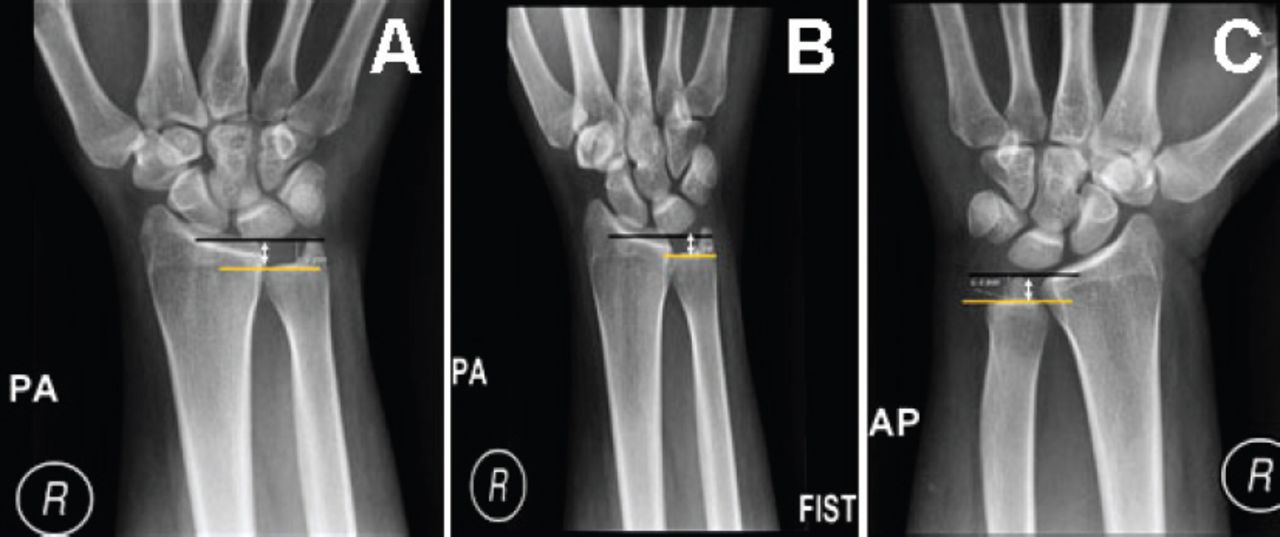
Variance was determined by drawing a transverse line at the level of the lunate fossa and a second one at the level of the ulnar head; the difference between both lines indicated the variance. Missing data were considered, and no imputation was performed. Complete wrist measurements included the ulnar variance of each side (right and left) on 3 radiographs, namely PA, PA clenched fist, and AP views (Figure 1)

Radiographs showing the A) posteroanterior (PA), B) PA clenched fist and C) anteroposterior (AP) views of the right wrist demonstrate ulnar variance: vertical distance between a line drawn parallel to the proximal surface of the lunate facet of the distal radius (black line) and a line parallel (yellow line) to the articular surface.
Statistical analysis
Our total sample size was 104, and as it is a preliminary study, the sample size was similar to other studies. Volunteers were randomly selected from the hospital’s health professional population. Data were collected using excel sheets, and the quantitative radiological measurements of the ulnar variance in different wrist positions were directly added to the sheet by a radiology resident.
Data was analyzed using the Statistical Package for Social Sciences (SPSS Inc., Chicago, IL, USA), version 17.1. Descriptive statistics were computed for variables such as age, gender, and dominant hand. The variables of interest were the PA, AP, and clenched fist measurements of both right and left wrists. These measurements varied from negative to positive values. The relationship between the dominant hand and PA view was investigated using the independent t-test by comparing 2 means. The test for homogeneity of variance was performed using Levene’s test for equality of variances. Statistical significance was set at p<0.05. Results are expressed as frequency (percentage) and mean ± standard deviation (SD).
Results
A total of 104 volunteers were screened for participation in the study. Of these, we included 83 adults (46 women and 37 men) aged on average 25 ± 2.5 years (range 22-35 years). Seventy-six (91.6%) of the participants were right-handed, and ulnar variance did not significantly differ by handedness. The mean ulnar variance in the standard position (PA) was -0.52 ± 2.1 mm on the right side as opposed to -0.34 ± 1.8 mm on the left side. Changes in ulnar variance on different radiographic views of the wrist are shown in Table 1.
Ulnar variance on various radiographic views of the wrist.
The correlation between right PA view and other views was strongly positive for fist view (p<0.001) and weakly to moderately positive for right AP (p=0.001), left AP (p=0.015), and left fist views (p=0.001). A significant positive correlation was also observed between left PA and right fist (p<0.001), left AP (p<0008), and left fist views (p=0.001). A total of 74 participants had complete wrists measurements, and these were analyzed to determine changes in ulnar variance obtained with the wrist in different positions. Of the 17 participants who had a negative ulnar variance on right PA views, a significant proportion of participants who had neutral, or positive values on right PA views maintained the same values on right fist views.
Table 2 shows the other changes of ulnar variance for the position of the right wrist.
Proportion of changes in ulnar variance relative to the position of the right wrist.a
Table 3 shows that a positive, negative, or neutral ulnar variance was maintained in a significantly high proportion of participants on left PA and left fist views (p<0.001), and the other changes in relative to the position of the left wrist. The absolute difference in magnitude of ulnar variance was significant between PA and AP views on both right and left sides (p<0.001), and not significant between PA and clenched fist views as shown in Table 4.
Proportion of changes in ulnar variance relative to position of the left wrist.a
Difference in magnitude of ulnar variance due to a change in wrist position.
Discussion
To assess ulnar variance, we studied 3 different views, including the PA, AP, and fist views. The mean ulnar variance in our sample varied with wrist position, with the most negative values observed on AP radiographs (-2.60 ± 2.2 for the right side and -1.96 ± 2.6 for the left side). Posteroanterior radiographs showed a positive ulnar variance on the right (68.7%) and left (72.3%) sides. Similarly, on AP views a negative ulnar variance was observed on the right (65.1%) and left (50.6%) sides. Because changes in ulnar variance have not been previously studied in a Saudi-based sample, our findings are compared with those of studies conducted abroad. Ulnar variance measurements vary with forearm rotation and fist.1 Dynamic ulnar variance is greater than static variance with an average of 0.9 mm under loading and gripping conditions.6 In a study, they reported a progressive increase of 1.34 mm of maximum ulnar variance that occurred with gripping in pronation compared to ulnar variance with the forearm in relaxed pronation.6
However, wrist position has been reported to be a major determinant of ulnar variance.1 Ulnar variance decreases with maximum forearm supination while it increases with maximum pronation.3,8 Previous studies3 showed that ulnar variance increased by an average of 2.5 mm using a pronated grip view. Also demonstrated a relative increase in ulnar variance with grip.3 Although most patients in our report maintained a positive, negative or neutral ulnar variance on different radiographic views, maximum supination of the forearm resulted in a significant decrease in ulnar variance on both right and left sides. The absolute differences in the magnitude of ulnar variance were 2.13 mm on the right and 1.63 mm on the left sides.
This study, besides having a relatively small sample size, has all the limitations inherent to cross-sectional studies. Another limitation is the use of a hospital-based sample. Thus, our results cannot be extrapolated to the population of Jeddah. However, our findings provide a preliminary insight by exploring ulnar variance changes in a sample of the Saudi population, and our results prove that the ulnar variance is variable in our population similar to other societies. We recommend conducting larger multicenter studies to make relevant conclusions.
In conclusion, our findings demonstrate that most of our participants had a neutral ulnar variance on PA views that was significantly affected by a change in forearm position to AP view. Conversely, there was no change in ulnar variance on PA grip views, similar to what has reported in the literature. However, this difference in magnitude of ulnar variance calls into the question the clinical significance of this difference among our sample, prompting us to recommend further studies with a larger sample to determine the implications of ulnar variance changes in our population.
Acknowledgment
We would like to acknowledge the help of the Clinical Research Unit and the Radiology Department of King Abdulaziz University Hospital, Jeddah, Saudi Arabia for their help and to the technicians of the Radiology Department as well as medical students, residents, and interns for their assistance.
Footnotes
Disclosure. Authors have no conflict of interests, and the work was not supported or funded by any drug company.
- Received December 6, 2015.
- Accepted June 1, 2016.
- Copyright: © Saudi Medical Journal
This is an open-access article distributed under the terms of the Creative Commons Attribution-Noncommercial-Share Alike 3.0 Unported, which permits unrestricted use, distribution, and reproduction in any medium, provided the original work is properly cited.
In this issue


Jump to section
Related Articles
Cited By...
- No citing articles found.