


Article Figures & Data
Figures
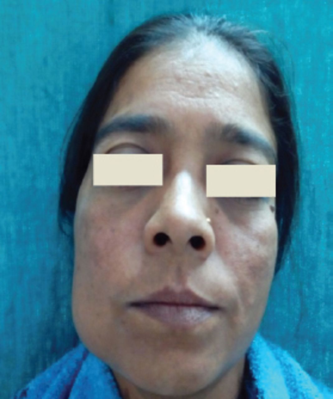
- Figure 1
Facial asymmetry of the right side with a well-defined dome-shaped swelling over the right body of mandible.
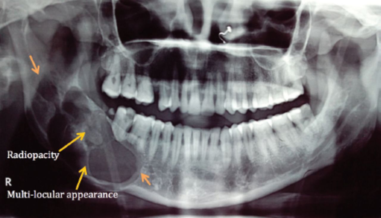
- Figure 2
Panoramic radiograph reveals a well-defined multilocular radiolucent lesion in the right mandibular body region extending up to the ramus, with expansion of cortical plate and scattered diffuse radiopacity inside the radiolucent lesion
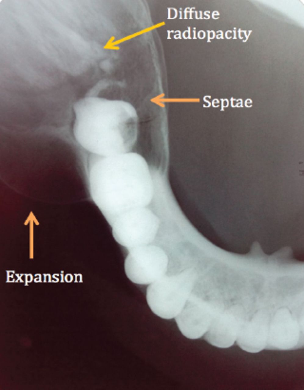
- Figure 3
Mandibular occlusal radiograph reveals a well-defined expansion of both the buccal and lingual cortical plates arising from lower right 1st molar region, with evidence of septa suggesting a multi-locular appearance and diffuse irregular radiopacity within the largely radiolucent lesion
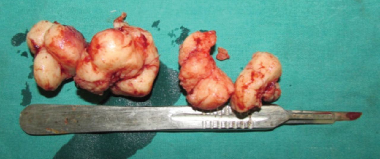
- Figure 4
Gross specimen after surgical enucleation.
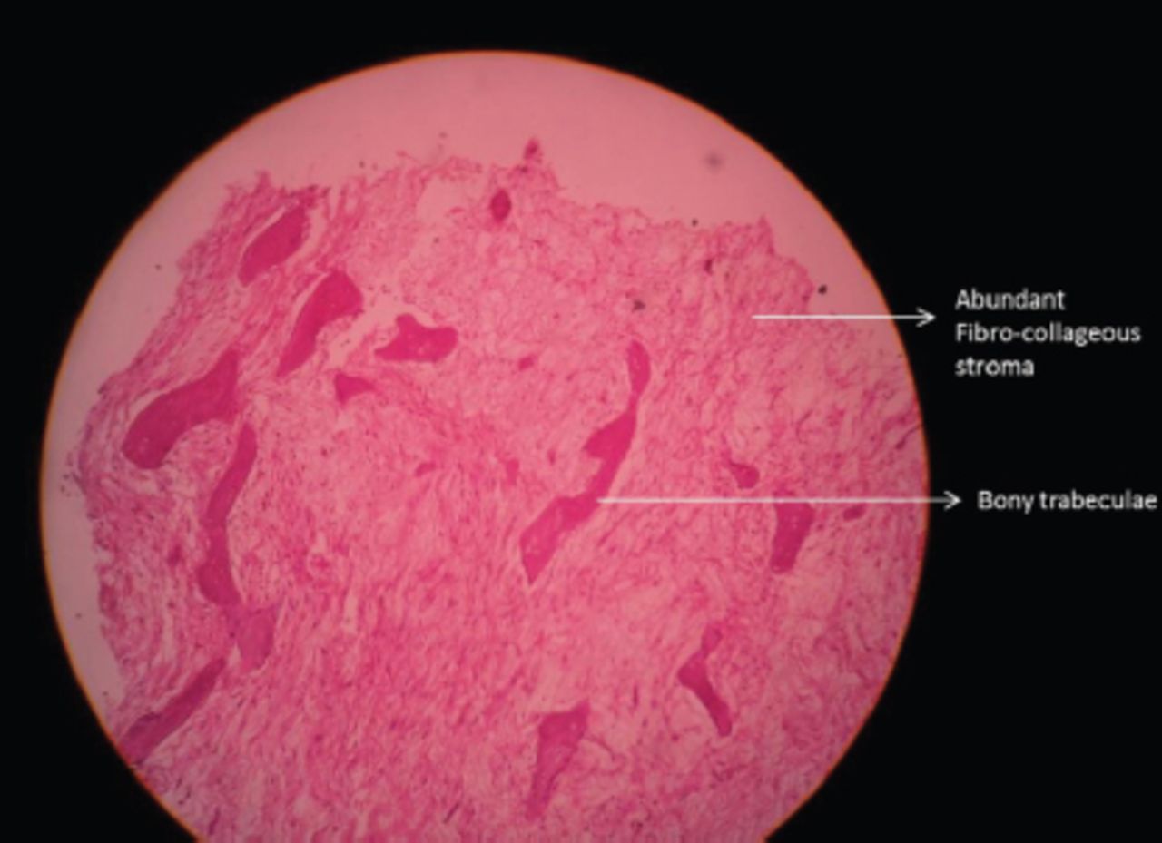
- Figure 5
Low power histopathological picture showing a fair number of bony trabecula admixed with abundant loose fibrocollagenous stroma.
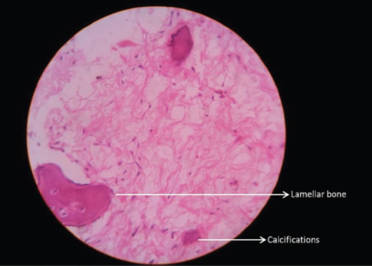
- Figure 6
High power histopathological picture showing lamellar bone with osteoblastic rimming and psammomatous-like nodules and calcifications in the fibrous stroma (haematoxylin-eosin x400).


In this issue


Jump to section
Related Articles
Cited By...
- No citing articles found.