


Abstract
Objectives: To evaluate and compare the shear bond strength (SBS) of Erbium laser etched enamel to acid etched and to detect morphological changes on laser etched enamel surface using scanning electron microscope (SEM). Moreover, Laser induced caries resistance is advantageous in Orthodontics.
Methods: This is an Ex vivo study between January 2016 and December 2017, which comprises a total of 50 human premolars, extracted for orthodontic purpose, were used in this study. The samples were randomly divided into 2 groups of 25 each. The first group was etched using 37% phosphoric for 30 seconds. As for the second group, enamel was treated by Er: YAG laser operating at wavelength 2.94µm, power 1.5W and repetition rate 15Hz. Five teeth from each group were selected for SEM evaluation and the study were continued on 20 teeth from each group. Teeth were subjected to shear bond strength test.
Results: It showed, no-significant difference between the mean of shear bond strength and of the etched groups (p=0.016).
Conclusion: It was concluded that, laser etched group (1.5W/15Hz) resulted in clinically accepted bond strength and could be an alternative to conventional acid etching.
The bonding of orthodontic brackets is mainly based on the mechanical interlocking of an adhesive to irregularities in the enamel surface created by previous enamel etching. Acid etching has been the conventional method of enamel conditioning since its development in 1955.1-3 Enamel acid etching improves retention by hydroxyapatite eroding and subsequently facilitates penetration, via the production of resin tags.4 However, it can result in unintentional demineralization of the most superficial layer of the enamel surface, initiate caries around the metal bracket, expose the acid to uninvolved enamel, and prolong clinical manipulation.2,5
Throughout the past years, many studies have focused on finding alternative methods to conventional acid etching that are less damaging to the tooth structure and simultaneously yield optimum bond strength.6-9
Lately, attention has been driven to the use of laser etching. It has attracted significant attention because it can remove the smear layer and results in an irregular surface pattern which is comparable to that of an acid etching pattern.10,11
It modifies the chemical and crystalline structure of the enamel and prevents caries. In addition, it is a painless procedure, it does not involve heat or vibration, and can be used without anesthesia. Moreover, it reduces chair time in the dental office.2,7,8,12 Since 1964, several types of lasers have been introduced for use in dentistry; for example, ruby, neodymium-doped yttrium aluminum garnet (Nd: YAG), carbon dioxide, and erbium lasers.13 Compared to the other lasers, the Erbium laser is the most effective for hard tissue ablation with minimal thermal effects on the pulp.7,14,15
However, previous conducted studies comparing enamel etching with acid to that of laser were controversial.16 This posed a question whether Erbium laser etching could be an alternative to that of acid. Furthermore, different laser irradiation setting or different operation mode were used.11,17-20
According to our knowledge, no standard irradiation settings of enamel etching using Erbium Laser have been reached. Consequently, another question regarding the optimum parameters that could achieve optimum bod strength and be comparable to acid etching was raised.
In view of the questions raised, the purpose of the present study was designed with different parameters to evaluate and compare the SBS of Erbium laser etched enamel to acid etched. Moreover, to detect morphological changes on laser etched enamel surface using SEM.
Methods
Selection criteria and preparation
This is an Ex vivo study between January 2016 and December 2017, 50 sounds extracted human premolar teeth for orthodontic purposes and collected from discarded teeth after surgical extraction were used in this study. The 4 tooth selection criteria were: intact uncracked buccal enamel surface, restoration material, and caries. Teeth were cleaned thoroughly using tap water, brushed and scaled from any calculus, and then sterilized using a cobalt-60 gamma source (GB50 Type B, Canada) at a dose of 25KGy for 6 hours at room temperature. The samples were stored in saline solution until ready for use. The buccal surface of the teeth was polished using a mixture of fluoride-free pumice powder and distilled water, with a rubber-polishing cup and low-speed hand piece for 15 seconds. Subsequently, all polished teeth were rinsed with water and dried with oil-free compressed air. To delineate the area for enamel conditioning, a piece of scotch tape with a pre-punched circular window of diameter 4 mm was adhered to the enamel surface of each tooth.
Enamel surface etching
The samples were allocated on random basis into 2 groups: (Group 1) The acid-etched group (control), which consisted of 25 premolar teeth which were etched using a 37% phosphoric acid gel (ExciTE F, Ivoclar Vivadent AG, FL-9494 Schaan/Liechtenstein) for 30 seconds, rinsed thoroughly with water, and dried in oil-free air until the enamel surface exhibited a frosty chalky appearance. (Group 2) The Laser-etched group, which consisted of 25 premolar teeth which were etched using Erbium-doped yttrium aluminum garnet (Er: YAG, Fotona, Light walker AT, Ljubljana, Slovenia, EU). The laser device was operated at wavelength of 2940nm at 1.5W, with an energy output of 100mJ, pulse duration of 140µm, frequency of 15Hz, and a water flow rate of 48mL/min. The beam was directed perpendicular to the enamel at a distance of 3 mm from the tooth surface to the laser tip and moved in an extensive fashion for 20 seconds under water spray. Then, an oil-free air was used to dry the tooth surface until the enamel surface exhibited a frosty chalky appearance.
Scanning electron microscopy analysis
Five teeth from each group were randomly selected to examine morphological changes on the enamel surface via SEM, while the other 20 teeth from each group underwent bracket bonding (JEOL JSM 5400, Tokyo, Japan).
Bonding techniques
Forty self-cured acrylic blocks (n=20 for each group) were made to mount teeth that would be subjected to the SBS test. Blocks were color-coded to differentiate between the 2 groups. A stainless-steel premolar bracket (Gemini series Roth, 3M Unitek, Monrovia, CA, USA) with a 0.022 in slot and a base area of 9.806 mm2, as per manufacturer’s specifications, were used in this study. The brackets were bonded to the anatomical crowns using a thin layer of no-mix composite (Transbond TM XT Light cure adhesive, 3M Unitek, Monrovia, CA, USA). After removing excess adhesive with a probe, the brackets were light-cured for 40 seconds.
Shear bond strength evaluation
The SBS of the samples was measured using an Instron universal testing machine (Hampshire Ltd, Bognor Regis, England). The machine blade was inserted between the resin and ortho-bracket and then a vertical occluso-gingival force was applied at a cross head speed of 1 mm/min. The measured bond strength was directly recorded in Newtons, and then converted into mega Pascal (MPa) units.
Statistical analysis
Data are presented as mean and standard deviation (SD). The data were analyzed for normality using Kolmogorov-Smirnov tests. An independent t-test was used to compare between the etching techniques. A p-value≤0.05 was considered significant. Statistical analysis was carried out using IBM® SPSS® Version 25 (IBM Corp., Armonk, NY, USA).
Ethical consideration
The Medical Research Ethics Committee of National Centre of Atomic Research and Technology approved our study; as all subjects were unknown a waiver of informed consent was accepted.
Results
Numerical data was explored for normality using Kolmogorov-Smirnov tests as shown in (Table 1). In addition to check the distribution curves.
Data explored for normality using Kolmogorov-Smirnov tests.
Shear bond strength
Shear bond strength showed normal parametric distribution. Analysis of variance using student’s t-test was used to compare between the 2 groups. A significant level was set at p≤0.05. Our results indicated no statistically significant difference (p=0.016) between the acid-etched (8.988±1.6194) and laser-etched (9.600±1.1072), (Table 2) groups.
Statistical tests indicated no statistically significant difference between the acid-etched and laser-etched groups.
Scanning electron microscope of enamel surface
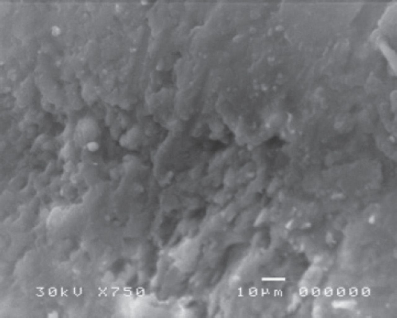
Scaning electron microscope was performed after acid etching using 37% phosphoric acid for 30 seconds, the resulting samples showed enamel surface roughness and preferential dissolution of the prism cores, resulting in a honeycomb-like appearance. (Figures 1 & 2).

Enamel surface roughness after conventional acid etching for 30 seconds (×750).

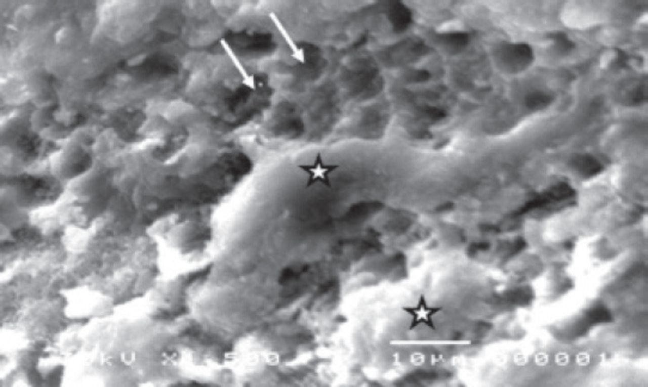
Enamel surface after conventional acid etching for 30 seconds (×1500). The enamel surface has the appearance of a characteristic regular honey comb (arrow), with areas of aprismatic enamel (star).
Enamel surface after Er: YAG laser irradiation
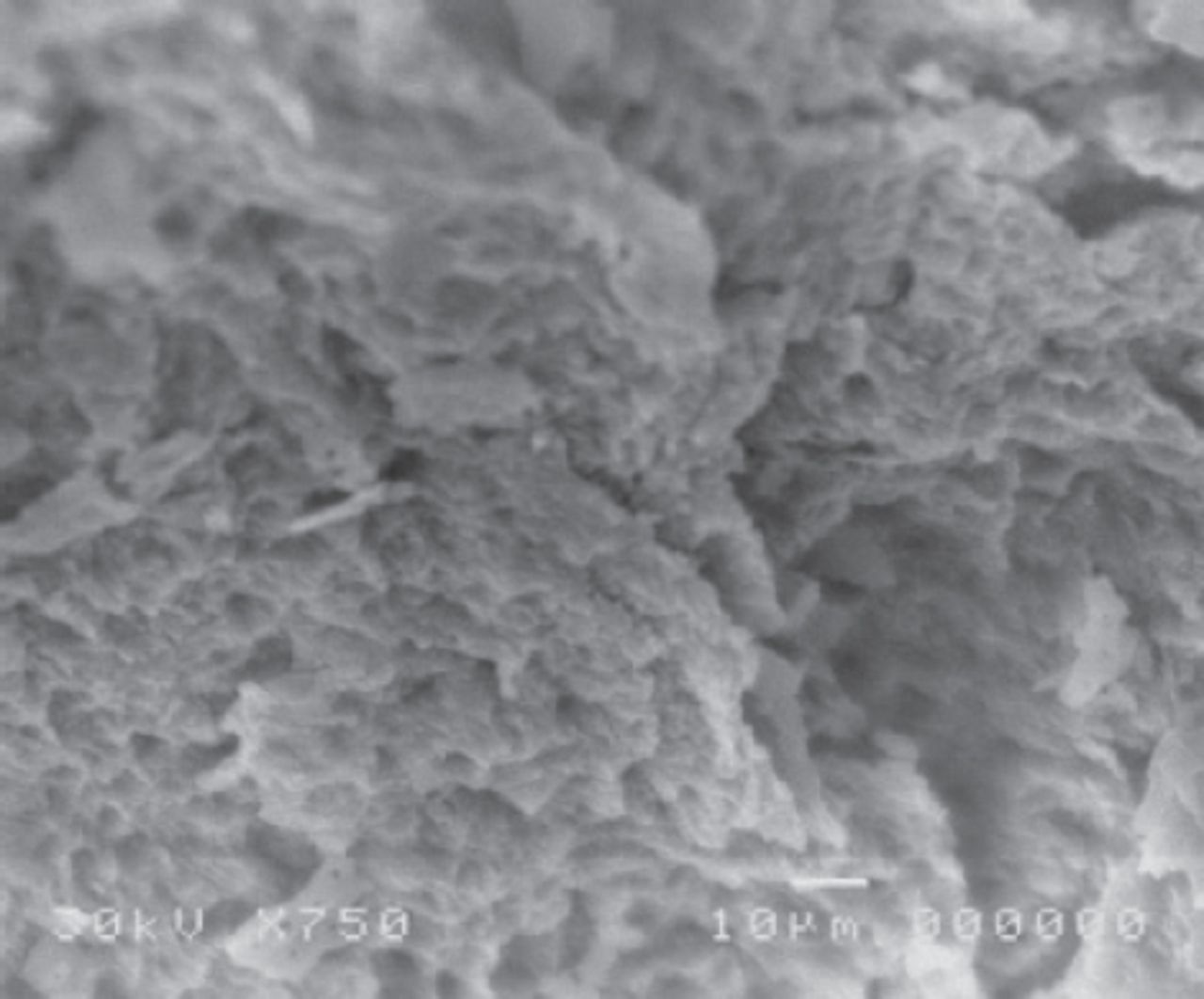
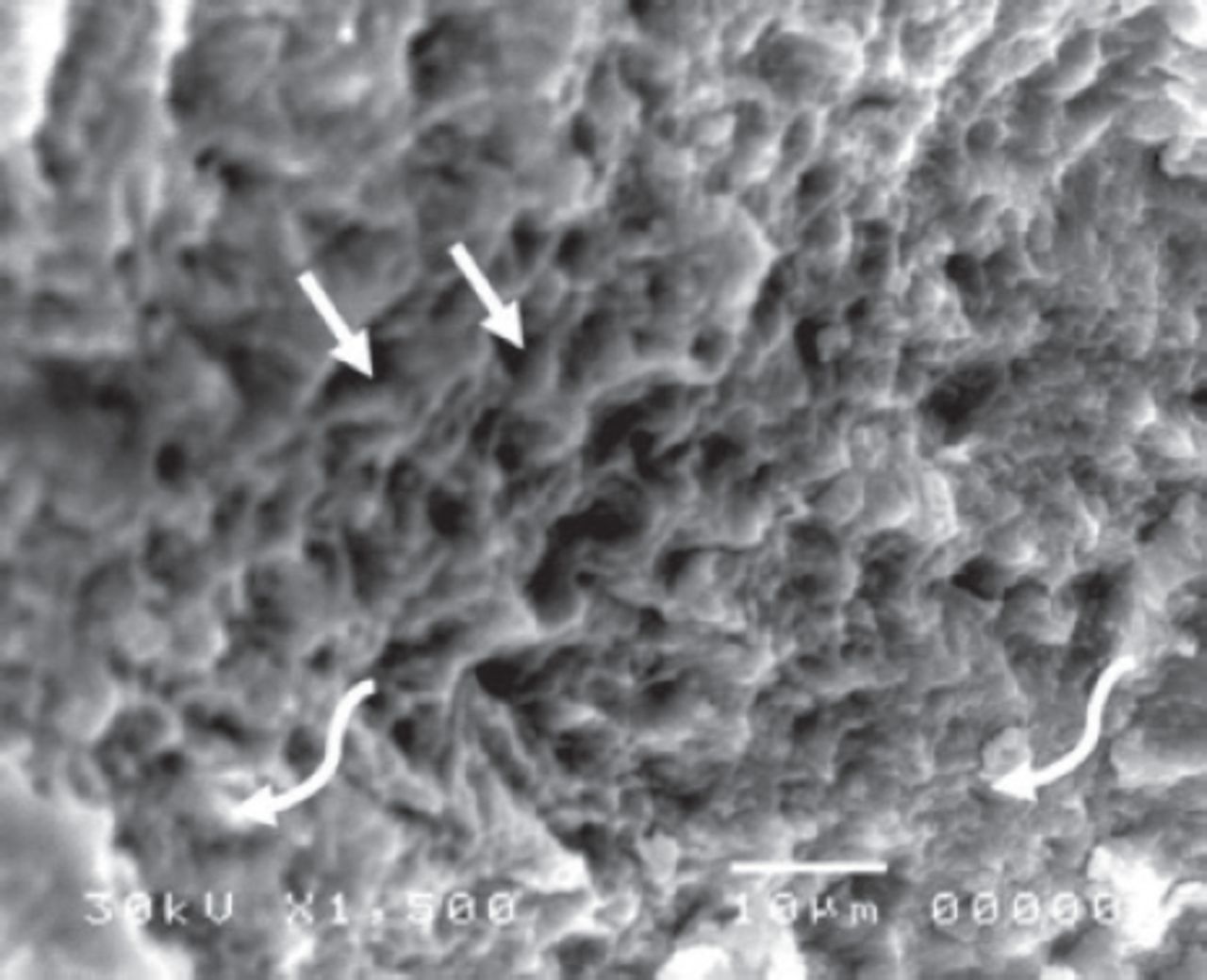
Specimens conditioned with a Er: YAG laser exhibited areas of ablated enamel surface, with a combination of preferential dissolution of prism cores and prism peripheries, areas of hollowed prism cores adjacent to areas of demineralized prism peripheries, and crater formation (Figures 3 & 4).

Enamel surface after Er:YAG laser irradiation (×750) showing surface roughness and crater formation (arrow).

Enamel surface roughness after Er: YAG laser irradiation (×1500). Resorption of the prism cores was observed when the peripheral boundaries were highly projected (arrows).
Discussion
The Erbium laser is one of the most widely used lasers for hard tissue ablation. Erbium lasers emit a wavelength of 2.94µm which matches –OH groups in hydroxyapatite 2.8 µm and the main absorption band of water (3 mm).21 Thus, the irradiation is absorbed strongly by water and increases its affinity for hydroxyl apatite with minimal thermal effect on the pulp.1,3,16 In this study, Erbium laser etching was compared to acid etching using 37% phosphoric acid as a control group, since it is the standard and most common etching method. Moreover, to preserve enamel integrity without decreasing bond strength, the etching time was set to 30 seconds, as suggested in several previous studies.22,23
Various studies have been performed to investigate laser etching using different irradiation settings. The parameters used in this study were chosen so that the energy output would not exceed 100 mJ (power 1.5W, pulse repetition rate 15Hz), so as not to destroy the enamel.24 Samples were irradiated using a short pulse duration (140 µm) to effectively modify the enamel surfaces with low thermal impact on the pulp.16 Moreover, an external water supply was used for safe and efficient ablation of the enamel.25
Scanning electron microscope imaging and SBS tests were conducted to evaluate the changes to the enamel after etching. Scanning electron microscope of the acid-etched group (group 1/control) resulted in a honey comb type I etching pattern.8
Interestingly, areas of non-etched enamel were observed despite the adequate SBS of the acid-etched group (8.988 MPa). This is likely because an ideal acid etching pattern is not necessarily correlated with or influences bond strength.8
On the other hand, SEM images of the laser-etched group (group 2) showed profound surface roughness with a type III Silverstone etching pattern (Figures 4 & 5). This finding was in agreement with several previous studies.20,21 The laser-etched group exhibited clinically acceptable SBSs of 13.59 MPa.
The comparison of SBS of the acid-etched and laser-etched groups revealed no statistically significant difference between SBSs. This is in agreement with previous studies which used the same radiation power (1.5W) and concluded that adequate etching of orthodontic bonding is achieved when irradiation with a 1.5W laser used.26,27
Similarly, it was concluded that low-power Er: YAG laser etching results in clinically acceptable SBS values.20 In addition, our results were in accordance with other literature that used different brackets materials.28,29
Furthermore, laser etching at 100mJ energy (the energy output used in this study) was found to be adequate for bond strength, and Er: YAG laser etching provides a viable alternate to regular acid-etching.1
Conversely, the results presented herein conflict with 2 previous studies that showed a significant difference in SBS between acid-etched and Er: YAG laser-etched groups.3,11
This conflict may be due to the different samples used (human/bovine teeth), different laser machines, different laser irradiation settings (power output, pulse repetition rate, pulse duration settings, energy output, and irradiation time), or different operation modes (contact or non-contact mode, external water cooling, and irradiation distance). Further studies are required to determine standard and optimal parameters for laser etching. Being an Ex-vivo study, as an In-vivo study is inapplicable because of ethical consideration.
In conclusion, Erbium laser etching imparts clinically acceptable shear bond strength of 13.59 MPa. There was no statistically significant difference between the shear bond strength of the laser-etched and acid-etched groups and there is no significant relationship between the bond strength and etching pattern.
Footnotes
Disclosure. Authors have no conflict of interests, and the work was not supported or funded by any drug company.
- Received April 24, 2018.
- Accepted July 19, 2018.
- Copyright: © Saudi Medical Journal
This is an open-access article distributed under the terms of the Creative Commons Attribution-Noncommercial-Share Alike 3.0 Unported, which permits unrestricted use, distribution, and reproduction in any medium, provided the original work is properly cited.
In this issue


Jump to section
Related Articles
Cited By...
- No citing articles found.