


Article Figures & Data
Figures
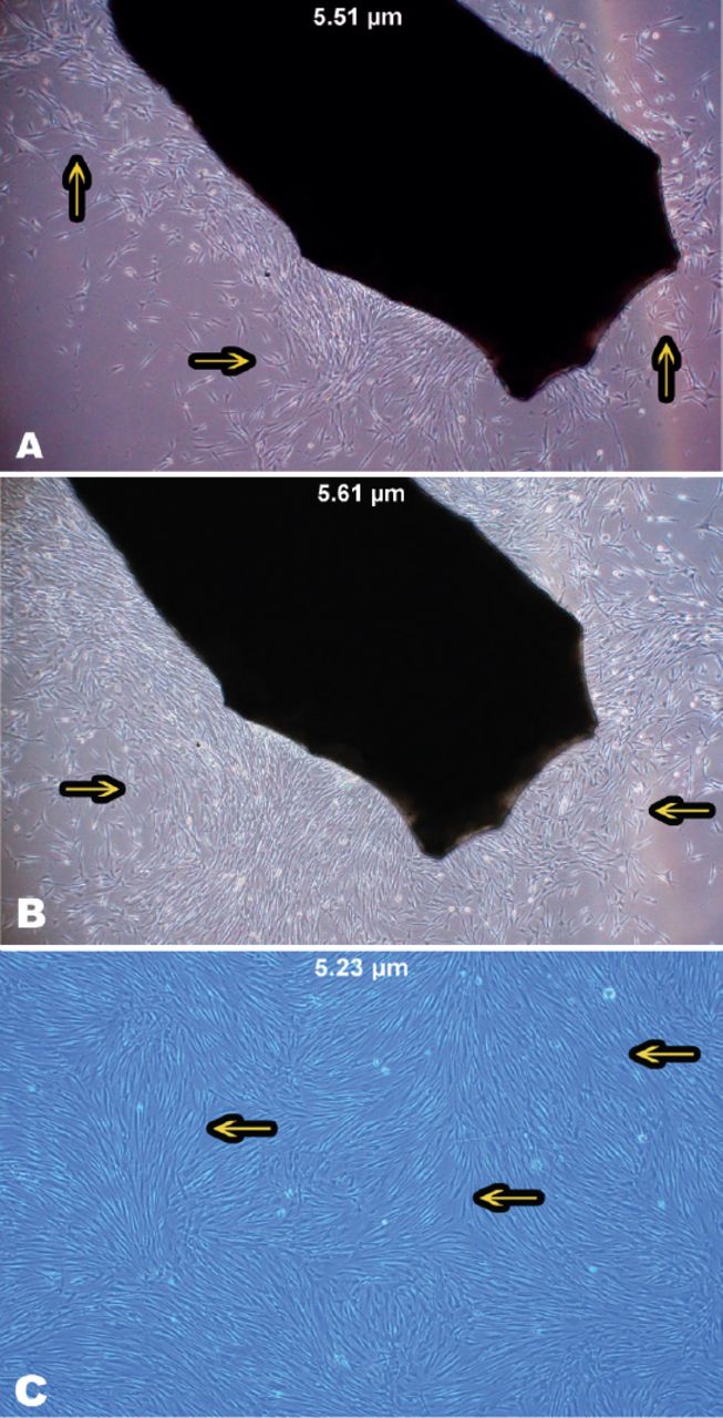
- Figure 1
Growth of human gingival fibroblast cells on a gingival biopsy. A) Morphology of spindle-shaped fibroblast cells. B) An 80% confluence of a full monolayer without overlapping cells was observed after 7-9 days. C) Human gingival fibroblast cell appeared radial or whorl-shaped after the first generation. Images of the cells were taken with OLYMPUS cellSens entry system, 40-fold magnification.
- Figure 2
Cell cultures were tested to support the modified methodology. A) Growth rate of human gingival fibroblast cells (HGFCs) was identified from the 5-13th day. B) MTT formazan absorbance played a role as a function of cellular proliferation for a range of cell concentrations. C) Cell viability test on HGFC with porphyromonas gingivalis (PG-LPS) at various concentrations (0.5, 1, 5, and 100 µg/mL) after 48 hours using cell counting kit-8. Based on the one-way analysis (ANOVA) the result showed that 100 µg/mL porphyromonas gingivalis treatment were significantly different compared to control. *** and ****p<0.0001
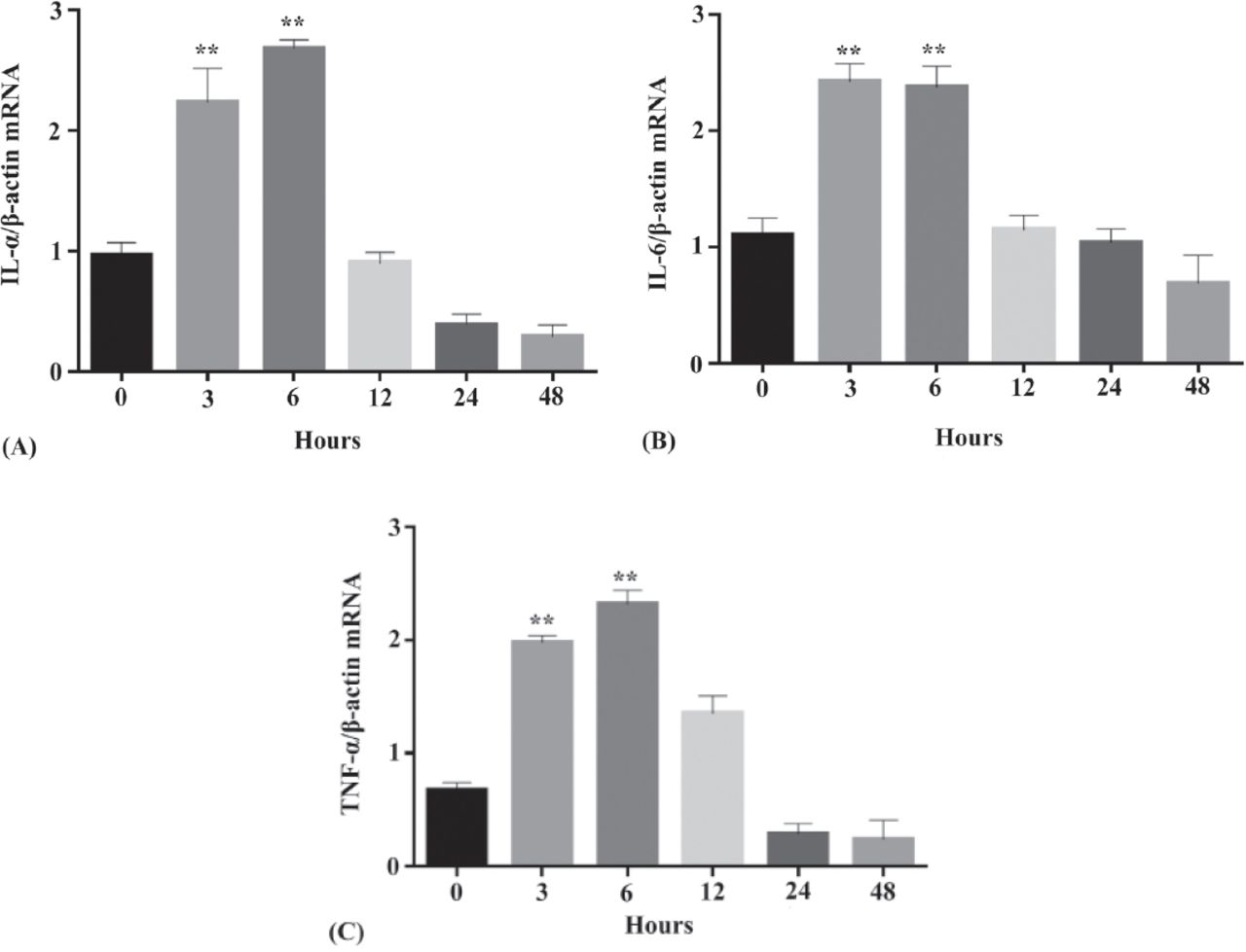
- Figure 3
Expression of the 3 recommended proinflammatory cytokines by human gingival fibroblast cells after incubation with one µg/mL PG-LPS at different time points (3, 6, 12, 24, and 48 hours) messenger ribonucleic acid (mRNA) expression level of A) IL-1β/β -actin mRNA, B) IL-6/β -actin, and C) TNF-α/β-actin. Based on the one-way analysis analysis of variance. **p<0.001 demonstrated a statistically significant difference as compared to experimental control. TNF-a - Tumor necrosis factor-alpha, IL-1b - interleukin-1beta, IL-6 - interleukin-6
- Figure 4
A) The released differential growth factors (PDGF-AA and TGF-β1) were quantified using Enzyme-linked Immunosorbent Assay method at various time points (1-3, 24, 48, 72 hours, and 10 days). B) Cumulative release of differential growth factors (PDGF-AA and TGF-β1) in term of concentration (pg/mL) at different time points. Based on the two-way analysis (ANOVA), **p<0.002 determines a statistically significant higher release than all the other groups, #p<0.002 determines a statistically significant lower release than all the other groups. PDGF-AA - platelet derived growth factor, TGF-b1 - transforming growth factors-beta 1, GF - growth factor
- Figure 5
Suppressive effects of L-PRF on the PG-LPS-induced proinflammatory cytokine releasants by HGFCs. The concentration of IL-1β, TNF-α, and IL-6 detected in the culture medium of PG-LPS challenged HGFCs in the presence or absence of L-PRF was quantified with ELISA method after 20 hours. Based on the two-way analysis (ANOVA), ***p<0.001 illustrated a statistically significant higher concentration from HGFCs alone, while ###p<0.001 demonstrated a statistically significant lower concentration as compared to PG-LPS challenged HGFCs. L-PRF - leukocyte-platelet rich fibrin, PG-LPS - lipopolysaccharide from porphyromonas gingivalis, TNF-a - tumor necrosis factor-alpha, IL-1b - interleukin-1beta, IL-6 - interleukin-6, HGFCs - human gingival fibroblast cells
- Figure 6
Macroscopic observation showing the comparison of the ulcerated area between the L-PRF-treated group and control group. A) The pictures show the more superior healing progress for the L-PRF group. B) The mucositis induced area was significantly smaller in the L-PRF-treated group than the control group by day 7 and day 14. L-PRF - leukocyte-platelet rich fibrin


In this issue


Jump to section
Related Articles
Cited By...
- No citing articles found.