


Article Figures & Data
Figures
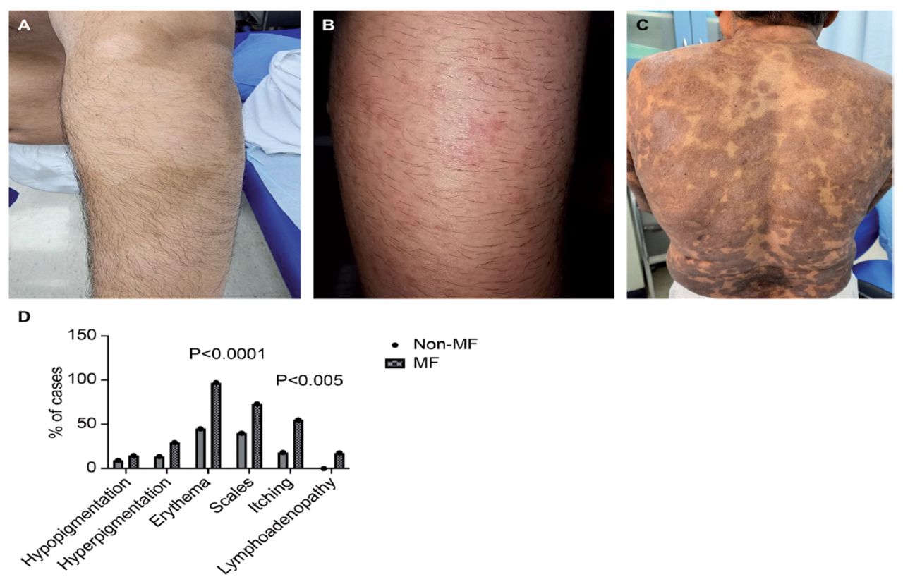
- Figure 1
- Clinical presentations and characteristics of lesions suspicious of MF. (A) Patches present for > 6 months in the upper arm and trunk that were associated with hypopigmentation; the lesions were suspicious of MF. (B) Multiple erythematous papules, accompanied by intense pruritus of the upper arms and H&E-confirmed MF diagnosis. (C) Multiple hyperpigmented scaly patches involving the trunk and H&E-confirmed MF diagnosis. (D) Clinical characteristics of lesions suspicious of MF and their presence in MF cases (n=34) and non-MF cases (n=22). The graph shows that erythema and itching were significantly more common in MF cases than in non-MF cases. H&E: hematoxylin and eosin staining, MF: mycosis fungoides
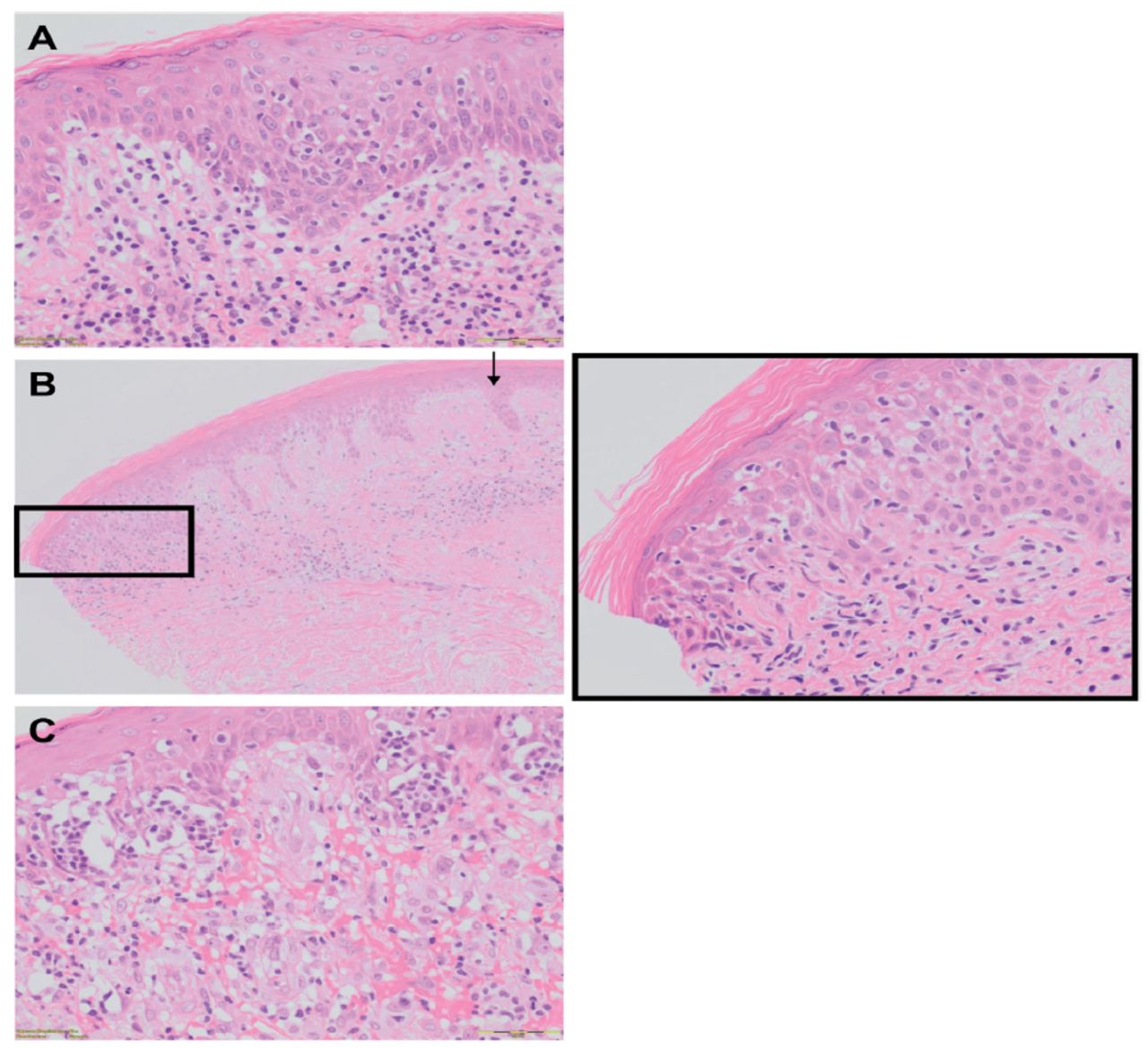
- Figure 2
- Histological features used in the study to distinguish mycosis fungoides (MF) from non-MF cases. A) Epidermotropism is significantly found in MF cases compared with non-MF cases. B) Spongiosis (Grade 1) is present in MF cases, where it is located at the epidermal sites invaded by atypical lymphocytes (black rectangle). Areas not invaded by atypical lymphocytes show no spongiosis (black arrow). C) Pautrier’s microabscesses are significantly found more in the plaque stage than in the patch stage of MF (original, ´200 all).
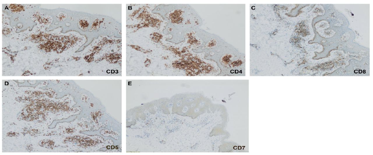
- Figure 3
- Immunohistochemical staining of mycosis fungoides cases. A) Positive expression of pan T-cell markers (CD3). B) Positive expression of CD4. C) Positive expression of CD8 with a ratio of 2.5:1 epidermally. D) Positive expression of CD5. (E) Loss of CD7. (original, ´200 all)


Tables
- Table 1
- Grading of histological parameters for the assessment of suspected mycosis fungoides.
Histological criteria Grade 0 Grade 1 Grade 2 Grade 3 Epidermotropism (-) Focal (1-5/HPF) Moderate (6-10/HPF) Marked (> 10/HPF) Pautrier’s microabscess (-) (+) NA NA Spongiosis* (-) Mild (focal) Moderate Severe Parakeratosis (-) Focal (<10%) Moderate (10-50%) Marked (>50%) Pigment incontinence (-) (+) NA NA Dermal infiltrates (-) Mild perivascular Lichenoid pattern Dense with adnexal structure Dermal fibrosis (-) (+) NA NA Atypical lymphocytes (-) 1–5/100 cells 6–10/100 cells >10/100 cells Large cerebriform lymphocytes (-) 1–3/10 HPF 4–5/10 HPF >5/100 HPF Adnexal involvement (-) (+) NA NA HPF: high-power field, NA: not applicable
↵* Spongiosis was assessed as follows: mild=degree of intercellular edema mildly greater than what would be typically expected for the interface change displayed within the specimen, moderate=moderate spongiotic change, severe=severe spongiotic change with vesicle or bullae formation.
Number of patients (N=56) Clinical presentation of the patients Lesions with hypopigmentation (n) Lesions with hyperpigmentation (n) Lesions with erythema (n) Lesions with scales (n) Association with itching (n) 4 Macules 1 3 0 1 1 20 Patches 4 3 14 9 5 22 Plaques 0 6 21 21 14 4 Nodules 0 0 3 0 0 2 Follicular eruption 0 1 1 1 1 2 Erythroderma 0 0 2 2 0 2 Poikiloderma 2 0 2 0 0 Number of patients (N=56) Association with lymphadenopathy (n) Confirmed MF cases (n) Diagnosis of non-MF cases (n) Average age of patients diagnosed with MF (years) Female-to-male ratio in MF cases 4 0 0 LP (1); PA (1); Inconclusive(1); PIH (1) 0 0 20 0 11 PA (1); DE (1); TC (2); Psoriasis (1); Inconclusive(4) 52 5:6 22 0 14 Psoriasis (3) SD (1); DE (1); TC (1); Inconclusive(2) 57 4:10 4 4 4 - 59 3:1 2 0 1 LP (1) 45 1:0 2 2 2 - 70 0:2 2 0 2 - 39 0:2 MF: mycosis fungoides, n: number of cases, PIH: post inflammatory hyperpigmentation, LP: lichen planus, PA: pityriasis alba, DE: discoid eczema, SD: statis dermatitis, TC; tinea corporis
- Appendix 1
- Histological feature analysis of the 56 suspected mycosis fungoides (MF) lesions involved in the study.
Case no. Diagnosis Clinical feature Epidermotropism Epidermotropism grade * Pautrier’s microabscess Spongiosis Spongiosis grade Parakeratosis 1 MF patch 1 1 1 1 1 1 2 MF patch 1 1 1 1 1 1 3 MF patch 1 1 0 1 1 1 4 MF patch 1 1 0 1 1 1 5 MF patch 1 1 0 1 1 0 6 MF patch 1 1 0 1 1 0 7 MF patch 1 2 0 1 1 1 8 MF patch 1 2 1 1 1 1 9 MF patch 0 0 0 1 1 0 10 MF patch 0 0 0 1 1 1 11 MF patch 0 0 0 1 1 1 12 MF plaque 0 0 1 1 2 1 13 MF plaque 1 1 1 1 1 1 14 MF plaque 1 1 1 1 1 1 15 MF plaque 1 1 1 1 1 1 16 MF plaque 1 1 1 1 1 1 17 MF plaque 1 1 1 1 1 1 18 MF plaque 1 1 0 1 1 0 19 MF plaque 1 1 0 1 1 0 20 MF plaque 1 1 0 1 1 0 21 MF plaque 1 2 1 1 1 1 22 MF plaque 1 2 1 1 1 1 23 MF plaque 1 2 1 1 2 1 24 MF plaque 1 1 0 1 1 1 25 MF plaque 1 1 1 0 0 1 26 MF nodule 1 1 1 1 1 1 27 MF nodule 1 1 1 0 0 0 28 MF nodule 0 0 0 1 1 0 29 MF nodule 0 0 0 0 0 0 30 MF erythroderma 1 1 0 1 1 1 31 MF erythroderma 0 0 0 1 1 1 32 MF poikiloderma 1 1 0 1 1 0 33 MF poikiloderma 0 0 0 0 0 0 34 MF follicular 1 1 1 1 1 0 35 non-MF patch 0 0 0 0 0 0 36 non-MF patch 0 0 0 1 1 1 37 non-MF patch 0 0 0 1 1 1 38 non-MF patch 0 0 0 1 1 0 39 non-MF patch 0 0 0 1 1 0 40 non-MF patch 0 0 0 1 1 0 41 non-MF patch 0 0 0 1 1 0 42 non-MF patch 0 0 0 1 1 0 43 non-MF patch 0 0 0 1 2 0 44 non-MF plaque 0 0 0 1 1 1 45 non-MF plaque 0 0 0 1 1 1 46 non-MF plaque 0 0 0 1 1 1 47 non-MF plaque 0 0 0 1 1 0 48 non-MF plaque 0 0 0 1 1 0 49 non-MF plaque 0 0 0 1 2 0 50 non-MF plaque 0 0 0 0 0 0 51 non-MF plaque 0 0 0 1 1 0 52 non-MF follicular 0 0 0 1 1 0 53 non-MF macule 0 0 0 0 0 0 54 non-MF macule 0 0 0 0 0 0 55 non-MF macule 0 0 0 0 0 0 56 non-MF macule 0 0 0 0 0 0 Case no. Parakeratosis grade Atypical lymphocytes Atypical lymphocytes grade Cerebriform lymphocytes Cerebriform lymphocytes grade Infiltrating lymphocytes Infiltration grade Pigment incontinence Adnexal involvement Dermal fibrosis 1 1 1 1 1 1 1 1 1 0 1 2 1 1 1 1 2 1 1 1 0 1 3 1 1 1 1 1 1 1 0 0 1 4 1 1 1 1 1 1 1 0 0 1 5 0 0 0 0 0 1 1 0 0 1 6 0 0 0 0 0 1 1 0 0 0 7 2 1 2 1 2 1 2 0 0 1 8 1 1 2 1 2 1 1 0 0 1 9 0 1 1 1 1 1 2 0 0 0 10 1 0 0 0 0 1 1 0 0 0 11 1 0 0 0 0 1 1 0 0 0 12 1 0 0 0 0 1 1 0 0 0 13 1 0 0 0 0 1 1 0 0 0 14 1 0 0 0 0 1 1 0 0 0 15 1 0 0 0 0 1 1 0 0 0 16 1 0 0 0 0 1 1 0 0 0 17 1 1 1 1 1 1 1 0 0 1 18 0 1 1 1 1 1 1 0 0 0 19 0 1 1 1 1 1 2 0 0 0 20 0 1 1 1 2 1 2 0 0 0 21 1 0 0 0 0 1 1 0 0 0 22 2 1 1 1 1 1 1 0 0 1 23 2 1 2 1 3 1 2 0 0 1 24 1 1 1 1 1 1 1 0 0 0 25 1 0 0 0 0 1 2 1 0 1 26 1 1 2 1 2 1 3 1 1 1 27 0 1 2 1 2 1 2 0 0 1 28 0 1 2 1 2 1 2 0 0 0 29 0 1 2 1 2 1 2 0 0 0 30 1 1 1 1 1 1 2 1 0 1 31 1 1 1 1 1 1 2 0 0 1 32 0 1 1 1 1 1 1 1 0 1 33 0 1 1 1 1 1 1 1 0 1 34 0 1 1 1 1 1 2 1 0 1 35 0 0 0 0 0 1 1 0 0 0 36 0 0 0 0 0 1 2 1 0 0 37 0 0 0 0 0 1 2 1 0 0 38 0 0 0 0 0 1 1 0 0 0 39 0 0 0 0 0 1 1 0 0 0 40 0 0 0 0 0 1 1 0 0 0 41 0 0 0 0 0 1 1 0 0 0 42 0 0 0 0 0 1 1 0 0 0 43 0 0 0 0 0 1 1 0 0 0 44 1 0 0 0 0 1 1 0 0 0 45 1 0 0 0 0 1 1 1 0 0 46 1 0 0 0 0 1 1 1 0 0 47 0 0 0 0 0 1 1 0 0 0 48 0 0 0 0 0 1 1 0 0 0 49 0 0 0 0 0 1 1 0 0 0 50 0 0 0 0 0 1 1 0 0 0 51 0 0 0 0 0 1 1 0 0 0 52 0 0 0 0 0 1 1 0 0 0 53 0 0 0 0 0 1 1 0 0 0 54 0 0 0 0 0 1 1 0 0 0 55 0 0 0 0 0 1 1 0 0 0 56 0 0 0 0 0 1 1 0 0 0 ↵* Traits were graded from 0 to 3 in epidermotropism, spongiosis, parakeratosis, dermal infiltrate, atypical lymphocytes, and large atypical cerebriform cells and graded from 0 to 1 in Pautrier’s microabscess, pigment incontinence, dermal fibrosis, and adnexal involvement. Analysis was performed using Fisher’s exact test with a Bonferroni-corrected threshold of significance = 0.005 = 0.05/10). If a significant association found, follow-up testing was performed to assess the grade severity of a trait vis-à-vis the diagnosis, using ordinal logistic regression (using maximum likelihood fitting), in 2 ways: first, by including zero values in the response variable (0, 1, 2, 3; 5 models fitted), and second, by considering only non-zero cases (such as including only those patients exhibiting the trait). When narrowing the testing to the patch and plaque groups only, 2×2 contingency tables (Fisher’s exact test, 2-tailed) were used. Likewise, it is pertinent to know whether the grade severity of a trait differentiating MF from a non-MF diagnosis was also more pronounced in patch versus plaque patients. To this end, non-zero cases were excised, and contingency tables were analysed (9 2×2 tables with Fisher’s exact test and one 3×2 table analysed with a likelihood ratio test of the chi-square). Finally, for either the patch or plaque subgroup, 10 goodness-of-fit tests were performed, 1 per histological trait, to determine where the observed counts of presence/absence deviated from those expected by chance (such as 50% frequency of occurrence).
In this issue


Jump to section
Related Articles
Cited By...
- No citing articles found.