


Abstract
Objectives: To investigate the relationship between dietary choices and brain choline (Cho) levels using magnetic resonance spectroscopy (MRS).
Methods: A total of 88 female students from the radiology department at King Abdullah bin Abdulaziz University Hospital, Riyadh, Saudi Arabia, participated in this study. Brain total choline (tCho) levels were estimated using MRS single volume sequence at a 3 Tesla field, with an echo time of 30 ms, repetition time of 2000 ms, voxel size of 15x15x15 mm, and water suppression bandwidth of 50 Hz. Participants’ food consumption habits were assessed using a dietary questionnaire to quantify the amount of protein in their daily servings, as protein intake affects Cho levels in the brain.
Results: Linear regression test applied using the Statistical Package for the Social Sciences, and the result showed significant impact of diet protein intake on the brain tCho level (p=0.000).
Conclusion: The study’s findings indicated that dietary choices significantly affect the levels of tCho in the brain. This research can serve as a baseline for health education, highlighting the close connection between dietary decisions and brain Cho levels. Understanding this relationship is essential for promoting a healthy lifestyle among younger generations.
The brain is an organ of the central nervous system, and it serves as the primary control center for gathering and correlating sensations, storing memories, making decisions, and initiating actions. Several nutrients affect the brain’s functions, whether by enhancing or sometimes by hindering its performance, one of which is choline (Cho), the precursor of the neurotransmitter acetylcholine. For instance, the loss of cholinergic neurons is linked to impaired cognitive function, particularly in Alzheimer’s disease (AD), and Cho is necessary for DNA synthesis, crucial to brain development and function. Overall, Cho has been proven remarkably important for optimal brain growth, development, and peak performance.1,2
Choline, a crucial dietary nutrient, is essential for optimal brain function; while the body can synthesize some Cho, but this amount often falls short of what is needed.3,4 Choline serves multiple purposes: it forms a major component of cell and organelle membranes, and it significantly impacts various physiological processes, including nerve insulation (myelination), signal transmission within cells (signal transduction), and DNA creation and regulation (histone methylation), highlighting the critical role Cho in maintaining a healthy brain.3
Choline is an element readily available in many foods consumed every day. Eggs stand out as a particularly rich source, significantly boosting overall Cho intake compared to other options. While plant-based options, such as cruciferous vegetables and some beans, offer Cho, they generally contain less per serving than animal products, emphasizing the importance of dietary diversity to ensure sufficient Cho consumption for optimal brain function.5
Changes in the brain’s Cho levels can signal various health issues and diseases.6 For example, elevated Cho levels are often observed in brain tumors, while decreased Cho production in the brain is linked to age-related cognitive decline. Choline deficiency can also affect multiple bodily systems, including the liver, muscles, and lymphocytes in humans, as well as the kidneys, pancreas, and brain and nervous system development. One of the earliest clinical signs of dietary Cho deficiency is fatty liver (hepatosteatosis) due to the lack of phosphatidylcholine required to package and export very-low-density lipoproteins. In rodents, prolonged Cho deficiency can lead to spontaneous hepatocarcinoma, making it the only nutrient deficiency known to cause spontaneous carcinoma. In humans, elevated serum aminotransferases can indicate liver damage from insufficient dietary Cho. In addition, elevated muscle enzymes, such as serum creatine phosphokinase, may develop due to Cho deficiency, indicating compromised muscle membrane integrity and potential cell death.7-9
A cholinesterase test can be used to measure Cho, where blood is collected from a vein using a needle and placed in an airtight vial or syringe. The sample is then sent to a laboratory to evaluate the enzymes acetylcholinesterase and pseudocholinesterase, which break down acetylcholine. Then, Cho levels can be measured in plasma samples, with concentrations as low as 1 μmol/L. The most common methods for locating Cho in biological samples include chromatographic techniques, radioenzymatic assays, enzyme-linked immunosorbent assay, and potentiometric methods. In addition, magnetic resonance spectroscopy (MRS) can measure Cho levels in vivo by detecting the characteristic MR signals of Cho in the brain.10-13
Magnetic resonance spectroscopy is a sophisticated, non-invasive method that complements magnetic resonance imaging (MRI) measurements. Single-voxel spectroscopy (SVS) is the preferred method for Cho analysis, utilizing 3 primary sequences: point resolved spectroscopy, stimulated echo acquisition mode, and chemical shift selective imaging sequence (CHESS). It is important to note that CHESS is not a component of SVS itself; rather, it is used in conjunction with SVS for water suppression. This function is particularly crucial as it selectively suppresses the strong water signal, enabling a clearer analysis of the weaker signals from fat and Cho, and thus allowing for a more accurate assessment of Cho levels within the tissue.14-19
The lifestyles of Saudi Arabians have changed recently, particularly among those aged 18-25 years, a period during which habits are easily influenced and frequently altered. As a result, there is instability in the choices carried out, leading to fluctuations in the variety of food groups consumed. In response, this research aims to investigate the relationship between female students’ dietary choices and their brain Cho levels by comparing their MRS results with their answers to a dietary questionnaire.20
This study can serve as a baseline for health education, highlighting the connection between dietary choices and brain Cho levels, which affect bodily functions and potentiate numerous diseases. It will also enhance our understanding of the impact of dietary decisions on daily life.
Methods
This cross-sectional study, carried out from January to June 2024 at King Abdullah bin Abdulaziz University Hospital, Riyadh, Saudi Arabia, (IRB registration number: HA-01-R-104; log number: 23-0189), aimed to examine the relationship between dietary choices and brain Cho levels among radiology department students. The study utilized MRI, MRS, and a dietary questionnaire to assess this relationship.
The inclusion criteria included: healthy Saudi female university students aged 18-21 years and the exclusion criteria included: non-Saudi students, male students, students with health issues, those with any MRI contraindications, and those younger than 18 years or older than 21 years.
Volunteers completed an MRI safety form before the exam and were positioned head-first in the supine position in a Siemens 3T MAGNETOM Vida scanner (Siemens Healthineers, Erlangen, Germany). The laser beam localizer was centered over the glabella, and the MRI and MRS procedure followed a specific sequence. Initially, a 3-plane brain localizer MRI was used to position and plan the sequences, typically utilizing T1-weighted low-resolution scans lasting less than 25 seconds. Subsequently, a T2-weighted scan (TSE sequence) was carried out, with axial slices planned on the sagittal plane and the block positioned parallel to the genu and splenium of the corpus callosum. The planning block was verified in the other 2 planes to ensure a suitable angle in the coronal plane, making it perpendicular to the midline of the brain and the fourth ventricle, with slices covering the entire brain from the vertex to the foramen magnum line. The T2-w TSE axial sequence parameters included a TR of 3,000 ms, TE of 100 ms, slice thickness of 4 mm, gap of 0, flip angle of 130, phase R>L, field of view (FOV) of 239, and NEX of 2.
The MRS single-voxel sequence was then planned based on the T2-w TSE image, with the signal obtained from the voxel at the basal ganglia on the axial plane. The SVS parameters included a voxel size of 15×15×15, TR of 2,000 ms, TE of 30 ms, and water suppression BW of 50 Hz. A short echo time (TE) of 30 ms was selected to allow for the detection of a broader range of metabolites, including total choline (tCho), enhancing the accuracy of metabolite quantification. To address the signal overlap associated with a short TE, and to achieve precise estimation of Cho amplitude, we employed LCModel for spectral fitting. This robust post-processing software effectively mitigates frequency drifts and compensates for minor motions, ensuring reliable and accurate quantification of metabolites. Additionally, we employed automated shimming techniques before acquisition to optimize magnetic field homogeneity within the voxel, contributing to consistent and high-quality spectra for accurate metabolite assessment.
In this study, we utilized a published questionnaire to assess food intake, with a particular emphasis on Cho-rich sources within various food groups, including eggs, milk, grains, vegetables, and seafood.20 Participants were asked specific questions regarding their consumption of these Cho-rich foods. For each item, the questionnaire provided options for participants to report how frequently a given portion size, measured in ounces, was consumed on a weekly basis. To calculate the Cho content from the amount of protein-rich food consumed, the portion size of each food item was multiplied by the frequency of consumption per week and then adjusted by the Cho content specific to that food. This Cho content information was obtained from reference databases, such as the United States Department of Agriculture choline content database.21 For example, if a person consumes 2 large eggs per day, the total Cho intake from this portion can be calculated by first taking the amount of Cho in one large egg, approximately 0.147 grams, and multiplying it by 2 to account for the 2 eggs consumed daily.21 This result is then multiplied by 7 to obtain the weekly Cho intake from eggs, reflecting the tCho consumption from this source per week.
Data were collected from volunteers’ MRS scans and their responses to the dietary questionnaire. The estimated Cho amplitude from the MRS spectrum represents tCho, which reflects the combined signal from phosphorylcholine and glycerophosphorylcholine, as these metabolites cannot be reliably distinguished using standard MRS at clinical field strengths.
Statistical analysis
The results were analyzed using a linear regression test in the Statistical Package for the Social Sciences, version 28.0 (IBM Corp., Armonk, NY, USA) to determine the impact of dietary Cho intake on brain Cho levels.
Results
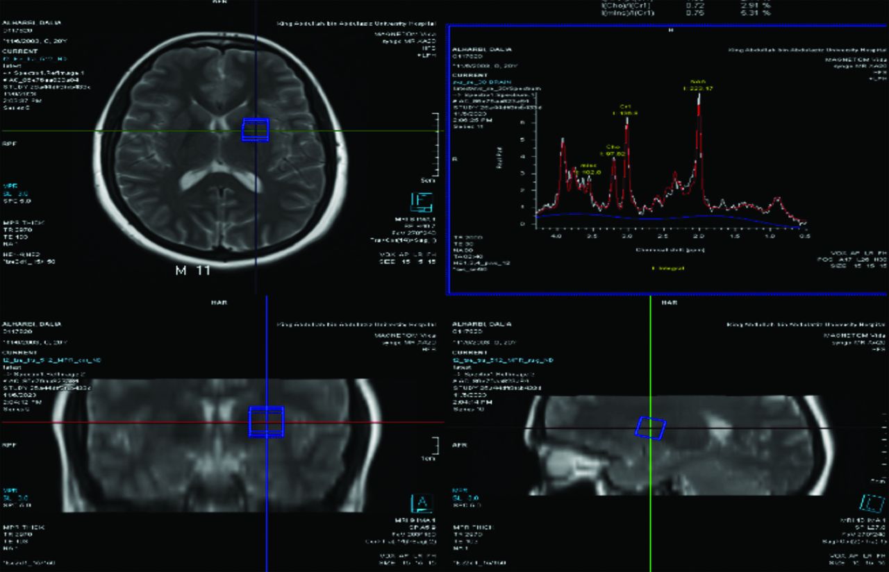
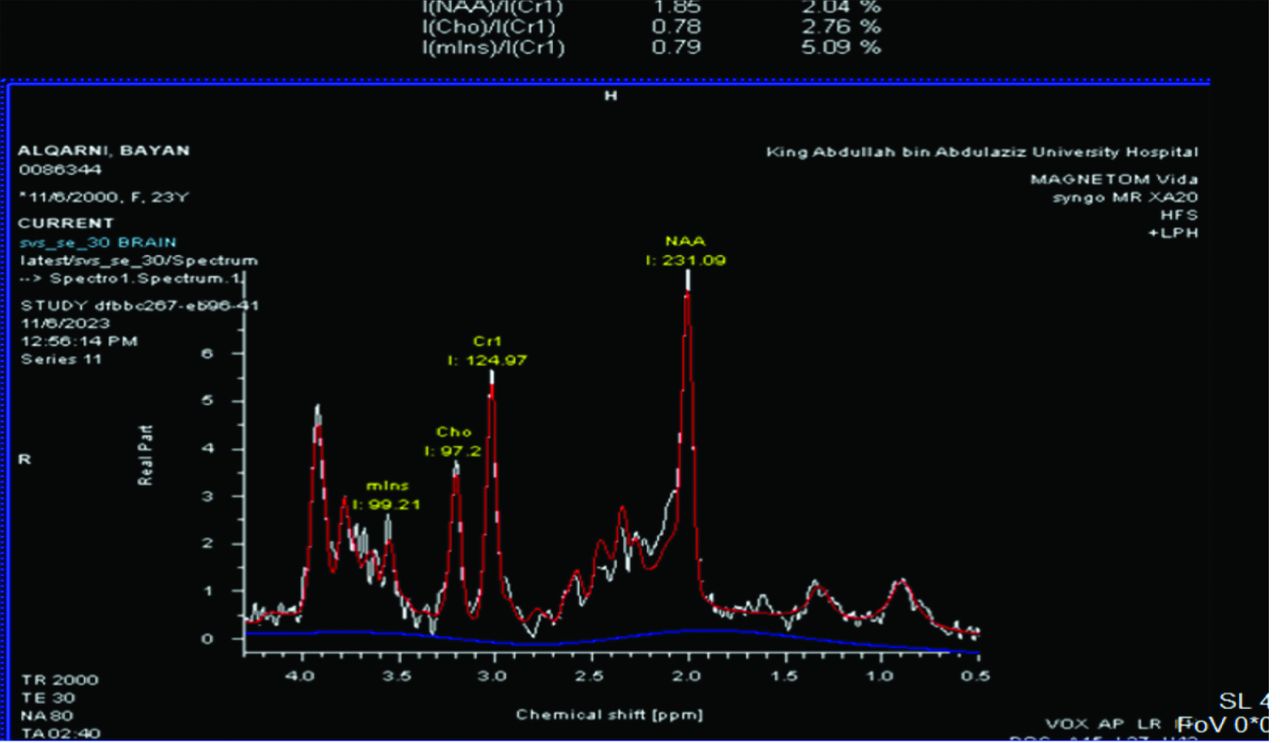
The data from the questionnaire were categorized, and Cho intake was calculated to assess its impact on brain Cho levels, estimated from the MRS spectrum. The linear regression test used, where dietary intake is the independent variable and brain Cho level the dependent variable. An example of a volunteer’ MRI brain planes and the FOV volume chosen to enquire the MRS data and the corresponding MRS spectrum are shown in Figure 1. In addition, the MRS full brain spectrum is shown in Figure 2, where the Cho peak at 3.02ppm.

-Magnetic resonance imaging of the brain and the corresponding brain spectrum. The blue box is the volume from which the magnetic resonance spectroscopy spectrum is obtained.

- Magnetic resonance imaging of the brain and the corresponding brain spectrum. In the brain spectrum, choline (Cho) peak appears at 3.02 ppm frequency. Cho: choline, Cr: creatinine, NAA: N-acetylaspartate
The linear regression results are summarized across 3 tables. Table 1 presents a descriptive analysis of the relative tCho concentration, estimated as the tCho/creatine (Cr) ratio from the MRS spectrum. This relative concentration is calculated by normalizing the tCho peak amplitude to the Cr peak amplitude, creating a unitless measure. This approach helps account for variations in signal intensity and tissue composition across scans, providing a consistent indicator of Cho levels relative to Cr within the tissue. Additionally, Cho intake data from the dietary questionnaire are included for comparison.
- Descriptive analysis of the relative total choline concentration, estimated as the total choline/creatine ratio from the magnetic resonance spectroscopy spectrum.
Table 2 displays the correlation between tCho relative concentration and dietary Cho intake, and Table 3 presents the regression coefficients for the relationship between dietary Cho intake (independent variable) and tCho relative concentration (dependent variable).
- The correlation between total choline relative concentration and dietary choline intake.
- The regression coefficients for the relationship between dietary choline intake and total choline relative concentration.
A p-value of 0.000 indicates a significant correlation between dietary Cho intake and tCho relative concentration in brain (Tables 2 & 3), and this result suggests a strong impact of diet on brain Cho levels.
Discussion
Magnetic resonance spectroscopy is an emerging method for estimating brain amino acids. Because it lacks widespread adoption, limited studies have been carried out on humans correlating brain Cho relative concentration estimated via MRS with dietary intake. For simplicity, we will refer to the relative tCho concentration as “brain Cho level” throughout this discussion.
A study by Poly et al22 focused on the relationship between Cho intake and brain functions, with 744 female subjects. The result showed that high Cho intake is related to better cognitive performance, such as verbal and visual memory, which agrees with the present study. In addition, a loss of cholinergic neurons and decreased Cho acetyltransferase activity are consistent features observed in AD. It is thus believed that Cho plays a role in the learning and memory impairments characteristic of this condition. The present study shows a significant correlation between diet and brain Cho levels, with a p-value of 0.000, as presented in Table 3.
Derbyshireet al23 emphasize the significance of early intake of Cho-rich nutrients for brain health, optimal neural development, and sustained cognitive function throughout life. They suggest that such dietary practices may help mitigate declines in cognitive functions. This recommendation is in line with the findings of the present study, which proves a significant correlation between dietary Cho consumption and Cho levels in the liver and brain among healthy young volunteers.
A previous study carried out by Schoen et al24 reported no significant association between dietary Cho consumption and brain function, contrasting the findings of the present study, which identified a significant correlation between Cho intake and cognitive performance. This contradiction may arise from differences in the studied groups, including variations in age and health conditions. In addition, methodological disparities could contribute to the divergent outcomes between the studies.
Moreover, the current study was carried out on healthy young adults, with a larger sample size compared to the earlier study by Schoen et al,24 which investigated a population with congenital disorders and involved a smaller sample (42 participants). This difference could significantly contribute to the contrasting findings on Cho intake and brain function. In addition, the current study employed a more direct approach, measuring brain Cho levels using MRS results and a dietary questionnaire to evaluate dietary Cho consumption.
As per the findings from Wallace et al,25 Cho deficiency in individuals, even when supplemented with sufficient folate and vitamin B12, can lead to health complications, such as fatty liver disease. This condition manifests through elevated levels of liver enzymes in the bloodstream, coupled with indications of muscle damage, as evidenced by increased circulating Cr phosphokinase levels. The results of Wallace et al’s study align with those of the present study, which revealed a significant correlation between dietary intake and liver Cho levels, as shown by a correlation coefficient of 0.000, as shown in Table 3.
Study limitations
The dietary intake data were collected using self-reported questionnaires, which may introduce minor variations due to recall bias or reporting accuracy. While these factors could slightly influence the precision of estimated dietary Cho intake, they are common to dietary studies and were carefully managed to maintain the robustness of our results.
In conclusion, this cross-sectional study investigated the effects of dietary choices on brain Cho levels among young female Saudi students who presented to the MRI department of King Abdullah bin Abdulaziz University Hospital, Riyadh, Saudi Arabia. The findings revealed that dietary intake, particularly of Cho, significantly influences brain Cho levels. The results suggest that young women should ensure consistent intake of Cho-rich foods to maintain optimal brain Cho levels.
Acknowledgment
This research was funded by the Deanship of Scientific Research at Princess Nourah bint Abdulrahman University, through the Research Funding Program, Grant No. (FRP-1444-17).
This article underwent English language editing services by Scribendi (www.scribendi.com).
Footnotes
Disclosure. This study was funded by the Deanship of Scientific Research at Princess Nourah bint Abdulrahman University, Riyadh, Saudi Arabia, through the Research Funding Program, grant No.: (FRP-1444-17).
- Received November 8, 2024.
- Accepted February 4, 2025.
- Copyright: © Saudi Medical Journal
This is an Open Access journal and articles published are distributed under the terms of the Creative Commons Attribution-NonCommercial License (CC BY-NC). Readers may copy, distribute, and display the work for non-commercial purposes with the proper citation of the original work.
References
In this issue


Jump to section
Related Articles
Cited By...
- No citing articles found.