
Main menu

  • Home
  • Content
    • Latest
    • Archive
    • home
  • Info for
    • Authors
    • Reviewers
    • Subscribers
    • Institutions
    • Advertisers
    • Join SMJ
  • About Us
    • About Us
    • Editorial Office
    • Editorial Board
  • More
    • Advertising
    • Alerts
    • Feedback
    • Folders
    • Help
  • Other Publications
    • NeuroSciences Journal

User menu

  • My alerts
  • Log in

Search

  • Advanced search
Saudi Medical Journal
  • Other Publications
    • NeuroSciences Journal
  • My alerts
  • Log in
Saudi Medical Journal

Advanced Search

  • Home
  • Content
    • Latest
    • Archive
    • home
  • Info for
    • Authors
    • Reviewers
    • Subscribers
    • Institutions
    • Advertisers
    • Join SMJ
  • About Us
    • About Us
    • Editorial Office
    • Editorial Board
  • More
    • Advertising
    • Alerts
    • Feedback
    • Folders
    • Help
  • Follow psmmc on Twitter
  • Visit psmmc on Facebook
  • RSS
Case ReportCase Report
Open Access

An eosinophilic variant granulomatosis with polyangiitis involving the dura, bilateral orbits, and mastoids

Hasan Al-Hakami, Abdurhman S. Al-Arfaj, Mohammed Al-Sohaibani and Najma A. Khalil
Saudi Medical Journal June 2016, 37 (6) 690-693; DOI: https://doi.org/10.15537/smj.2016.6.14718
Hasan Al-Hakami
From the Department of Medicine (Hakami, Al-Arfaj, Khalil), Rheumatology Division, the Department of Pathology (Al-Sohaibani), College of Medicine, King Saud University, Riyadh, Kingdom of Saudi Arabia
BSc, ChB, MD
  • Find this author on Google Scholar
  • Find this author on PubMed
  • Search for this author on this site
Abdurhman S. Al-Arfaj
From the Department of Medicine (Hakami, Al-Arfaj, Khalil), Rheumatology Division, the Department of Pathology (Al-Sohaibani), College of Medicine, King Saud University, Riyadh, Kingdom of Saudi Arabia
FRCP, FACP
  • Find this author on Google Scholar
  • Find this author on PubMed
  • Search for this author on this site
  • For correspondence: [email protected]
Mohammed Al-Sohaibani
From the Department of Medicine (Hakami, Al-Arfaj, Khalil), Rheumatology Division, the Department of Pathology (Al-Sohaibani), College of Medicine, King Saud University, Riyadh, Kingdom of Saudi Arabia
BSc, MD
  • Find this author on Google Scholar
  • Find this author on PubMed
  • Search for this author on this site
Najma A. Khalil
From the Department of Medicine (Hakami, Al-Arfaj, Khalil), Rheumatology Division, the Department of Pathology (Al-Sohaibani), College of Medicine, King Saud University, Riyadh, Kingdom of Saudi Arabia
MSc, MPhil
  • Find this author on Google Scholar
  • Find this author on PubMed
  • Search for this author on this site
  • Article
  • Figures & Data
  • eLetters
  • Info & Metrics
  • References
  • PDF
Loading

Article Figures & Data

Figures

  • Figure 1
    • Download figure
    • Open in new tab
    • Download powerpoint
    Figure 1

    Photograph showing: A) Axial CT sections without contrast through orbits showing varying levels of ill-defined soft tissue thickening surrounding the lateral rectus muscle on left side and the superior recti muscles. B) Coronal CT sections without contrast through orbits show varying levels of ill-defined soft tissue thickening around the lateral aspects of the left orbit surrounding the lateral rectus muscle and also extending into intraconal fat, superior rectus muscle and lacrimal gland.

  • Figure 2
    • Download figure
    • Open in new tab
    • Download powerpoint
    Figure 2

    Photograph showing: A) Coronal high resolution T2 weighted image (1) demonstrating increased signal intensity in the soft tissue along lateral aspect of the left orbit. T1 weighted image (2) shows corresponding decreased signal intensity. Post contrast T1 weighted images (3,4) show enhancement in the abnormal orbital soft tissue. Minimal similar abnormalities are seen in the right orbit as well. There is also abnormal meningeal thickening and enhancement along the base and lateral aspect of the cerebral hemisphere. B) Axial post contrast T1 weighted images (5,6) show enhancement of the abnormal soft tissue as well as abnormal thickening and enhancement of the dura on left side. Axial pre contrast T1 weighted image (7) shows ill-defined soft tissue thickening around the lateral aspect of the left orbit.

  • Figure 3
    • Download figure
    • Open in new tab
    • Download powerpoint
    Figure 3

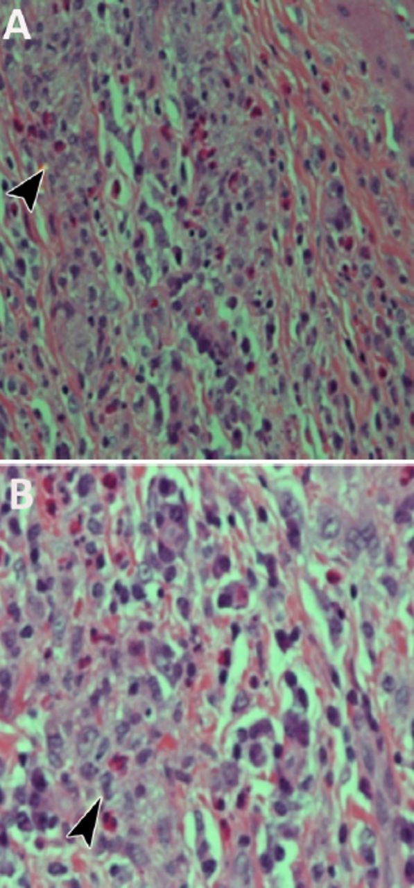
    Photograph showing: A) Lacrimal gland biopsy shows granulation, medium sized vessel wall infiltrated by eosinophils (arrow head) and plasma cells. The lumen of the vessel is obliterated by similar cells and there are newly formed vessels (hematoxylin and eosin stain X400). B) Lacrimal gland biopsy shows eosinophilic granuloma. The arrow head points to an aggregate of histiocytes. There are numerous plasma cells at the periphery (hematoxylin and eosin stain X600).

  • Figure 4
    • Download figure
    • Open in new tab
    • Download powerpoint
    Figure 4

    Axial (top row) and coronal (bottom row) CT sections without contrast through orbits show remarkable resolution of the ill-defined soft tissue thickening around the lateral aspects of the left orbit in the axial (top row figures 1-3) and coronal (bottom row figures 4-6).

PreviousNext
Back to top

In this issue

Saudi Medical Journal: 37 (6)
Saudi Medical Journal
Vol. 37, Issue 6
1 Jun 2016
  • Table of Contents
  • Cover (PDF)
  • Index by author
Print
Download PDF
Email Article

Thank you for your interest in spreading the word on Saudi Medical Journal.

NOTE: We only request your email address so that the person you are recommending the page to knows that you wanted them to see it, and that it is not junk mail. We do not capture any email address.

Enter multiple addresses on separate lines or separate them with commas.
An eosinophilic variant granulomatosis with polyangiitis involving the dura, bilateral orbits, and mastoids
(Your Name) has sent you a message from Saudi Medical Journal
(Your Name) thought you would like to see the Saudi Medical Journal web site.
Citation Tools
An eosinophilic variant granulomatosis with polyangiitis involving the dura, bilateral orbits, and mastoids
Hasan Al-Hakami, Abdurhman S. Al-Arfaj, Mohammed Al-Sohaibani, Najma A. Khalil
Saudi Medical Journal Jun 2016, 37 (6) 690-693; DOI: 10.15537/smj.2016.6.14718

Citation Manager Formats

  • BibTeX
  • Bookends
  • EasyBib
  • EndNote (tagged)
  • EndNote 8 (xml)
  • Medlars
  • Mendeley
  • Papers
  • RefWorks Tagged
  • Ref Manager
  • RIS
  • Zotero
Share
An eosinophilic variant granulomatosis with polyangiitis involving the dura, bilateral orbits, and mastoids
Hasan Al-Hakami, Abdurhman S. Al-Arfaj, Mohammed Al-Sohaibani, Najma A. Khalil
Saudi Medical Journal Jun 2016, 37 (6) 690-693; DOI: 10.15537/smj.2016.6.14718
Twitter logo Facebook logo Mendeley logo
  • Tweet Widget
  • Facebook Like
  • Google Plus One
Bookmark this article

Jump to section

  • Article
    • Abstract
    • Case Report
    • Discussion
    • Footnotes
    • References
  • Figures & Data
  • eLetters
  • References
  • Info & Metrics
  • PDF

Related Articles

  • No related articles found.
  • PubMed
  • Google Scholar

Cited By...

  • No citing articles found.
  • Google Scholar

More in this TOC Section

  • Cutaneous metastasis of signet ring cell adenocarcinoma of the colon
  • Giant left gastric artery aneurysm with intrathoracic extension
  • A rare case of a horseshoe kidney with a single left-sided ureter presented with recurrent urinary tract infection
Show more Case Report

Similar Articles

CONTENT

  • home

JOURNAL

  • home

AUTHORS

  • home
Saudi Medical Journal

© 2025 Saudi Medical Journal Saudi Medical Journal is copyright under the Berne Convention and the International Copyright Convention.  Saudi Medical Journal is an Open Access journal and articles published are distributed under the terms of the Creative Commons Attribution-NonCommercial License (CC BY-NC). Readers may copy, distribute, and display the work for non-commercial purposes with the proper citation of the original work. Electronic ISSN 1658-3175. Print ISSN 0379-5284.

Powered by HighWire