


Abstract
Objectives: To determine the dental arch dimensions and arch forms in a sample of Saudi orthodontic patients, to investigate the prevalence of Bolton anterior and overall tooth size discrepancies, and to compare the effect of gender on the measured parameters.
Methods: This study is a biometric analysis of dental casts of 149 young adults recruited from different orthodontic centers in Jeddah, Saudi Arabia. The dental arch dimensions were measured. The measured parameters were arch length, arch width, Bolton’s ratio, and arch form. The data were analyzed using IBM SPSS software version 22.0 (IBM Corporation, New York, USA); this cross-sectional study was conducted between April 2015 and May 2016.
Results: Dental arch measurements, including inter-canine and inter-molar distance, were found to be significantly greater in males than females (p<0.05). The most prevalent dental arch forms were narrow tapered (50.3%) and narrow ovoid (34.2%), respectively. The prevalence of tooth size discrepancy in all cases was 43.6% for anterior ratio and 24.8% for overall ratio. The mean Bolton’s anterior ratio in all malocclusion classes was 79.81%, whereas the mean Bolton’s overall ratio was 92.21%. There was no significant difference between males and females regarding Bolton’s ratio.
Conclusion: The most prevalent arch form was narrow tapered, followed by narrow ovoid. Males generally had larger dental arch measurements than females, and the prevalence of tooth size discrepancy was more in Bolton’s anterior teeth ratio than in overall ratio.
Dental arch dimensions, including dental arch width, length, and form, are important values for the diagnosis, treatment, planning, and treatment outcomes concerning patients who are seeking orthodontic treatment in all age groups.1 Different ethnic groups and populations display variable dental arch measurements and characteristics.2 It is well-known that dental arch dimensions continue changing throughout growth and development, but during adulthood, the changes decrease.1 This explains why many researchers were interested in investigating the changes in dental arch dimensions during each stage of growth and development.1-7 It is well documented in the literature that using preformed arch wires for orthodontic patients, regardless of their arch form, will lead to post-treatment instabilities in the form of relapse.8 Accordingly, there have to be shifts from using preformed arch wires routinely for all patients to selecting specific arch wires for individual patients, depending on his or her arch form and malocclusion adaptability. Several researchers had been trying to classify the dental arch forms. It is accepted that the dental arch is shaped and confined by the supporting bone configurations, and it is affected by the eruption of teeth and the surrounding muscular forces.6 Chuck,9 in 1934, made the first classification for the dental arch in 3 forms, namely: ovoid, tapered, and square shape. He also emphasized the importance of individualizing the arch wire form to each patient rather than using the same arch wire form for all patients. Dental crowding occurs mainly due to tooth size versus arch length discrepancy. Moreover, post-orthodontic stability greatly depends on lower arch form maintenance; thus, dental arch dimensions are important values to be investigated.10 Several arch forms were documented in the literature. The most popular were the Ricketts pentamorphic arch forms, which considered factors such as arch correlation, size, and length11 (Figure 1). He classified them into in 5 forms to fit most facial forms and patients. Growth and development of the dental arches differ between males and females.4 The 6 keys of occlusion by Andrew12 describe the feature of normal occlusion; any significant deviation from the normal occlusion will cause malocclusion. Tooth size is another factor to achieve normal occlusion with good intercuspation of teeth. It has been considered the seventh key of occlusion.13 Some studies had described different methods for measuring tooth size discrepancy;14,15 the most recognized method is Bolton’s tooth size ratio. Bolton had concluded that the normal ratio for normal occlusion is an overall ratio of 91.3±1.91 and an anterior ratio of 77.2±1.65.
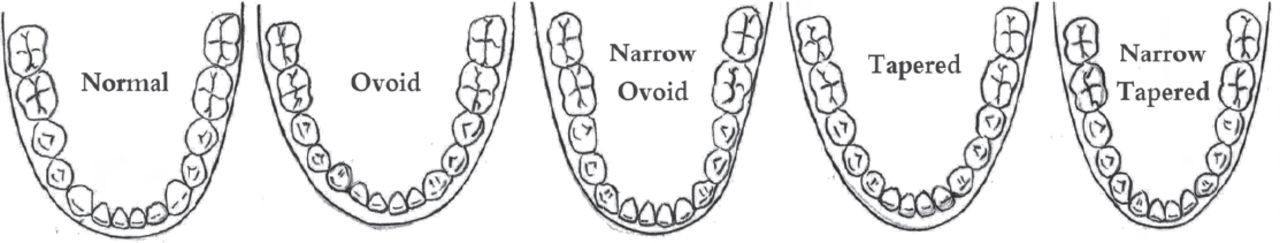
Pentamorphic arch forms (illustrative drawing).
Much research has been carried out to document dental arch dimensions, arch forms, and tooth size discrepancy in several populations, but very few were performed in the western region of Saudi Arabia. Therefore, the aim of this study was to determine the dental arch dimensions and arch forms in a sample of patients seeking orthodontic treatment, to investigate the prevalence of Bolton anterior and overall tooth size discrepancies and to compare the effect of gender on the measured parameters in this sample.
Methods
Our sample was collected from several orthodontic centers across Jeddah city in the western region of Saudi Arabia between April 2015 and May 2016. Twenty orthodontic models were excluded from the 169 original samples due to poor quality. The sample used in this study consisted of the total number of included 149 (85 females and 64 males) subjects’ pretreatment maxillary and mandibular dental casts of young adults (Table 1).
Characteristics of the 149 study samples.
The inclusion criteria were: 1) Sound pretreatment orthodontic models of adult patients. 2) Permanent dentition. 3) Class I, II, and III malocclusion. 4) Full dentition in both arches, excluding third molars.
The exclusion criteria were: 1) Casts from children with mixed or primary dentition. 2) Missing teeth. 3) Dental anomalies. 4) Posterior crossbite. 5) Retained deciduous teeth. 6) Casts with severe transverse arch discrepancies. 7) Casts with severe crowding.
A digital caliper was used to measure the following parameters: i) Arch length: from the center of the palatal incisal papilla to the middle point on a line drawn between the right and left first molars. ii) Arch circumference: the sum of 3 measurements, from the mesial of the first molar to the mesial of the canine, from the mesial of the canine to the mesial of the contralateral canine and from the mesial of the canine to the mesial of the first molar. iii) Inter-molar distance: the distance measured from the buccal groove along the occlusal surface of the first molar to the contralateral first molar. iv) Inter-premolar distance: from the buccal cusp tip of the first premolar to the contralateral. v) Inter-canine distance: from the cusp tip of the canine to the contralateral. vi) Width of all teeth: mesial to distal of each tooth from the first permanent molar across the arch. vii) Arch form: referring to the Ricketts pentamorphic arch form templates, narrow ovoid, ovoid, narrow tapered, tapered, and normal forms.11 The preceding parameters were measured on the selected dental cast models in a random order by one examiner. An intra-examiner calibration was also conducted. Data were analyzed using IBM SPSS version 22.0 (IBM Corporation, New York, USA). Simple descriptive statistics were used to define the characteristics of the study variables through a form of counts and percentages for the categorical and nominal variables; continuous variables were presented by mean and standard deviations. To establish relationship between the categorical variables, a Chi-square test was used. To compare 2 group means and more than 2 group means, an independent t-test and one-way analysis of variance with least significant difference (LSD) as a post hoc test, respectively, were used. These tests were carried out with the assumption of normal distribution. Otherwise, for nonparametric distribution, Welch’s t-test for 2 group means and Games Howell for multiple group means were used as an alternative of the LSD test. Conventional p-value <0.05 was the criterion for significance.
Results
Dental arch measurements
The mean inter-canine distances in the upper arch was 34.99±3.8 mm and inter-molar was 35.97 ± 4.6; whereas in the lower arch, the results were 26.83±2.1 mm and 32.51±3.7 mm, respectively. All dental arch measurements were significantly increased in male cases when compared with female cases, except that in the upper arch length, the increase was not significant (Table 2).
Comparison of arch measurements between male and female.
Dental arch form
In this study, the lower arch was the reference for arch form. The most prevalent arch forms were narrow tapered (50.3%) and narrow ovoid (34.2%). The most prevalent arch form in males (36%) and females was also narrow tapered (61.2%). In Class I, Class II, and Class III cases, the most prevalent arch form was narrow tapered, followed by narrow ovoid (Table 3). In some cases, the upper arch form did not match the lower arch form, especially in Class II and Class III cases.
Distribution of dental arch forms in lower arch.
Bolton’s tooth size ratio
The mean Bolton’s anterior teeth ratio in all cases was 79.81% ± 5.42, whereas the mean Bolton’s overall ratio was 92.21% ± 3.66 (Table 4). The prevalence of tooth size discrepancy in all cases was 43.6% for anterior ratio and 24.8% for overall ratio (Table 5). There was no significant difference in the means of Bolton’s tooth size ratios for male and female samples. Moreover, there was no significant difference between males and females in each group of malocclusion. The mean value of Bolton’s ratios in this study was significantly increased when compared to normal Bolton’s ratios (p<0.05) (Table 6).
Comparison of Bolton’s ratios between male and female.
Prevalence of tooth size discrepancy in several studies.
Comparison of Bolton’s ratios between original Bolton study and present study.
Discussion
Measuring arch dimensions and determining arch forms before orthodontic treatment are essential steps for proper diagnosis, treatment planning, treatment strategy, and post-treatment stability.7-9 Angle pointed out the arch form in his classification as the line of occlusion, which he considered as an important criterion for ideal occlusion. Since then, researchers had emphasized the importance of determining the prevalence of the different arch forms among populations.
In the Western region of Saudi Arabia, very few studies were conducted to assess dental arch forms and tooth mass ratio of lower to upper dentitions. Unlike the previous studies, the sample of the present study was collected from different private practices in Jeddah, Saudi Arabia to be representative for the patients seeking treatment in that city.
In our study, arch dimensions, forms and Bolton ratios were assessed directly on models of patients seeking orthodontic treatment and who had different types of malocclusion, which makes the sample more representative of Jeddah’s population. The results provide new base line data about arch form and dimensions as well as the Bolton ratios, which can be used clinically in diagnosis and treatment planning of patients living in the city of Jeddah.
In this study, arch width measurements were significantly greater in male subjects than in female subjects. This result supports the findings of several previous studies, Bishara et al,7 Al-Khateeb and Abu Alhaija,10 and Uysal et al.17 Even longitudinal studies found that the arch width in males was greater than in females.18 In this Saudi sample, the mean arch widths were narrower when compared to a Turkish sample,19 a North American sample, a South American sample, a Korean sample,20 and an Egyptian sample;21 on the other hand, it was close to a Malay sample.22 These ethnic groups’ differences in arch dimensions explain the need for specific orthodontic arch wire for each patient, based on the initial arch form.
Several more studies had been carried out on the lower arch rather than on the upper arch to find out the prevalence of arch forms, especially in different malocclusion groups19-26 because, usually, in normal-occlusion cases, the upper arch form will follow the lower arch form, and the lower arch is considered the reference for which arch wires to be used in each patient. In addition, post-orthodontic stability depends greatly on the maintenance of lower inter-canine distance during treatment, without significant expansion.
The Ricketts pentamorphic 5 arch forms can fit most of the dental arches and facial forms. These 5 arch forms had been concluded from 12 original arch forms, which had been identified from Ricketts’s different studies.11 The 5 arch forms (normal, ovoid, tapered, narrow ovoid, and narrow tapered) can describe the dental arch measurements by more than just using 3 arch forms (ovoid, tapered, and square). In our study, the Ricketts pentamorphic 5 arch forms were used to determine the prevalence of arch form in a Saudi sample. The most common arch forms were narrow tapered followed by narrow ovoid in both males and females and in all the 3 Classes of malocclusion. This showed a narrower arch form than normal in this specific Saudi sample, which should be considered in the selection of the preformed orthodontic arch wires in the treatment of orthodontic patients in the city of Jeddah. These results disagree with the findings of Murshid,39 who found that the most common arch form was the ovoid arch form in both males and female. They also found a significant difference in the prevalence of the different arch forms among the different Angle classes of malocclusion; Ovoid was the most common in Class I and Class II and square was the most common in Class III malocclusion. The differences could be due to the use of different simpler visual method to describe the arch form, which include: ovoid, tapered and square. In addition, the sample used in that study was collected from only one treatment center in Jeddah.39
The original article by Wayne Bolton in 1958 studied a sample of 55 females with excellent occlusion to detect the ratio of tooth size discrepancy.16,27 Afterward, several studies tried to detect any significant differences in Bolton’s ratios between both genders, and other studies measured the differences in Bolton’s ratios of various malocclusion groups.28-32
In our study, there was no significant differences between the means of male and female groups for the overall and anterior Bolton’s ratios. This supports the finding of Othman and Harradine,29 O’Mahony et al,30 and Asiry and Hashim31 in the Saudi sample, in which they found no significant gender dimorphism on Bolton’s ratio. Other studies, such as Bishara et al32 and Strujic et al,33 found that within different ethnic groups, samples showed a significant gender difference in Bolton’s ratio, especially in anterior ratio.
From the original study of Bolton,27 2 standard deviations from the Bolton’s ratio were considered a tooth size discrepancy. The prevalence of tooth size discrepancy in this study was 20% in males and 28% in females for overall ratio and 40% in males and 46% in females for anterior ratio. This increased percentage in tooth size discrepancy in our sample led to a significant increase in the means of Bolton’s ratio when compared to the normal ratio of the Bolton study. On the other hand, the increased tooth size discrepancy of anterior ratio in our sample supports the finding of other studies, such as Crosby and Alexander,34 Freeman et al,35 Santoro et al,36 Bernabé et al,37 O’Mahony et al,30 and Araujo and Souki,38 which found that the percentage of tooth size discrepancy in anterior ratio was more than the overall ratio (17.5% to 37.9% anterior ratio, 5.4% to 13.5% overall ratio).
A possible limitation of the present study was the sample size. Although it was equivalent to many previous studies, we believe that it was not adequate to draw strong conclusions about the differences in the prevalence of tested parameters between males and females and between the different Classes of malocclusions.
In conclusion, the most prevalent arch form in this Saudi sample was the narrow tapered form, followed by the narrow ovoid form. Males generally had significantly larger dental arch measurements than females. The prevalence of tooth size discrepancy was 43.6% for anterior ratio and 24.8% for overall ratio. The tooth size discrepancy in Class III cases was the most prevalent. There was no significant difference in the means of Bolton’s tooth size ratios for males and females. These data should be considered with caution when treating orthodontic patients in the city of Jeddah.
Footnotes
Disclosure
Authors have no conflict of interests, and the work was not supported or funded by any drug company.
- Received August 16, 2017.
- Accepted December 13, 2017.
- Copyright: © Saudi Medical Journal
This is an open-access article distributed under the terms of the Creative Commons Attribution-Noncommercial-Share Alike 3.0 Unported, which permits unrestricted use, distribution, and reproduction in any medium, provided the original work is properly cited.
References
In this issue


Jump to section
Related Articles
Cited By...
- No citing articles found.