


Abstract
This clinical case report presents fixed orthodontic treatment of a patient with moderately crowded teeth. It was performed with a new technique called ‘discision’. Discision method that was described for the first time by the present authors yielded predictable outcomes, and orthodontic treatment was completed in a short period of time. The total duration of orthodontic treatment was 4 months. Class I molar and canine relationships were established at the end of the treatment. Moreover, crowding in the mandible and maxilla was corrected, and optimal overjet and overbite were established. No scar tissue was observed in any gingival region on which discision was performed. The discision technique was developed as a minimally invasive alternative method to piezocision technique, and the authors suggest that this new method yields good outcomes in achieving rapid tooth movement.
The main objective of the orthodontic treatment is to provide dentofacial function, as well as ideal smile aesthetics. In order to achieve these objectives, orthodontic treatment should be completed within a certain period of time. Recent studies have revealed that an orthodontic treatment takes a long period, 2 to 3 years on average, and this period poses the risks of caries, apical root resorption, and reduced patient cooperation.1-3 From this perspective, many techniques have been developed and implemented to accelerate orthodontic tooth movement and shorten the duration of orthodontic treatment.4-6 Being one of the most recent ones of these techniques, corticotomy is used to increase the velocity of orthodontic tooth movement.3 While this technique shortens the duration of the treatment by increasing the velocity of orthodontic tooth movement, it also has some disadvantages. Since mucoperiosteal flap is elevated in this technique, the patient’s comfort becomes significantly impaired after the operation.7 As an alternative to this technique, piezocision technique has been developed that uses a piezoelectric knife, which increases the velocity of orthodontic tooth movement without flap elevation.8 However, this device is not available in all orthodontics clinics, and this technique also causes some patient discomfort.8,9 In this case report, fixed orthodontic treatment of a patient with moderately crowded teeth was performed with a new technique called ‘discision’.
Case Report
Patient information
A 17-year-old female patient presented to the Department of Orthodontics, Faculty of Dentistry at Ordu University seeking orthodontic treatment. She requested to complete her treatment within a short period of time due to unaesthetic appearance of the labial orthodontics brackets. The patient’s general health status was good. In the extraoral examination, it was found that she had ideal convex profile and she had harmonious facial proportions. Moreover, no symptom was found in the clinical, radiographic and anamnestic examination of the temporomandibular joint.
Clinical findings
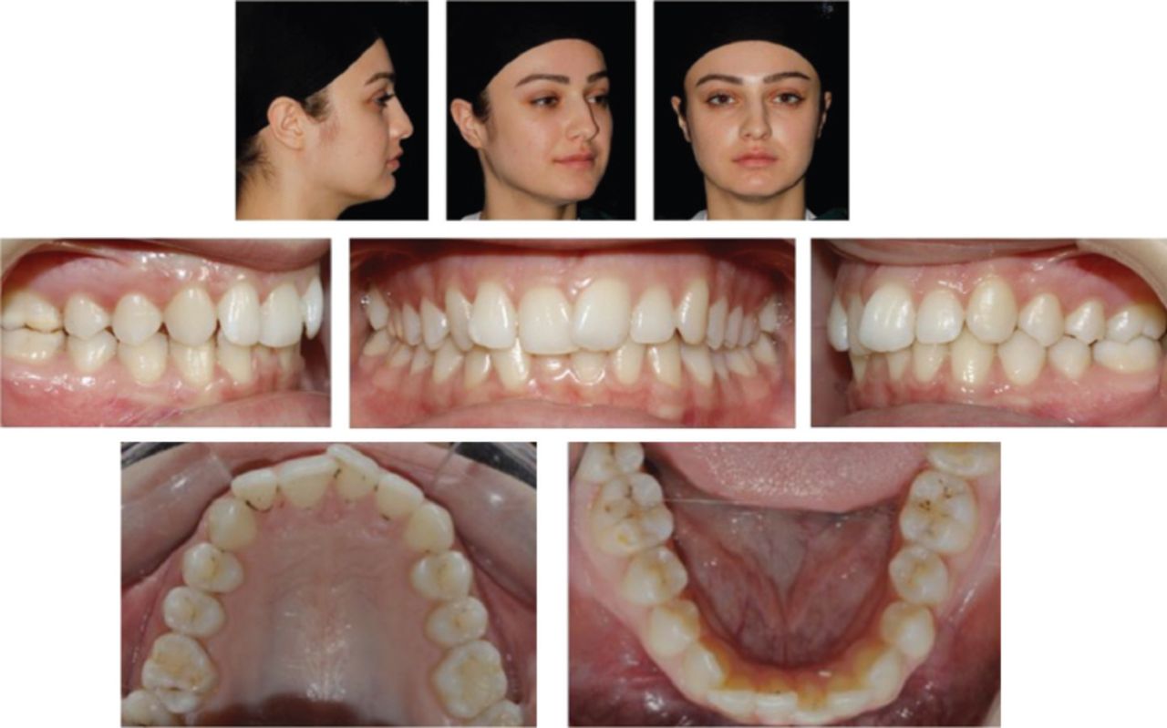
Maxillary midline was in harmony with the facial midline, and mandibular midline was shifted to the right side by 1 mm relative to the facial midline. In dental examination, it was observed that she had class I molar and canine relationships on the right side, class I molar and canine relationships on the left side, and 3 mm overjet and 5 mm overbite. Moderate crowding according to Little’s irregularity index was observed in both arches (Figures 1 & 2).

Pretreatment facial and intraoral photographs.

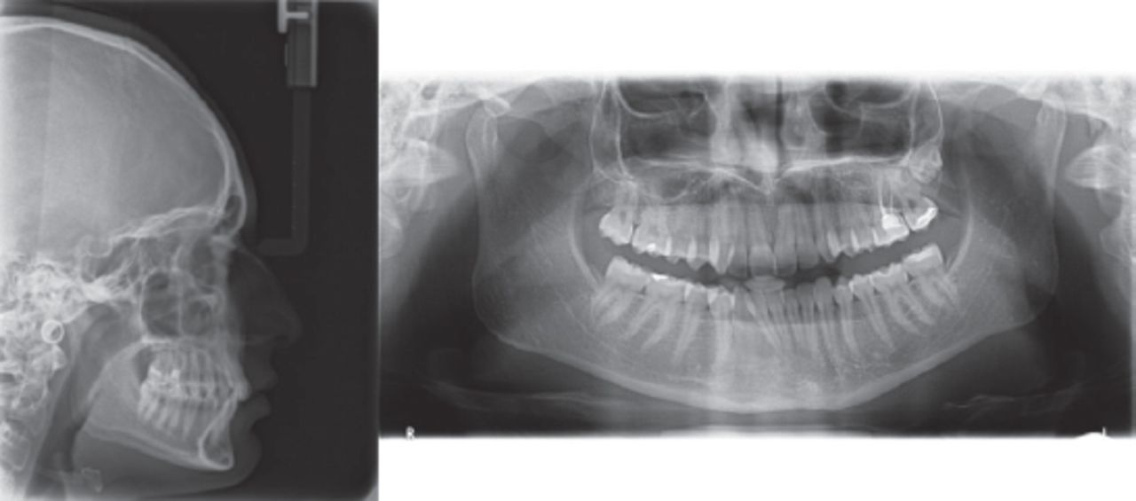
Pretreatment cephalometric and panoramic radiographs.
Diagnostic assessment
In cephalometric examination, it was observed that the patient had skeletal Class I relationship and horizontal growth (Table 1, Figure 2). When panoramic radiography was examined, no tooth cavity, apical root resorption, and no pathological mandibular or maxillary findings were observed (Figure 2). The patient was periodontally healthy. Bleeding on probing, probing pocket depth and gingival index were evaluated using a periodontal probe. Also, plaque index was recorded (Table 2). The measurements were obtained before the orthodontic treatment, one month after the discision procedure, and at the end of the orthodontic treatment. In addition, pain level after discision procedure was assessed using VAS.
Pretreatment and posttreatment of lateral cephalometric measurements.
Mean PPD values and BOP rates in the maxilla (Mx) and mandibular (Md) arches before oral surgery (T0), at 1 Mo, (T1) and at the end of treatment (T2).
The objective of the treatment is to correct crowding while shortening the duration of the orthodontic treatment, and achieve a balanced and stable dental occlusion by providing appropriate overjet and overbite. The patient was offered 3 different treatment alternatives.
The first option offered to the patient was the application of standard pre-adjusted orthodontic brackets without performing further procedures. However, the patient rejected this option, as she wanted to complete her orthodontic treatment in a short period of time.
The second option offered to the patient was corticotomy-assisted orthodontic treatment in order to shorten the duration of the orthodontic treatment. However, the patient rejected this option, too, as corticotomy is an invasive procedure and there are risks of postoperative pain, swelling and infection.
Another option offered to the patient was performing the method called ‘discision’. The patient accepted this less invasive method (Table 3). The advantages and disadvantages of this method were explained to the patient. The patient was informed that the duration of treatment using this method would be shorter compared to average duration of the conventional standard orthodontic treatment. A written informed consent was obtained from the patients’ parent.
The progress of the patient case from initial to diagnosis, treatment and follow-up.
Therapeutic intervention
Orthodontic treatment was started with the bonding of mandibular and maxillary arches by using self-ligating brackets (In-ovation, GAC International, Islandia, NY). As initial arch wire, 0.014-inch nickel-titanium arch wires were placed on both dental arches. Discision procedure was performed on both arches 1 week after placement bonding brackets. After local anesthesia, starting from teeth number 6 in both sides of the maxillary and mandibular arch, guiding incisions were performed using a scalpel in both jaws to the sites corresponding to interdental regions under interdental papillae by also considering the dental roots. Afterwards, incisions with a depth of approximately 3 mm were performed on these marked regions with the aid of the disc saw (Osstem Implant, Esset KIT-Saw, Seoul, Korea) (Figure 3). No sutures were placed to the incision areas. After the operation, the patient was advised to brush her teeth at least twice daily, and perform mouthwash for one week with chlorhexidine-containing solutions. Due to regional acceleratory phenomenon (RAP), the patient was seen at 3-4 week intervals. At her next orthodontics appointment, 0.016 × 0.022-inch nickel-titanium arch wires were placed in the lower and upper arches. In the next session, 0.017 × 0.025 stainless steel arch wires were placed in both dental arches. In order to correct Class II canine relationship, vertically acting Class II elastics were used bilaterally. The total orthodontic treatment was completed in a very short period of time (4 months). Fixed lingual retainers were placed in both dental arches. The patient was also provided with Essix plates to wear all times except during meals.
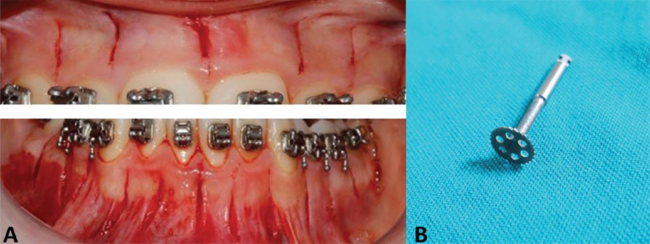
A) Discision on both arches. B) ‘Disc Saw’
Follow-up and outcomes
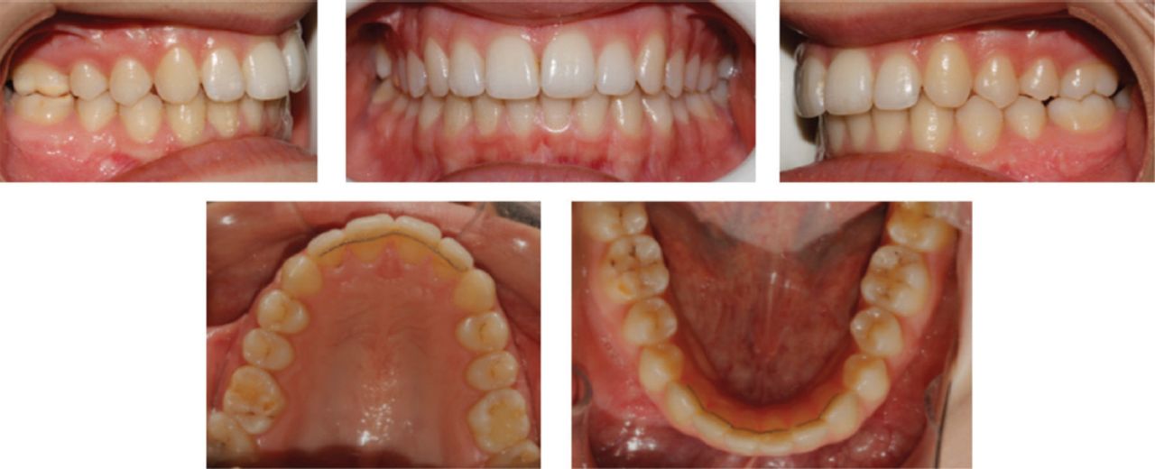
The total duration of orthodontic treatment was 4 months. Class I molar and canine relationships were established at the end of the treatment. Moreover, crowding in the mandible and maxilla was corrected, and optimal overjet and overbite were established. No scar tissue was observed in any gingival region on which discision was performed. Also, there was no resorption or pathology in the apical regions of the teeth in the panoramic radiographic examination that was obtained after the completion of orthodontic treatment. The examination of periodontal health showed that highly acceptable outcomes have been obtained (Figures 4 & 5). The patient was followed-up for 6 months post-treatment, and the occlusion was perfect without any relapse and no root resorptions were detected on the 6-months posttreatment radiographs (Figure 6).
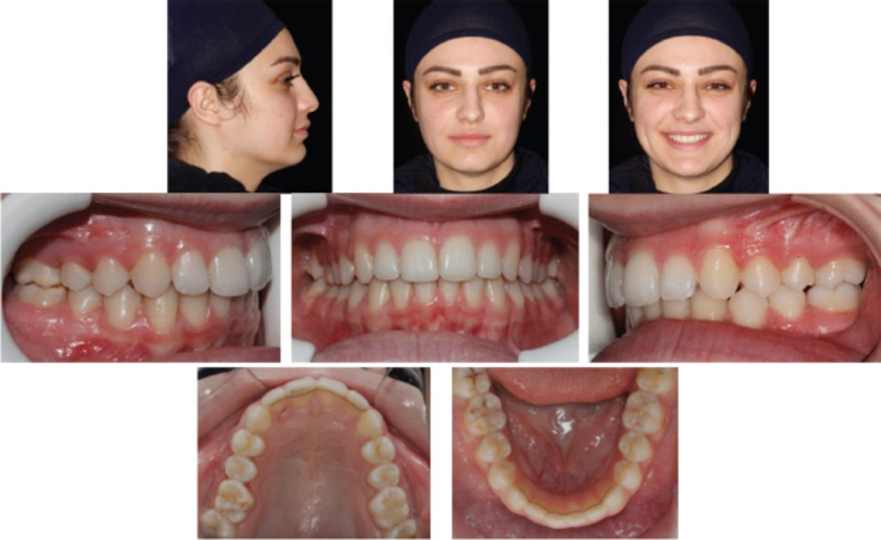
Posttreatment facial and intraoral photographs.
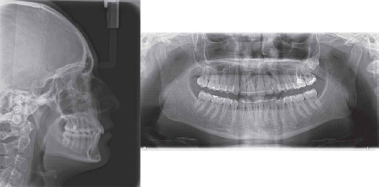
Posttreatment cephalometric and panoramic radiographs.

Posttreatment intraoral photographs at 6 months.
Discussion
Shortening the duration of the orthodontic treatment is desired by both orthodontists and patients. In this case report, the orthodontic treatment was performed using self-ligating brackets and in shorter period than normal by a method called discision. Discision method that was described for the first time by the present authors yielded predictable outcomes, and orthodontic treatment was completed in a short period of time.
Normally, when a traumatic force was applied on a bone, a biological process is initiated in the bone, and the bone turnover increases, and regional bone density decreases.9,10 This biological process was defined as regional acceleratory phenomenon (RAP) by Harold Frost. Following wound healing in the cortical bone, RAP allows remodeling of hard and soft tissues by strengthening tissue reorganization.8-10 In a study on RAP that was performed on the mandibular bone, it was observed that mucoperiosteal flaps re-adapt without suturing. The authors stated that RAP emerges a few days after surgery in humans, peaks at 1-2 months, and persists 6 to 24 months in the underlying structure.8 Moreover, the studies report that the decrease in bone density and bone calcium level observed upon initiation of RAP following surgery reduce the resistance of the cortical bone and provide a more rapid tooth movement.9 When the process of RAP is considered, orthodontic tooth movement started one week before the discision procedure, and orthodontic follow-up visits were scheduled at 3- to 4-week intervals to apply active force.
Wilcko et al9 referred to the advantages of corticotomy and alveolar augmentation in their method. In this method, full-thickness mucoperiosteal flap was elevated and decortication was performed on alveolar bone, and they reshaped the supporting alveolar bone using bone grafts, wherever deemed necessary. They reported a reduction in dehiscence and fenestrations, and their method also increased the bone thickness. Although this method initiates RAP process and it was recognized as a successful method by many authors, the method also has disadvantages such as being an invasive procedure as full-thickness flap is elevated, post-operative pain, avascular necrosis and low acceptance rate by patients.9
Due to morbidity risks associated with corticotomy, Dibart et al8 has introduced an alternative method using piezosurgery without flap elevation.In this technique, corticotomy was performed using only piezosurgery knife with the depth of 3 mm on the gingiva adhered to the buccal side one week after the placement of orthodontic brackets bonding. As being a minimally invasive procedure, this technique has gained wider acceptance by the patients. However, this method requires an expensive device, a piezosurgery device, and it is not possible for all clinics to have this device available. In our case report, a minimally invasive method which can be readily applied on every orthodontics clinic using disc saw was used, and the patient’s orthodontic treatment was completed in a short period of time.
The increase in the duration of the orthodontic treatment increases the risk of tooth decay and root resorption, and more importantly decreases patient cooperation.1 The method described in the present report and named by the present authors was developed as an alternative method to piezocision technique, and this new method offers a minimally invasive procedure that can be performed in any orthodontics clinics. In this technique, incisions with the depth of 3 mm were performed on the adhered gingiva by preserving the dental roots using disc drills. With the initiation of RAP process, velocity of tooth movement was increased, and no problematic periodontal condition was encountered at the end of the treatment. Consequently, the study achieved successful orthodontic treatment outcomes in a short period of time.
There are a limited number of limitations with this novel method. The depth of incision cuts could be examined by using cone beam computed tomography. However, we did not prefer cone beam computed tomography because we do not want to give extra radiation dose to the patient. Also, the future clinical studies should be performed in a larger number of patients, with greater crowding conditions comparing piezocision technique in order to support our novel surgical approach.
Patient perspective
The patient was satisfied with the duration and results of the orthodontic treatment.
In conclusion, the discision technique was developed as a minimally invasive alternative method to piezocision technique, and the authors suggest that this new method yields good outcomes in achieving rapid tooth movement. The efficacy of this newly developed technique should be proved by performing controlled clinical trials comparing this technique with the other tooth movement acceleration methods.
Footnotes
Disclosure. Authors have no conflict of interests, and the work was not supported or funded by any drug company.
- Received September 6, 2017.
- Accepted November 29, 2017.
- Copyright: © Saudi Medical Journal
This is an open-access article distributed under the terms of the Creative Commons Attribution-Noncommercial-Share Alike 3.0 Unported, which permits unrestricted use, distribution, and reproduction in any medium, provided the original work is properly cited.
In this issue


Jump to section
Related Articles
Cited By...
- No citing articles found.