


Article Figures & Data
Figures
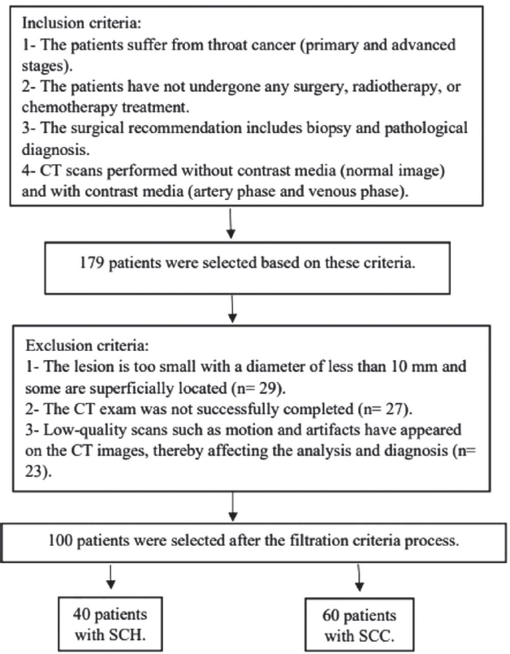
- Figure 1
- Flowchart representing the patient selection criteria process. SCH: squamous cell hyperplasia, SCC: squamous cell carcinoma (SCC)
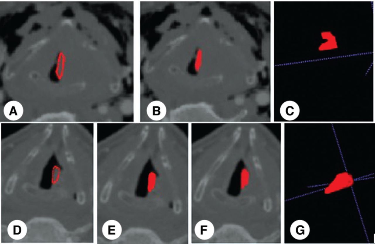
- Figure 2
- The segmentation process by ITK-SNAP software for squamous cell carcinoma showing A-C) malignant and D-G) the squamous cell hyperplasia benign.
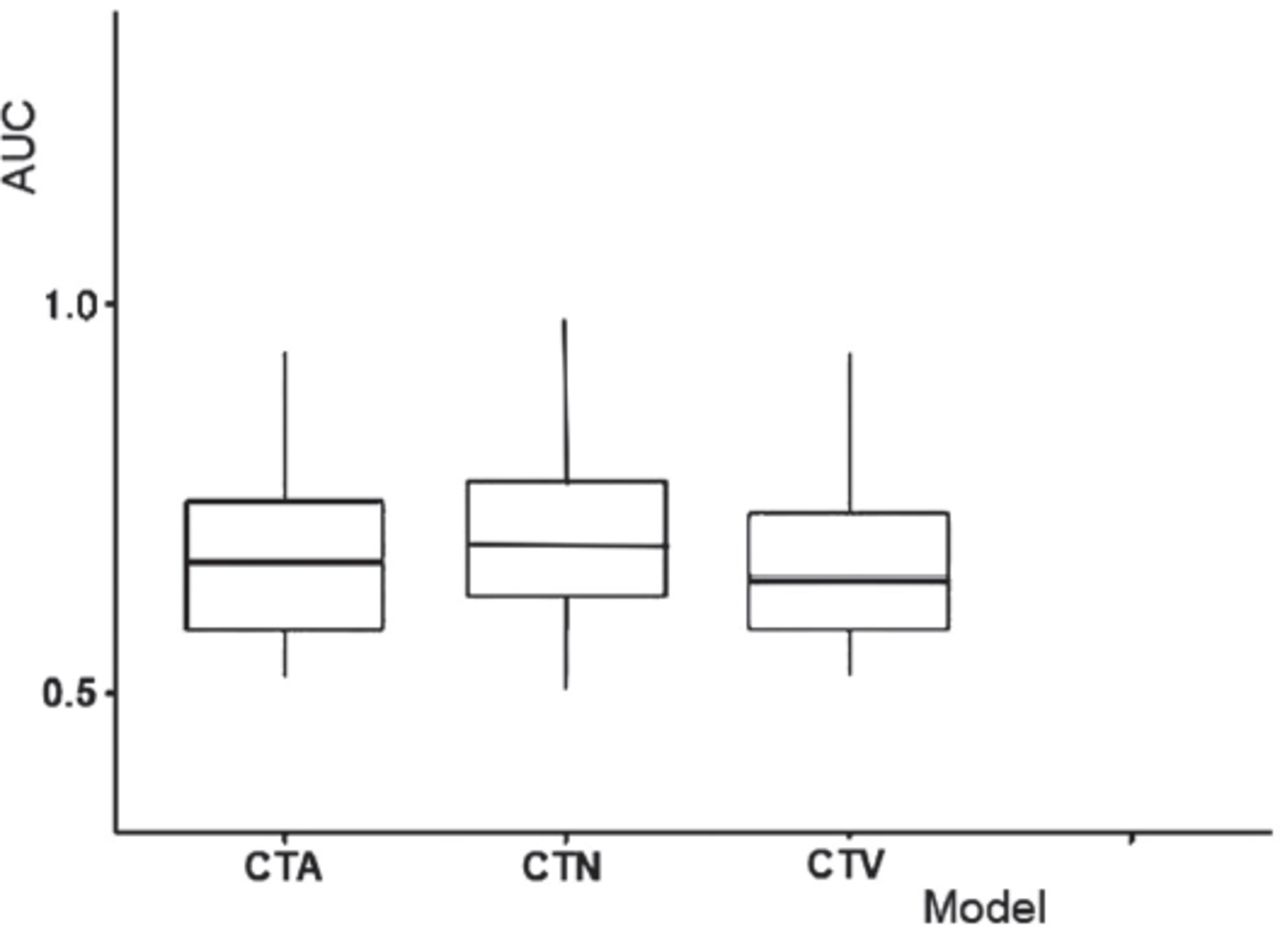
- Figure 3
- Box plot of area-under-the-curve (AUC) values for throat cancer illustrating the higher values of computed tomography normal (CTN) parameters as compared to computed tomography artery (CTA) and computed tomography venous (CTV).
- Figure 4
- The least absolute shrinkage and selection operator (LASSO) features after univariate logistic regression and removing redundancies, for each parameter was used in our study. A) The right and left image of compared to computed tomography artery (CTA), B) the right and left image of computed tomography normal (CTN), C) the right and left image of computed tomography venous (CTV).


Tables
CT parameters Training data Testing data Acc. Sen. Spe. PPV. NPV. Acc. Sen. Spe. PPV. NPV. CTA 0.91 0.92 0.9 0.93 0.9 0.78 0.93 0.0 0.83 0.0 CTN 0.93 0.93 0.933 0.95 0.90 0.84 0.94 0.33 0.88 0.50 CTV 0.92 0.97 0.87 0.91 0.96 0.78 0.75 1 1 0.43 CTA: computed tomography artery, CTV: computed tomography venous, CTN: computed tomography normal, Acc: accuracy, Sen: sensitivity, Spe: specificity, PPV: positive predictive value, NPV: negative predictive value
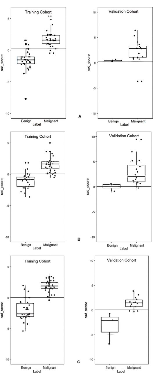
- Table 2
- The statistically significant for least absolute shrinkage and selection operator (LASSO) features selected of throat cancer for each parameter used in this study.
Model Training cohort (p-value) Validation cohort (p-value) CTA <0.000* 0.014* CTN <0.000* <0.008* CTV <0.000* <0.002* ↵* p-value is determined by the Spearman’s rank correlation test and p<0.05 is statistically significant. CTA: computed tomography artery, CTV: computed tomography venous, CTN: computed tomography normal
In this issue


Jump to section
Related Articles
Cited By...
- No citing articles found.