
Main menu

  • Home
  • Content
    • Latest
    • Archive
    • home
  • Info for
    • Authors
    • Reviewers
    • Subscribers
    • Institutions
    • Advertisers
    • Join SMJ
  • About Us
    • About Us
    • Editorial Office
    • Editorial Board
  • More
    • Advertising
    • Alerts
    • Feedback
    • Folders
    • Help
  • Other Publications
    • NeuroSciences Journal

User menu

  • My alerts
  • Log in

Search

  • Advanced search
Saudi Medical Journal
  • Other Publications
    • NeuroSciences Journal
  • My alerts
  • Log in
Saudi Medical Journal

Advanced Search

  • Home
  • Content
    • Latest
    • Archive
    • home
  • Info for
    • Authors
    • Reviewers
    • Subscribers
    • Institutions
    • Advertisers
    • Join SMJ
  • About Us
    • About Us
    • Editorial Office
    • Editorial Board
  • More
    • Advertising
    • Alerts
    • Feedback
    • Folders
    • Help
  • Follow psmmc on Twitter
  • Visit psmmc on Facebook
  • RSS
Brief CommunicationBrief Communication
Open Access

Association between sex hormones and migraine in young Saudi females

Lubna I. Al Asoom, Modhi S. Alajmi, Reem R. Alsudairi, Abeer A. AlShamlan, Amal A. Almomaten, Asma A. Alqarni, Manal H. Alshammari, Nazish Rafique, Rabia Latif, Ahmed A. Alsunni, Dana A. Almohazey, Hind S. Alsuwat, Sayed Abdul Azeez and Francis J. Borgio
Saudi Medical Journal July 2021, 42 (7) 793-797; DOI: https://doi.org/10.15537/smj.2021.42.7.20210110
Lubna I. Al Asoom
From the Department of Physiology (Al Asoom, Rafique, Latif, Alsunni); from the College of Medicine (Alajmi, Alsudairi, AlShamlan, Almomaten, Alqarni, Alshammari); from the Department of Stem Cell Research (Almohazey); and from the Department of Genetic Research (Alsuwat, AbdulAzeez, Borgio), Institute for Research and Medical Consultations, Imam Abdulrahman Bin Faisal University, Dammam, Kingdom of Saudi Arabia.
MBBS PhD
  • Find this author on Google Scholar
  • Find this author on PubMed
  • Search for this author on this site
  • For correspondence: [email protected]
Modhi S. Alajmi
From the Department of Physiology (Al Asoom, Rafique, Latif, Alsunni); from the College of Medicine (Alajmi, Alsudairi, AlShamlan, Almomaten, Alqarni, Alshammari); from the Department of Stem Cell Research (Almohazey); and from the Department of Genetic Research (Alsuwat, AbdulAzeez, Borgio), Institute for Research and Medical Consultations, Imam Abdulrahman Bin Faisal University, Dammam, Kingdom of Saudi Arabia.
MBBS
  • Find this author on Google Scholar
  • Find this author on PubMed
  • Search for this author on this site
Reem R. Alsudairi
From the Department of Physiology (Al Asoom, Rafique, Latif, Alsunni); from the College of Medicine (Alajmi, Alsudairi, AlShamlan, Almomaten, Alqarni, Alshammari); from the Department of Stem Cell Research (Almohazey); and from the Department of Genetic Research (Alsuwat, AbdulAzeez, Borgio), Institute for Research and Medical Consultations, Imam Abdulrahman Bin Faisal University, Dammam, Kingdom of Saudi Arabia.
MBBS
  • Find this author on Google Scholar
  • Find this author on PubMed
  • Search for this author on this site
Abeer A. AlShamlan
From the Department of Physiology (Al Asoom, Rafique, Latif, Alsunni); from the College of Medicine (Alajmi, Alsudairi, AlShamlan, Almomaten, Alqarni, Alshammari); from the Department of Stem Cell Research (Almohazey); and from the Department of Genetic Research (Alsuwat, AbdulAzeez, Borgio), Institute for Research and Medical Consultations, Imam Abdulrahman Bin Faisal University, Dammam, Kingdom of Saudi Arabia.
MBBS
  • Find this author on Google Scholar
  • Find this author on PubMed
  • Search for this author on this site
Amal A. Almomaten
From the Department of Physiology (Al Asoom, Rafique, Latif, Alsunni); from the College of Medicine (Alajmi, Alsudairi, AlShamlan, Almomaten, Alqarni, Alshammari); from the Department of Stem Cell Research (Almohazey); and from the Department of Genetic Research (Alsuwat, AbdulAzeez, Borgio), Institute for Research and Medical Consultations, Imam Abdulrahman Bin Faisal University, Dammam, Kingdom of Saudi Arabia.
MBBS
  • Find this author on Google Scholar
  • Find this author on PubMed
  • Search for this author on this site
Asma A. Alqarni
From the Department of Physiology (Al Asoom, Rafique, Latif, Alsunni); from the College of Medicine (Alajmi, Alsudairi, AlShamlan, Almomaten, Alqarni, Alshammari); from the Department of Stem Cell Research (Almohazey); and from the Department of Genetic Research (Alsuwat, AbdulAzeez, Borgio), Institute for Research and Medical Consultations, Imam Abdulrahman Bin Faisal University, Dammam, Kingdom of Saudi Arabia.
MBBS
  • Find this author on Google Scholar
  • Find this author on PubMed
  • Search for this author on this site
Manal H. Alshammari
From the Department of Physiology (Al Asoom, Rafique, Latif, Alsunni); from the College of Medicine (Alajmi, Alsudairi, AlShamlan, Almomaten, Alqarni, Alshammari); from the Department of Stem Cell Research (Almohazey); and from the Department of Genetic Research (Alsuwat, AbdulAzeez, Borgio), Institute for Research and Medical Consultations, Imam Abdulrahman Bin Faisal University, Dammam, Kingdom of Saudi Arabia.
MBBS
  • Find this author on Google Scholar
  • Find this author on PubMed
  • Search for this author on this site
Nazish Rafique
From the Department of Physiology (Al Asoom, Rafique, Latif, Alsunni); from the College of Medicine (Alajmi, Alsudairi, AlShamlan, Almomaten, Alqarni, Alshammari); from the Department of Stem Cell Research (Almohazey); and from the Department of Genetic Research (Alsuwat, AbdulAzeez, Borgio), Institute for Research and Medical Consultations, Imam Abdulrahman Bin Faisal University, Dammam, Kingdom of Saudi Arabia.
MBBS, PhD
  • Find this author on Google Scholar
  • Find this author on PubMed
  • Search for this author on this site
Rabia Latif
From the Department of Physiology (Al Asoom, Rafique, Latif, Alsunni); from the College of Medicine (Alajmi, Alsudairi, AlShamlan, Almomaten, Alqarni, Alshammari); from the Department of Stem Cell Research (Almohazey); and from the Department of Genetic Research (Alsuwat, AbdulAzeez, Borgio), Institute for Research and Medical Consultations, Imam Abdulrahman Bin Faisal University, Dammam, Kingdom of Saudi Arabia.
MBBS, PhD
  • Find this author on Google Scholar
  • Find this author on PubMed
  • Search for this author on this site
Ahmed A. Alsunni
From the Department of Physiology (Al Asoom, Rafique, Latif, Alsunni); from the College of Medicine (Alajmi, Alsudairi, AlShamlan, Almomaten, Alqarni, Alshammari); from the Department of Stem Cell Research (Almohazey); and from the Department of Genetic Research (Alsuwat, AbdulAzeez, Borgio), Institute for Research and Medical Consultations, Imam Abdulrahman Bin Faisal University, Dammam, Kingdom of Saudi Arabia.
MBBS, PhD
  • Find this author on Google Scholar
  • Find this author on PubMed
  • Search for this author on this site
Dana A. Almohazey
From the Department of Physiology (Al Asoom, Rafique, Latif, Alsunni); from the College of Medicine (Alajmi, Alsudairi, AlShamlan, Almomaten, Alqarni, Alshammari); from the Department of Stem Cell Research (Almohazey); and from the Department of Genetic Research (Alsuwat, AbdulAzeez, Borgio), Institute for Research and Medical Consultations, Imam Abdulrahman Bin Faisal University, Dammam, Kingdom of Saudi Arabia.
MSc, PhD
  • Find this author on Google Scholar
  • Find this author on PubMed
  • Search for this author on this site
Hind S. Alsuwat
From the Department of Physiology (Al Asoom, Rafique, Latif, Alsunni); from the College of Medicine (Alajmi, Alsudairi, AlShamlan, Almomaten, Alqarni, Alshammari); from the Department of Stem Cell Research (Almohazey); and from the Department of Genetic Research (Alsuwat, AbdulAzeez, Borgio), Institute for Research and Medical Consultations, Imam Abdulrahman Bin Faisal University, Dammam, Kingdom of Saudi Arabia.
BSc
  • Find this author on Google Scholar
  • Find this author on PubMed
  • Search for this author on this site
Sayed Abdul Azeez
From the Department of Physiology (Al Asoom, Rafique, Latif, Alsunni); from the College of Medicine (Alajmi, Alsudairi, AlShamlan, Almomaten, Alqarni, Alshammari); from the Department of Stem Cell Research (Almohazey); and from the Department of Genetic Research (Alsuwat, AbdulAzeez, Borgio), Institute for Research and Medical Consultations, Imam Abdulrahman Bin Faisal University, Dammam, Kingdom of Saudi Arabia.
MSc, PhD
  • Find this author on Google Scholar
  • Find this author on PubMed
  • Search for this author on this site
Francis J. Borgio
From the Department of Physiology (Al Asoom, Rafique, Latif, Alsunni); from the College of Medicine (Alajmi, Alsudairi, AlShamlan, Almomaten, Alqarni, Alshammari); from the Department of Stem Cell Research (Almohazey); and from the Department of Genetic Research (Alsuwat, AbdulAzeez, Borgio), Institute for Research and Medical Consultations, Imam Abdulrahman Bin Faisal University, Dammam, Kingdom of Saudi Arabia.
MSc, PhD
  • Find this author on Google Scholar
  • Find this author on PubMed
  • Search for this author on this site
  • Article
  • Figures & Data
  • eLetters
  • Info & Metrics
  • References
  • PDF
Loading

Abstract

Objectives: To assess the sex hormone levels in young Saudi female migraineurs during a migraine attack and during pain-free periods and compare them with control subjects.

Methods: A case-control study involving 14 Saudi female migraineurs and 21 control subjects was conducted between December 2019 and March 2020. Demographic and disease history data were collected through participant interviews. Blood samples were drawn during the migraine attack and pain-free periods.

Results: Follicular (30.00±19.60; p<0.001) and luteal (39.79±11.45; p=0.037) estrogen levels were significantly higher in patients with non-menstrual related migraine (NMM), while luteal testosterone levels (1.10±0.31; p=0.023) were significantly higher in patients with menstrually related migraine (MM). Body mass index (BMI) was higher in patients with NMM (25.77±6.53; p=0.013), and it was found to be associated with follicular estrogen (p=0.016), progesterone (p=0.018), and pain intensity (p=0.042). Luteal estrogen level was significantly lower (13.96±7.88; p=0.036) in patients with luteal onset of attack.

Conclusion: High estrogen levels were found to mediate NMM, their effect being more pronounced with increase in BMI; whereas low luteal estrogen levels mediated MM. Young females with MM might have high luteal testosterone levels, and a compensatory protective role could be surmised accordingly.

  • migraine
  • migraineur
  • estrogen
  • progesterone
  • testosterone

Migraine is a debilitating disease manifested by recurrent headache attacks and associated symptoms.1 Migraine commonly affects individuals between the ages of 18-44 years and is more common in females than in males.2 A recent epidemiological survey in the Kingdom of Saudi Arabia (KSA) reported the overall prevalence of migraine as 26.97%, with the incidence in females being 3 times as high as that in males.3 Migraine is classified into 2 main types: migraine without an aura and migraine with an aura. The aura is a neurological symptom that precedes the headache attack.4 Migraine in females can be classified into menstrual migraine, menstrually related migraine (MM), and non-menstrual migraine (NMM). These types are defined in the International classification of headache disorders, 3rd edition (ICHD-3).5

Some reports have postulated the role of sex hormones in the pathophysiology of migraine. This notion emerged due to the higher prevalence of migraine in females than in males. Furthermore, the rate of incidence of this disease changes throughout the female life. While migraine prevalence is identical in male and female children, the incidence rises rapidly after puberty in females, reaching approximately 2-3 times than seen in males. The course of the disease also changes during pregnancy, perimenopausal, and menopausal periods.6 Therefore, some studies have estimated the level of sex hormones in migraineurs, particularly females. Estrogen is the most studied hormone in this regard, and multiple theories have been postulated correlating estrogen to the pathophysiology of migraine.7 However, there is still a lack of consensus regarding the role of estrogen in migraine pathology, and this could be attributed to the multiple mechanisms involved in the disease pathophysiology as well as the presence of diverse sub-classes. Progesterone and testosterone are less investigated in this regard, but few available data propose a protective effect of these hormones in some cases.8

Therefore, in the present study we aimed to explore the possible association between sex hormones and migraine attacks in Saudi females. We planned to compare the serum levels of sex hormones, including estrogen, progesterone, and testosterone, between Saudi female migraineurs and healthy controls. The hormone levels were assessed on 2 occasions; once during the attack and once during an attack-free period.

Methods

This case-control study was carried out in the Department of Physiology, College of Medicine, Imam Abdulrahman Bin Faisal University (IAU), Dammam, Saudi Arabia. A total of 35 Saudi females (14 migraineurs and 21 controls) aged 18-30 years (mean age 21.91±2.78 years) were recruited by convenience sampling during the period from December 2019 to March 2020.

The target sample size was evaluated using G-power 3.1 software and the following values: power of the test (1-β) = 80%, type I error (α) = 5%, mean luteal estradiol level in female migraineurs = 127.71±59.9 pg/mL, and mean luteal estradiol level in normal controls = 189.26±42.21 pg/mL.9 Finally, the sample size calculation revealed the need for 11 subjects per group.

The participants were selected according to the following criteria: a young female (18-35 years) with a regular menstrual cycle who had a migraine diagnosed by a neurologist and fulfilled the ICHD-3 criteria for the same.5 We excluded those females who had undiagnosed headache, had disorders affecting sex hormones, were lactating or pregnant, or were on hormonal contraceptives.

All participants signed an informed written consent for participation in the study. The study followed the principles of Helsinki Declaration and received approval from the Institutional Review Board of IAU (IRB-UGS-2019-01-293). The participants were screened using the ICHD-3 diagnostic tool. Then the eligible candidates were asked to fill a data to collect information on demographic data such as age, body weight, height, and body mass index (BMI). It also included migraine-related questions such as type of migraine, onset of the disease, relation to menstruation, duration, number of attacks per month, severity of the attacks, family history of the disease, and triggers of pain. The migraineurs were divided into 2 groups: MM group, wherein the migraine attacks occurred during last 2 luteal days to day 2 or 3 of menstruation in at least 2 out of 3 menstrual cycles, but could also occur during other periods of the cycle, and the NMM group, wherein the attacks did not meet the MM criteria.5

The first blood samples were drawn when the participants visited during a migraine attack. At this time, they were asked to indicate the severity of pain using a visual analogue scale (score: 1-10) and mention the day of the menstrual cycle. The second blood samples were collected from the patients when they were in a pain-free period and in the opposite menstrual cycle phase compared to that during the first blood collection, that is, each migraineur had one follicular (day 1-10) and one luteal phase (day 18-27) blood sample. Similarly, 2 blood samples were collected from the controls during follicular and luteal phases. The migraineurs were also classified according to the onset of the actual attack into follicular or luteal actual onset of migraine pain.

Serum levels of estrogen, progesterone, and testosterone were estimated on 2 occasions (follicular and luteal samples). The analyses were performed using the following enzyme-linked immunosorbent assay kits: AccuDiag™ Estradiol, AccuDiag™ Progesterone, and AccuDiag™ Testosterone (Diagnostic Automation & Controls Private Limited, Calabasas, California 91302 USA) at the Institute for research and medical consultations, IAU, according to the manufacturers’ instructions, and readings were obtained with a Synergy Neo2 Hybrid Multi-Mode Reader (BioTek, Germany). All analyses were conducted in duplicate.

Statistical analysis

Data was analyzed using the Statistical Package for Social Sciences (SPSS) statistics for Windows, version 21 (IBM Corp., Armonk, NY, USA). The data was compared between the control subjects and migraineurs using an independent-sample student’s t-test. Analysis of variance (ANOVA) and least significant difference (LSD) post hoc tests were used to compare the hormonal levels in the luteal and follicular phases among the following groups: MM, NMM, and controls. The hormone levels were further compared between the groups of follicular and the luteal actual onset of migraine pain using an independent-sample student’s t-test. Correlation of hormone levels to the migraine features were performed using a Pearson correlation analysis. A p-value<0.05 was considered significant.

Prior related research was obtained from the PubMed database using the following key words: migraine and sex hormones, estrogen, progesterone, testosterone, menstruation.

Results

The general characteristics and hormonal levels were compared between the controls and migraineurs, and no significant difference was found in any of the tested variables (data are not shown). Then the same parameters were compared between MM, NMM, and control subjects using ANOVA and LSD post hoc test; the results are presented in Table 1. Body mass index was found to be significantly correlated with follicular estrogen (p=0.016) and progesterone levels (p=0.018) and the visual score of migraine intensity (p=0.042), (Figure 1).

Figure 1
  • Download figure
  • Open in new tab
  • Download powerpoint
Figure 1

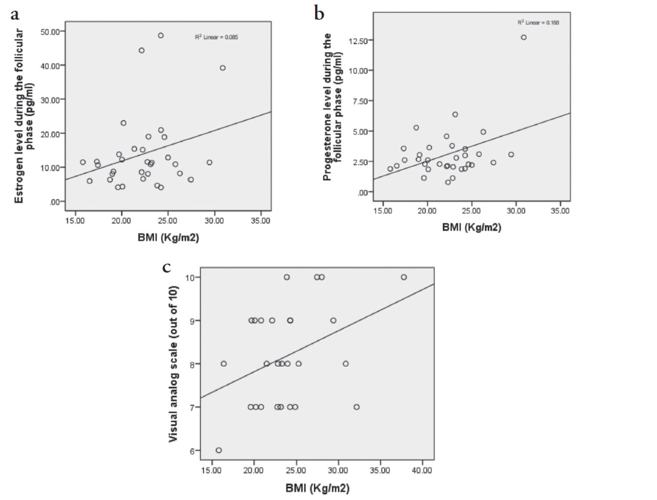
- Linear regression lines demonstrating the correlation of BMI with (a) estrogen during the follicular phase, (b) progesterone during the follicular phase, and (c) Migraine pain intensity by the visual analog scale.

View this table:
  • View inline
  • View popup
Table 1

- Comparison of the general characteristics and the hormonal levels between the migraineurs of MM, NMM migraine types and the controls during the follicular and luteal phases using ANOVA and LSD post hoc test.

The comparison of the hormonal levels between the groups of follicular versus luteal migraine onset is shown in Table 2.

View this table:
  • View inline
  • View popup
Table 2

- Comparison of the sex hormone levels during the actual attacks of follicular versus luteal onset. (using independent samples student t-test).

Discussion

The current study aimed to identify the possible associations between sex hormones and migraine attacks in young Saudi females.

The comparison of sex hormone levels during the 2 phases of menstrual cycle, namely, the follicular and luteal phases, between the migraineurs and controls revealed no significant difference. A prior study that tracked the urine levels of sex hormones, including estrogen and progesterone, on a daily basis found no difference between migraineurs and controls.10 However, this finding was not sufficient to overlook the influence of these hormones in migraine pathology. When the hormones were compared between the different classes of migraineurs, a dissimilarity in their sex hormone profiles was detected accordingly. Therefore, our data can shed light on the complexity of the role of the sex hormones, particularly estrogen, in the pathophysiology of migraine. Estrogen levels during the 2 phases of the menstrual cycle were significantly higher in the NMM group than in the control group. Therefore, the estrogen level might be a triggering or facilitating factor for the pain attacks in NMM. Some studies indicated that in female migraineurs, a high estrogen level might contribute to cortical spreading depression (CSD) and modulation of serotonin levels.11 Experimentally, an estradiol supplement administered to oophorectomized female rats induced CSD.12

The NMM group showed significant higher body weight and BMI compared to the other groups. Concurrently, a significant correlation was found between estrogen level and body weight. Estrogen can be stored in the adipose tissue, which acts as an extraovarian source of the hormone and contributes to the non-controlled elevation of blood estrogen level. Thus, increased body weight in this group of female migraineurs can aggravate the migraine pathology. In line with this finding, a clinical study on 110 overweight or obese females with migraine showed that the women responded positively to a weight loss program and demonstrated reduction in migraine pain and related disability.13

On the other hand, a significant lower luteal estrogen level was demonstrated in the migraineurs with a luteal attack onset than in those with a follicular attack onset. A migraine attack during the luteal phase is most probably an MM, as defined by ICHD-3.5 Nevertheless, the significantly lower estrogen level during the luteal phase that coincides with pain attack plausibly concurs with the explanation of the withdrawal theory. A sudden drop in estrogen level during the late luteal phase can activate an inflammatory response and facilitate the release of proinflammatory markers such as calcitonin gene-related protein, neurokinin, apolipoprotein E, and substance P.11

Furthermore, the MM group demonstrated a significantly higher luteal testosterone level compared to that of controls, with no significant difference seen with the testosterone level of the NMM group. In contrast, Rustichelli et al,14 demonstrated no difference in the level of testosterone between MM patients and controls; however, the subjects of Rustichelli et al’s study were older (19-45 years). Testosterone is believed to have a protective effect against the pathogenesis of migraine. It was found that testosterone levels negatively correlate with migraine disability assessment questionnaire scores (MIDAS) and the frequency of pain attacks.9 Thus, the elevated level of the luteal testosterone in our young MM patients might be a protective physiological mechanism and explain the progression of the disease and the increase in the pain frequency and intensity in older females who demonstrated a decrease in testosterone level, as reported by Li et al.9,15 Progesterone levels were not different in any of the studied groups during follicular as well as luteal phases of the menstrual cycle. Our findings were consistent with those reported by Rustichelli et al,14 and Li et al,9 who found no difference in progesterone levels between patients with MM and controls.

Study limitations

The current study might be limited by its small sample size. However, the narrow age group of our sample minimized the effect of confounding factors and highlighted the significant differences in the hormonal profiles of the studied groups.

In conclusion, we can conclude from our study that female migraineurs had a distinguished sex hormonal profile, that is not only differed from that of healthy controls but also differed between the types of migraine, MM and NMM. The latter presented with high estrogen level during both the follicular and luteal phases of the menstrual cycle, while MM was associated with low luteal estrogen levels. High estrogen level in NMM might culminate in CSD, whereas low luteal estrogen might justify the notion of the withdrawal theory and the inflammatory consequences of sudden drop of estrogen in MM. Our young MM migraineurs exhibited high luteal testosterone level that might act as a mitigation factor against headache attacks in young females.

Acknowledgment

The authors gratefully acknowledge Mr. Ranilo M. Tumbaga from the Department of Genetic Research, Institute for Research and Medical Consultations (IRMC), Imam Abdulrahman Bin Faisal University, Dammam, Kingdom of Saudi Arabia for his assistance in the enzyme-linked immunosorbent assay analysis, and we would like to thank Editage (www.editage.com) for English language editing.

Footnotes

  • Disclosure. This study was funded by the College of Medicine, Imam Abdulrahman Bin Faisal University, Damman, Kingdom of Saudi Arabia.

  • Received February 17, 2021.
  • Accepted May 30, 2021.
  • Copyright: © Saudi Medical Journal

This is an Open Access journal and articles published are distributed under the terms of the Creative Commons Attribution-NonCommercial License (CC BY-NC). Readers may copy, distribute, and display the work for non-commercial purposes with the proper citation of the original work.

References

  1. 1.↵
    1. Rafique N,
    2. Al-Asoom LI,
    3. Latif R,
    4. Alsunni AA,
    5. Salem AM,
    6. Alkhalifa ZH, et al.
    Prevalence of migraine and its relationship with psychological stress and sleep quality in female university students in Saudi Arabia. J Pain Res 2020; 13: 2423–2430.
    OpenUrl
  2. 2.↵
    1. Vosoughi K,
    2. Stovner LJ,
    3. Steiner TJ,
    4. Moradi-Lakeh M,
    5. Fereshtehnejad SM,
    6. Farzadfar F, et al.
    The burden of headache disorders in the Eastern Mediterranean Region, 1990-2016: findings from the global burden of disease study 2016. J Headache Pain 2019; 20: 40.
    OpenUrl
  3. 3.↵
    1. Muayqil T,
    2. Al-Jafen BN,
    3. Al-Saaran Z,
    4. Al-Shammari M,
    5. Alkthiry A,
    6. Muhammad WS, et al.
    Migraine and headache prevalence and associated comorbidities in a large Saudi sample. Eur Neurol 2018; 79: 126–134.
    OpenUrl
  4. 4.↵
    1. Vgontzas A,
    2. Burch R.
    Episodic migraine with and without aura: key differences and implications for pathophysiology, management, and assessing risks. Curr Pain Headache Rep 2018; 22: 78.
    OpenUrl
  5. 5.↵
    Headache Classification Committee of the International Headache Society (IHS). The International Classification of Headache Disorders, 3rd ed. Cephalalgia 2018; 38: 1–211.
    OpenUrlCrossRefPubMed
  6. 6.↵
    1. Vetvik KG,
    2. MacGregor EA.
    Sex differences in the epidemiology, clinical features, and pathophysiology of migraine. Lancet Neurol 2017; 16: 76–87.
    OpenUrlPubMed
  7. 7.↵
    1. Pavlović JM.
    Evaluation and management of migraine in midlife women. Menopause 2018; 25: 927–929.
    OpenUrl
  8. 8.↵
    1. Martin VT,
    2. Allen JR,
    3. Houle TT,
    4. Powers SW,
    5. Kabbouche MA,
    6. O’Brien HL, et al.
    Ovarian hormones, age and pubertal development and their association with days of headache onset in girls with migraine: an observational cohort study. Cephalalgia 2018; 38: 707–717.
    OpenUrl
  9. 9.↵
    1. Li W,
    2. Diao X,
    3. Chen C,
    4. Li C,
    5. Zhang Y,
    6. Li Y.
    Changes in hormones of the hypothalamic-pituitary-gonadal axis in migraine patients. J Clin Neurosci 2018; 50: 165–171.
    OpenUrl
  10. 10.↵
    1. Pavlović JM,
    2. Allshouse AA,
    3. Santoro NF,
    4. Crawford SL,
    5. Thurston RC,
    6. Neal-Perry GS, et al.
    Sex hormones in women with and without migraine: evidence of migraine-specific hormone profiles. Neurology 2016; 87: 49–56.
    OpenUrl
  11. 11.↵
    1. Delaruelle Z,
    2. Ivanova TA,
    3. Khan S,
    4. Negro A,
    5. Ornello R,
    6. Raffaelli B, et al.
    Male and female sex hormones in primary headaches. J Headache Pain 2018; 19: 117.
    OpenUrl
  12. 12.↵
    1. Chauvel V,
    2. Multon S,
    3. Schoenen J.
    Estrogen-dependent effects of 5-hydroxytryptophan on cortical spreading depression in rat: modelling the serotonin-ovarian hormone interaction in migraine aura. Cephalalgia 2018; 38: 427–436.
    OpenUrl
  13. 13.↵
    1. Farris SG,
    2. Thomas JG,
    3. Kibbey MM,
    4. Pavlovic JM,
    5. Steffen KJ,
    6. Bond DS.
    Treatment effects on pain catastrophizing and cutaneous allodynia symptoms in women with migraine and overweight/obesity. Health Psychol 2020; 39: 927–933.
    OpenUrl
  14. 14.↵
    1. Rustichelli C,
    2. Bellei E,
    3. Bergamini S,
    4. Monari E,
    5. Baraldi C,
    6. Castro FL, et al.
    Serum levels of allopregnanolone, progesterone and testosterone in menstrually-related and postmenopausal migraine: a cross-sectional study. Cephalalgia 2020; 40: 1355–1362.
    OpenUrl
  15. 15.↵
    1. Al-Hassany L,
    2. Haas J,
    3. Piccininni M,
    4. Kurth T,
    5. Maassen Van Den Brink A,
    6. Rohmann JL.
    Giving researchers a headache - sex and gender differences in migraine. Front Neurol 2020; 11: 549038.
    OpenUrl
PreviousNext
Back to top

In this issue

Saudi Medical Journal: 42 (7)
Saudi Medical Journal
Vol. 42, Issue 7
1 Jul 2021
  • Table of Contents
  • Cover (PDF)
  • Index by author
Print
Download PDF
Email Article

Thank you for your interest in spreading the word on Saudi Medical Journal.

NOTE: We only request your email address so that the person you are recommending the page to knows that you wanted them to see it, and that it is not junk mail. We do not capture any email address.

Enter multiple addresses on separate lines or separate them with commas.
Association between sex hormones and migraine in young Saudi females
(Your Name) has sent you a message from Saudi Medical Journal
(Your Name) thought you would like to see the Saudi Medical Journal web site.
Citation Tools
Association between sex hormones and migraine in young Saudi females
Lubna I. Al Asoom, Modhi S. Alajmi, Reem R. Alsudairi, Abeer A. AlShamlan, Amal A. Almomaten, Asma A. Alqarni, Manal H. Alshammari, Nazish Rafique, Rabia Latif, Ahmed A. Alsunni, Dana A. Almohazey, Hind S. Alsuwat, Sayed Abdul Azeez, Francis J. Borgio
Saudi Medical Journal Jul 2021, 42 (7) 793-797; DOI: 10.15537/smj.2021.42.7.20210110

Citation Manager Formats

  • BibTeX
  • Bookends
  • EasyBib
  • EndNote (tagged)
  • EndNote 8 (xml)
  • Medlars
  • Mendeley
  • Papers
  • RefWorks Tagged
  • Ref Manager
  • RIS
  • Zotero
Share
Association between sex hormones and migraine in young Saudi females
Lubna I. Al Asoom, Modhi S. Alajmi, Reem R. Alsudairi, Abeer A. AlShamlan, Amal A. Almomaten, Asma A. Alqarni, Manal H. Alshammari, Nazish Rafique, Rabia Latif, Ahmed A. Alsunni, Dana A. Almohazey, Hind S. Alsuwat, Sayed Abdul Azeez, Francis J. Borgio
Saudi Medical Journal Jul 2021, 42 (7) 793-797; DOI: 10.15537/smj.2021.42.7.20210110
Twitter logo Facebook logo Mendeley logo
  • Tweet Widget
  • Facebook Like
  • Google Plus One
Bookmark this article

Jump to section

  • Article
    • Abstract
    • Methods
    • Results
    • Discussion
    • Acknowledgment
    • Footnotes
    • References
  • Figures & Data
  • eLetters
  • References
  • Info & Metrics
  • PDF

Related Articles

  • No related articles found.
  • PubMed
  • Google Scholar

Cited By...

  • No citing articles found.
  • Google Scholar

More in this TOC Section

  • Successful management of human parainfluenza virus-3 outbreak in a tertiary neonatal intensive care unit
  • Experience of pediatric liver disease at a university hospital in Western Saudi Arabia
  • Risk of malignancy in thyroid nodules Bethesda III sub classification into nuclear atypia and architectural atypia. A retrospective study
Show more Brief Communication

Similar Articles

Keywords

  • migraine
  • migraineur
  • estrogen
  • progesterone
  • testosterone

CONTENT

  • home

JOURNAL

  • home

AUTHORS

  • home
Saudi Medical Journal

© 2025 Saudi Medical Journal Saudi Medical Journal is copyright under the Berne Convention and the International Copyright Convention.  Saudi Medical Journal is an Open Access journal and articles published are distributed under the terms of the Creative Commons Attribution-NonCommercial License (CC BY-NC). Readers may copy, distribute, and display the work for non-commercial purposes with the proper citation of the original work. Electronic ISSN 1658-3175. Print ISSN 0379-5284.

Powered by HighWire