


Article Figures & Data
Figures
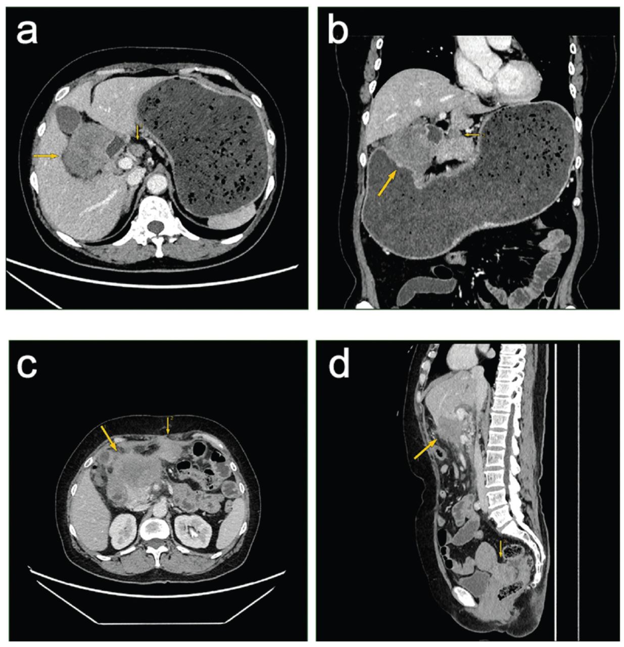
- Figure 1
- Results of first (before surgery) and second (12 months post-surgery) pelvic enhanced computed tomography (CT) scan. a & b) The first contrast-enhanced axial CT scan of the upper abdomen revealed an irregular soft tissue mass (thick arrows) on the lesser curvature side of the gastric antrum, measuring approximately 5.3 × 6.3 cm. The mass showed moderate and uneven enhancement. The gastric antrum lumen was compressed, leading to gastric retention and dilation. Additionally, a lymph node enlargement of approximately 1.7cm was observed in the hepatogastric ligament (thin arrows). c & d) The second contrast-enhanced axial CT scan of the upper abdomen and pelvic regions showed (c) an irregular, slightly lower-density mass (thick arrows) below the left lobe of the liver, measuring approximately 6.5 × 6.8 cm. The mass displayed moderate and uneven enhancement, with unclear demarcation from the lower edge of the liver, gallbladder, duodenum, and pancreas. An irregular peritoneal metastatic soft tissue mass (thin arrows) was found beneath the anterior abdominal wall in the left upper quadrant, measuring approximately 3.3 × 3.5cm. d) The contrast-enhanced sagittal CT scan of the abdominal and pelvic regions on the same date showed the same irregular, slightly lower density mass (thick arrows) below the left lobe of the liver, with similar size and enhancement characteristics. The mass had unclear demarcation from the lower edge of the liver, gallbladder, duodenum, and pancreas. Multiple scattered peritoneal metastatic soft tissue masses were observed in the abdominal cavity, mainly concentrated in the Douglas cul-de-sac (rectouterine pouch) in the pelvic floor (thin arrows), measuring approximately 3.9 × 5.4cm.
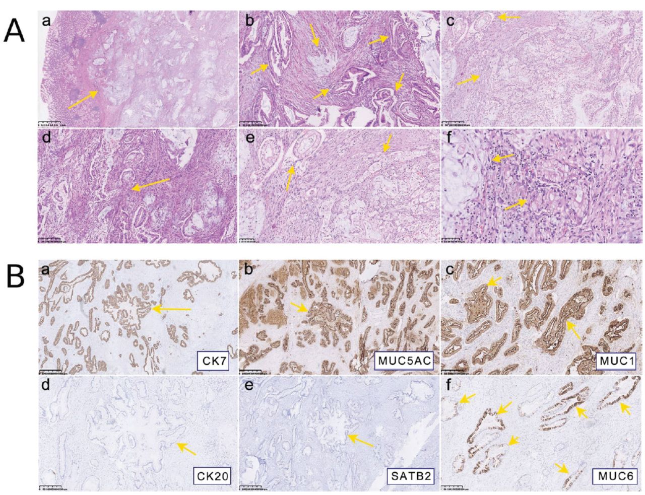
- Figure 2
- The A) histological morphology of hematoxylin and eosin (HE) (bar = 500 μm). a) Tumor located under the mucosa; glands of the mucosal layer were normal (HE × 25). b) In some areas, the tumor tissues are arranged in the shape of glandular tubes, and the glands are angled deformities (HE × 100). c) In some areas, the neoplastic glands are arranged in a back-to-back manner (HE × 100). d) Proliferative smooth muscle bundles, fibrous tissue, and inflammatory cell infiltration can be seen around the tumor tissue (HE × 100). e) Tumor cells are rich in the cytoplasm (HE ×200). f) The nucleus of tumor cells was abnormal, and the nuclear membrane was irregular (HE × 400). B) Immunohistology staining (EnVision, original magnification, × 50; bar = 500 μm). a) CK7 (+); b) MUC5AC (+); c) MUC1 (+); d) CK20 (-); e) SATB2 (-); and f) the tumor tissue was positive in some areas of MUC6, and the positive staining was located in the cytoplasm. a-c) The positive expression was localized in the cytoplasm. The CK7, MUC1, and MUC5AC are intensely positive in infiltrating adenocarcinoma tissue, with staining in the cytoplasm and cell membrane. The MUC6 exhibits strong positive expression in some infiltrating adenocarcinoma tissues, with a patchy distribution and strong intensity, also in the cytoplasm and cell membrane. However, CK20 and SATB2 are negative in infiltrating adenocarcinoma tissue.


Tables
Dates Patient’s information June 2, 2021 A 59-year-old female patient presented with abdominal discomfort with acid regurgitation. She was in good health and had no history of external injury or surgeries. Summaries from initial and follow-up visits Diagnostic testing (including dates) Interventions May 31, 2021 - Pyloric obstruction and thickening of the gastric wall in the gastric antrum.
- Mucosal congestion, swelling in the anterior pyloric area, and pyloric canal stenosis.
A computed tomography (CT) enhanced scan of the entire abdomen and pelvis Gastroscopy / June 9, 2021 - Deviation of 10 cm towards the lesser curvature and an extension of 22 cm towards the greater curvature.
- A 5.5 x 5.4 cm round-like mass was found during surgery.
/ Gastrointestinal surgery June 18, 2021 Gastric and pancreatic heterotopic tissue (type III) with adenocarcinoma with epithelial carcinogenesis. - Histology examination
- Immunohistology examination
July 30, 2021 6 rounds of chemotherapy and antibiotics (gemcitabine and tigecycline) June 20, 2022 - One year after the surgical treatment of pancreatic cancer originating from the ectopic pancreas in the lesser curvature of the gastric antrum, irregular and slightly lower-density tumor recurrence was observed in the surgical area.
- Multiple scattered metastatic soft tissue masses of varying sizes were present in the abdominal cavity, mainly concentrated in the Douglas cul-de-sac (rectouterine pouch) in the pelvic floor.
- The second contrast-enhanced axial CT scan of the upper abdomen and pelvic regions
- The contrast-enhanced sagittal CT scan of the abdominal and pelvic regions
In this issue


Jump to section
Related Articles
Cited By...
- No citing articles found.