


Abstract
Objectives: To evaluate belinostat’s (PXD101) activity on MCF-7 breast cancer stem cells (CSCs) via Wnt, Notch, and Hedgehog.
Methods: This research study was carried out at the Department of Medical Biology, Necmettin Erbakan University, Konya, Turkey, from June 2017 to July 2019. The effect of PXD101 on MCF-7 cell viability was determined by cell proliferation kit (XTT). Following belinostat treatment, CD44+/CD24- MCF-7 CSCs were isolated by FACS. Ribonucleic acid isolation and copy-deoxyribonucleic acid synthesis were carried out using HEK-293 cells, MCF-7 cells, and MCF-7 CSCs. Expression changes of metastasis-related genes, Wnt, Hedgehog, Notch, and stem cell markers were analysed by quantitative polymerase chain reaction. The IC50 in MCF-7 cancer cells was 5 μM for 48 hours. The FACS analysis indicated that 2% of the MCF-7 cancer cells were CSCs. Following belinostat treatment, the MCF-7 cell count decreased by 44%, and the MCF-7 CD44+/CD24- CSC count decreased by 66%.
Results: Belinostat treatment reduced the expression of metastasis, Wnt, Notch, Hedgehog, and stem cell marker genes.
Conclusion: Belinostat has a potential effect on the differentiation and self-renewal of breast CSCs.
Cancer is characterized by abnormal cells that become differentiated and proliferate. It is the leading cause of death globally, second only to cardiovascular disease. According to the American Cancer Society, there are expected to be 1,918,030 new cancer cases and 609,360 cancer deaths in the United States in 2022.1 The global incidence of cancer is increasing, and new approaches are needed to treat the disease effectively. Cancer is caused by a group of cells that are called cancer stem cells (CSCs).2 Cancer stem cells have been shown to be resistant to cytotoxic drugs as well as to radiation. It has been shown that this resistance may be due to an increased ability to recognize and repair DNA damage, decreased activity in apoptotic pathways, inadequate intake of chemotherapeutic agents into cells, and increased excretion of these agents out of the cells.3 The CSCs have cell surface markers, embryonic signaling pathways and detoxification enzyme systems.4 It aims to shrink the tumor in the treatment methods used against cancer. However, the cancer can repeat itself as long as the CSCs survive.5 Therefore, it is critical to develop new treatments targeting CSCs.6 The CSC treatments include targeting CSC surface markers with specific antibodies, blocking pathways that provide resistance to chemotherapy and radiotherapy, and blocking signal pathways involved in self-renewal. Monoclonal antibody blocking of the Wnt, Hedgehog, and Notch self-renewal signaling pathways is an example of CSC therapy.7
Breast cancer is the most common form of cancer in women around the world and represents a major global health problem.8,9 In 2020, 685,000 women died globally because of breast cancer. Despite the development of new methods of diagnosing and treating breast cancer, metastasis, drug resistance, and relapse remain important problems. However, it is thought that breast cancer recurrence and metastasis can be treated by targeting breast cancer stem cells (BSCSs).10 Approaches targeting BCSCs are quite remarkable in terms of reducing metastasis and recurrence, reducing the remission rate, and improving the quality of life of patients.11 Histone deacetylase inhibitors change the acetylation of histone and nonhistone proteins. Regulation of cellular events, such as cell life, differentiation and apoptosis, and anti-cancer effects have been observed in different pathways with gene expression changes. Thus, histone deacetylase inhibitors are considered potential anti-cancer drugs.12
Belinostat is a histone deacetylase inhibitor. It was approved by the Food and Drug Administration (FDA) in 2014 for the treatment of peripheral T-cell lymphoma. Belinostat has the molecular formula C15H14N2O4S.13 Clinical studies of the effects of belinostat and other histone deacetylases on cancer and nonmalignant diseases continue to appear.14,15 Histone deacetylase inhibitors have been examined as anti-cancer drugs in the literature.16,17
Therefore, in this study, we aimed to investigate the antiproliferative activity and anti-cancer effects of belinostat on MCF-7 and MCF-7 CSCs by evaluating markers of self-renewal pathways.
Methods
The research study was carried out at the Department of Medical Biology, Necmettin Erbakan University, Konya, Turkey, from June 2017 to July 2019. This study was approved by the Ethics Committee of Necmettin Erbakan University, Faculty of Medicine, Non-Pharmaceutical and Medical Device Research (date: June 16, 2017, issue: 2017/962).
Belinostat was obtained from Med Chem Express. Belinostat was dissolved in dimethyl sulfoxide (Sigma-Aldrich). The cells were treated with different concentrations of belinostat for different periods of time. The MCF-7 and HEK-293 cell lines were obtained from the American Type Culture Collection (Rockville, Maryland, 20852, USA). Cell lines were cultured in RPMI 1640 medium. The medium was added 10% fetal bovine serum (Sigma-Aldrich, St. Louis, MO) and 1% antibiotic-penicillin solution (Sigma-Alrich, St. Louis, MO). The cells were cultured at 37oC in a humid atmosphere containing of 5% CO2.
To establish the IC50 dosage of belinostat, approximately 104 cells were seeded into a 96-well plate, then incubated for 24 hours. Selected concentrations of belinostat (0, 0.25, 0.5, 0.75, 1, 3, 5, 15, and 25 µM) were administered to the wells. The cells were grown for a period of 24, 48, and 72 hours. In every dosage group, a well was cultured under identical circumstances to the control group, where belinostat administration was not present. At 24, 48, and 72 hours after treatment, cell cytotoxicity was assessed in each well treated with belinostat at various concentrations using the Cell Proliferation Kit (XTT, BI Biological Industries). The assay was carried out according to the manufacturer’s instructions. A microplate reader (BioTek, Biotek Winooski, Vermont, USA) was then used to measure absorbance at 450 nm.18
The MCF-7 and HEK-293 cells were incubated with 5 µM of belinostat for 48 hours. Approximately 10,000-15,000 cells were removed using trypsin-ethylenediamine tetraacetic acid (EDTA) and centrifuged at 1500 rpm for 5 minutes for stem cell isolation. The cellular pellets of MCF-7 cells were incubated with CD44+ and CD24- antibodies (BD Bioscience) in the dark for half an hour in 1X HBSS solution prepared with 2% FBS medium. Controls were diluted with 1% HBSS supplemented with 2% FBS. The CSCs (CD44+/CD24-) were sorted by FACS (FACS Aria III - BD Pharmingen, US). The FACS DivaTM version 6.1.3. software was used for analysis flow cytometry.19
The CSCs were incubated in serum-free medium. After 13 days, the spherical formations appeared. Thus, stem cells were confirmed through the sphere formation technique.20
Total cellular RNA was isolated using the TRIzol (Sigma Aldrich®), chloroform, and ethanol method. After removal of ethanol, RNA was dissolved in RNase-free water. The cDNAs were synthesized by cDNA Synthesis Kit (iScript™ cDNA Synthesis Kit) according to the kit protocol. The qPCR was carried out in triplicate using CFX ConnectTM real-time PCR instrument. The primer sequences were summarized in Table 1. The expression levels of the NANOG, OCT4, and SOX2 genes were analysed. This was carried out to determine the effects of belinostat on cell differentiation.
- Primer pairs nucleotide sequences.
Statistical analysis
The comparative ΔCT method was used to determine relative gene expression. The RT² Profiler™ PCR Array Data Analysis software was used for statistical analysis (housekeeping gene: GAPDH and CYPA). The decrease or increase in gene expression of 2-fold was considered significant (p<0.05).
Results
Cell viability was assessed by the XTT assay. Results indicate that belinostat inhibited the cell proliferation of MCF-7 cells. The IC50 dose of belinostat was determined as 5 μM for 48 hours.
The amount of MCF-7 CSCs was determined by flow cytometry (FACS) using CD44+/CD24- surface markers. According to FACS results, MCF-7 stem cell ratio in MCF-7 total cell was found to be 2%. After 48 hours treatment of 5 µM belinostat, a 44% decrease in MCF-7 cells and a 66% decrease in MCF-7 CSCs was observed comparing to the control group (Figure 1).
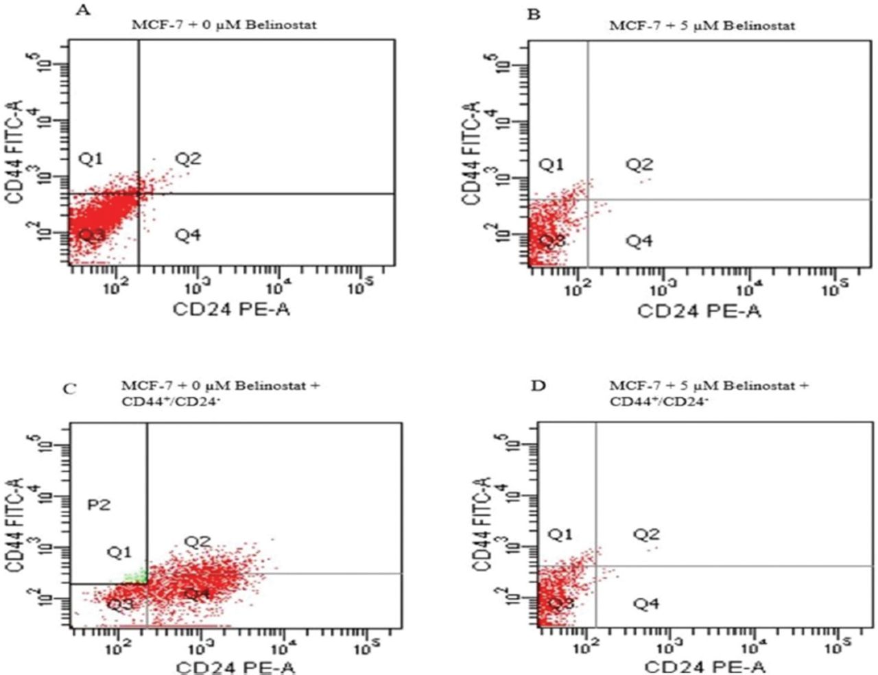
- The FACS analysis. A) The amount of MCF-7 total cell without belinostat. B) The amount of MCF-7 total cell with belinostat applied. C) Amount of CD44+/CD24- antibody treated MCF-7 stem cell without belinostat applied. D) Amount of CD44 +/CD24- antibody applied MCF-7 stem cells with belinostat applied. Q1: CD44+/CD24-, Q2: CD44+/CD24+, Q3: CD44-/CD24-, Q4: CD44-/24+
Isolation of CSCs was carried out by flow cytometry using CD44+/CD24- surface markers from MCF-7 cells. The isolated CSCs were cultured in serum free media and tumorsphere formation was observed from the 13th day and presence of isolated CSCs was confirmed. The MCF-7 CD44+/CD24- CSCs at the 13th, 21st, and 34th days of microscope views are shown in Figure 2.
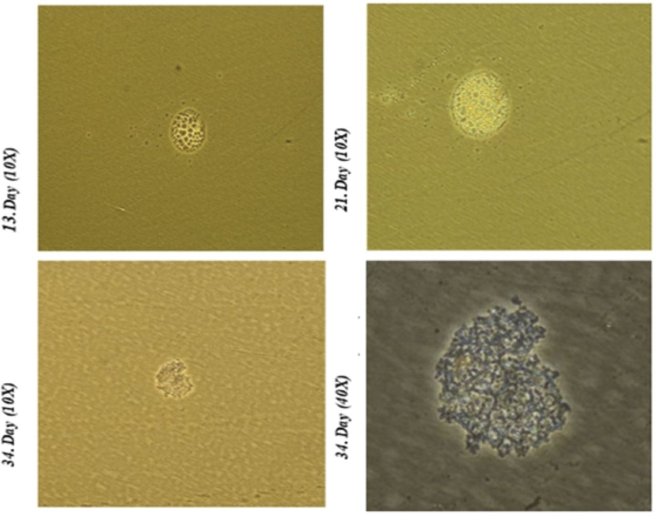
- Tumorsphere formation.
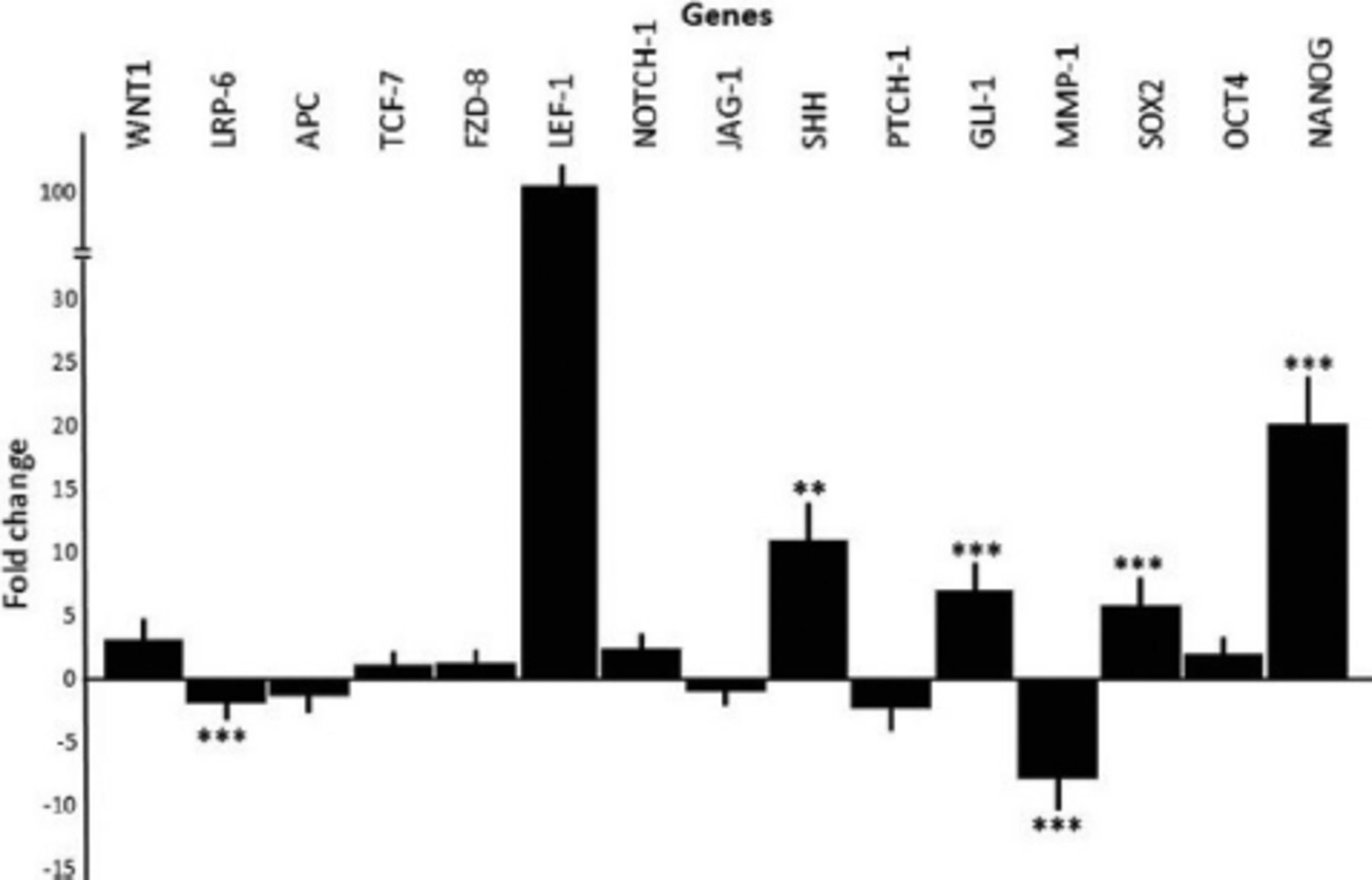
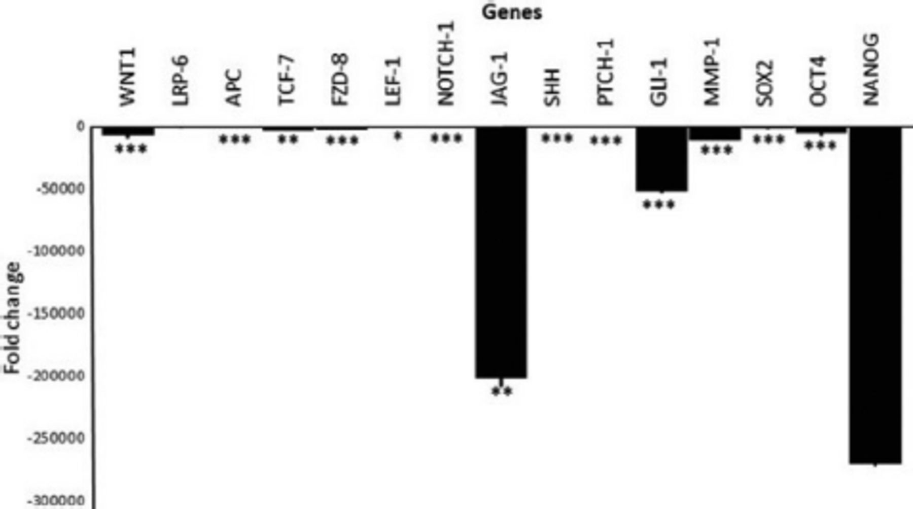
Belinostat administration down regulated genes in general in Wnt, Notch, and Hedgehog pathway in MCF-7 cells. For Wnt-related genes, LRP-6, APC, FZD-8, and LEF-1 expression levels were decreased (p<0.05). Both NOTCH-1 and JAG-1 was down-regulated but NOTCH-1 expression was not statistically important. The PTCH-1, Hedgehog pathway gene, expression was upregulated. The MMP1 gene expression level was downregulated. Gene expression levels of stem cell markers SOX2 was decreased but NANOG was increased MCF-7 cell lines. Results summarized in Figure 3. In this study, we also evaluated the gene expression levels of Wnt, Hedgehog, Notch, and stem cell markers in HEK-293 cell line (Figure 4). According to the results, gene expression levels of LRP-6 and MMP-1 were decreased. The SHH and GLI-1 were upregulated in HEK-293 cells. The SOX2 and NANOG gene expressions were found to be increased. Figure 5 shows expression levels of Wnt, Hedgehog, Notch, and stem cell markers in MCF-7 CD44+/CD24-SCSs. All genes were found to be downregulated. However, decreases observed in LRP-6 and NANOG were statistically unimportant (p>0.05).
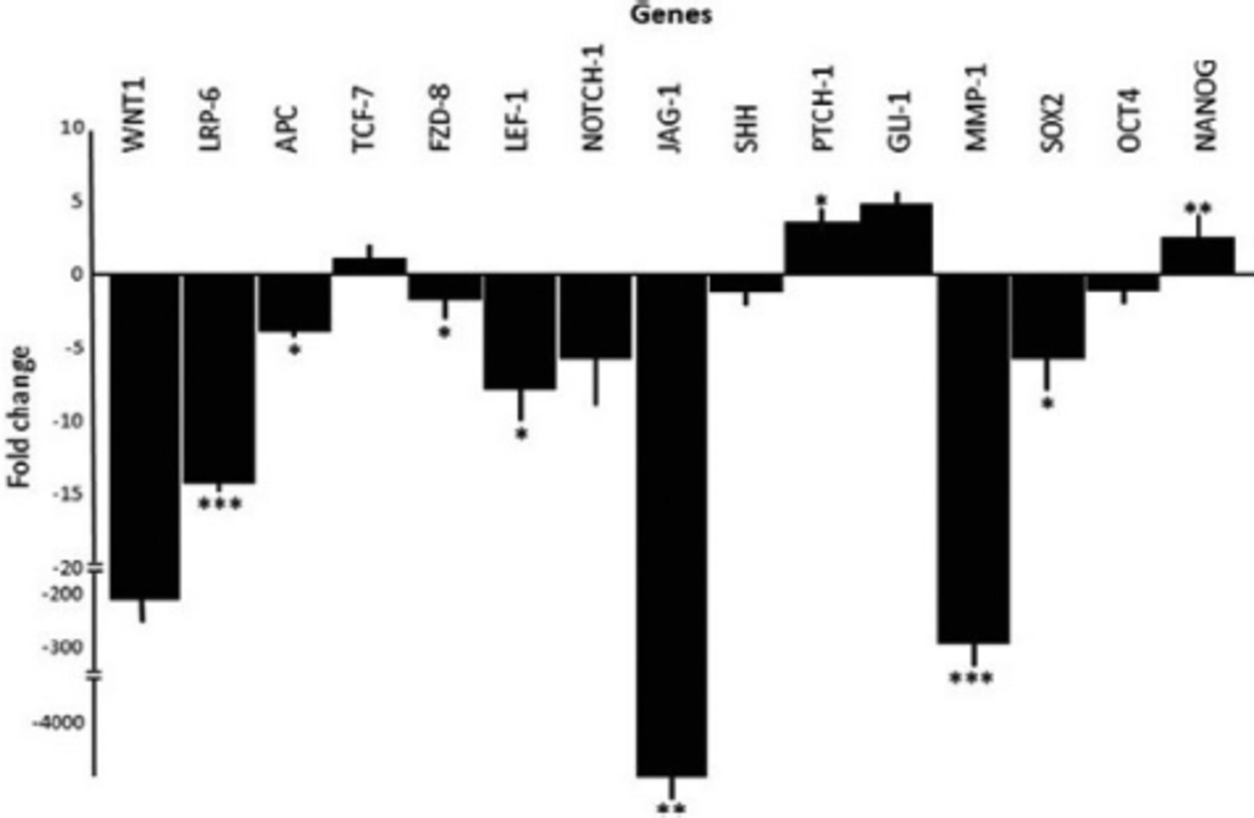
- Gene expression levels of Wnt, Hedgehog, Notch, and stem cell markers in MCF-7 cells.

- Gene expression levels of Wnt, Hedgehog, Notch, and stem cell markers in HEK-293 cells.

- Gene expression levels of Wnt, Hedgehog, Notch, and stem cell markers MCF-7 cells in MCF-7 CD44+/CD24-cancer stem cells.
Discussion
The mechanism of epigenetic modifications is crucial for tumor evolution and CSC viability.21 The HDAC inhibitors (HDACi) are used to develop anti-cancer drugs and their mechanisms of action are being investigated.22 The most remarkable findings in our study are based on the data we obtained as a result of FACS analysis and real time PCR. The findings revealed that the epigenetic drug was effective on cancer and CSCs by suppressing the self-renewal pathways.
In the present study, we demonstrated the belinostat effect on MCF-7 and CD44+/CD24-MCF-7-CSC. According to XTT result, IC50 was found 5µM for 48 hours on MCF-7 cells. Previous literature also reported that in vitro treatment of different cell lines with HDACi inhibited proliferation.23 Determined IC50 dose for MCF-7 was applied to HEK-293 cells and observed under inverted microscope. Belinostat did not affect cell morphology on HEK-293 as much as MCF-7 cells.
After belinostat treatment, gene expression of important genes in self-renewal pathways on MCF-7 and MCF-7 CD44+/CD24-cell line was suppressed more than HEK-293 cells. The fact that belinostat does not have the same effect on cancer cells but on healthy cells, supports the idea that belinostat is a suitable candidate anti-cancer agent and that it is the right approach to use therapeutically in cancer.
The rate of CSCs in tumors has been reported to vary between 0.1-30% depending on the type of cancer.24 As a result of FACS analysis, the amount of MCF-7 CD44+/CD24-stem cells isolated from MCF-7 was determined as 2%. These findings were found to be compatible with the findings obtained from the Fillmore et al’s study.24
In other studies, it was found that HDACi (abexinostat, vorinostat, and valporic acid) significantly reduced the amount of breast CSCs.25,26 A recent study demonstrated that HDACi is a strong candidate as therapeutic options when applied in combination with chemotherapeutics in BCSCs.27 In this study, belinostat treatment decreased 66% of CD44+/CD24-CSCs isolated from MCF-7. The findings of this study are similar to those carried out with the effects of HDACi on BCSCs.
The presence of stem cells obtained as a result of FACS analysis was confirmed by the tumorsphere formation assay. Stem cells have been reported to survive by forming tumorsphere under serum free suspension conditions.28 In this study, CD44+/CD24- CSCs isolated from MCF-7 cells were observed to form tumorsphere in serum-free medium. Therefore, it was confirmed that the isolated cells were stem cells. A previous study demonstrated that BCSCs obtained from SUM159 and MCF-7 cells were also capable of forming tumorsphere.29
The self-renewal feature of CSC is regulated by Wnt/β-catenin, Notch, and Hedgehog pathways.30 These pathways play a role in CSC development and progression, and abnormal activation of these pathways has been associated with various tumor types. Therefore, treatment methods that suppress these pathways are being investigated.31
In this study, the effectiveness of Wnt, Notch, and Hedgehog pathways in MCF-7 cell line and MCF-7 CD44+/CD24-CSCs were investigated. A decrease in the expression of Wnt pathway genes was detected in the MCF-7 cell line after administration of belinostat. Our study is compatible with studies reporting that Wnt signaling pathway inhibition prevents cancer development.32,33 The development of agents targeting the Wnt signaling pathway carries hope for diseases characterized by the abnormal activity of this pathway. The HDAC3 inhibition was reported to decrease CSC proliferation in TNBC cells by causing beta-catenin degradation, and these findings are consistent with the results of our study.34 It was found that expression of Notch pathway genes decreased MCF-7 cells and MCF-7 CD44+/CD24-CSCs after belinostat application in a similar study, the reduction of CSC population in the TNBC cell line after application of AR-42 and SAHA HDACi was associated with a decrease in NOTCH1 gene expression.35 It has been reported that the Hedgehog signal pathway plays a role in breast cancer continuity and the expression of GLI-1, one of the genes involved in this pathway, increases in breast cancer cells.36 Therefore, it is aimed to prevent cancer progress by inhibiting this pathway. In this study, a significant decrease in expression of PTCH-1, SHH, and GLI-1 from the Hedgehog pathway was detected in MCF-7 CD44+/CD24-CSCs after belinostat application. In a similar study, 4SC-202 HDACi has been reported to suppress the Hedgehog signal pathway in basal cell carcinoma.37 In another similar study, it was shown that the synergistic use of SHH and HDAC inhibitors inhibited proliferation of liver cancer cells, thus, it could be an effective therapeutic strategy for liver cancer.38
According to the results of this study, belinostat caused a decrease in the amount of MCF-7 CD44+/CD24- stem cells. Among the mechanisms that may be effective in reducing the amount of stem cells are the ability to suppress and differentiate self-renewal (Wnt, Notch, and Hedgehog) pathways and stem cell markers. Indeed, in this study, belinostat has been shown to cause a significant decrease in the expression of SOX-2 and OCT-4 genes which are accepted as stem cell markers, in MCF-7 CD44+/CD24-CSCs. It is thought that belinostat supports the differentiation of stem cells by suppressing self-renewal factors and causes a decrease in the amount of CSCs. However, there is a need to investigate other biological pathways that may be the cause of a decrease in the amount of CSCs.
The MMP1’s abnormal expression has been associated with tumor metastasis. A previous study reported that MMP1 expression was significantly higher in breast cancer tissues by immunohistochemistry analysis.39 This study shows, belinostat caused a decrease in MMP1 expression on MCF-7 and MCF-7 CD44+/CD24- stem cells. Thus, MMP1 expression status has become an important prognostic indicator for breast cancer and is a potential drug target.
Study strengths & limitations
In order to have a higher level of scientific evidence, cancer could be formed on experimental animals and cytotoxic effects on cancer cells could be observed after application of belinostat, but cell culture study was carried out because our budget was very limited. As an evidence-based medicine step, studies on experimental animals can be repeated in future studies. The strength of this study is that it contributes to the literature on epigenetic treatments on cancer and CSCs.
In conclusion, in this study, it was found that the application of belinostat suppressed the expression of genes that are subject to study, especially in CD44+/CD24-CSCs. It is stated that the anti-cancer activity of HDACi varies according to the dose and cell line, HDACi class, and other factors defined yet.40 The HDACi effects can be influenced by different cell signaling, apoptosis, cell cycle, cell differentiation, angiogenesis and metastasis, and more. The recognition of epigenetics and its physiopathology at the molecular level enables the development of new generation drugs and alternative therapies.
Acknowledgment
The authors gratefully acknowledge Scribendi Inc. for English language editing.
Footnotes
Disclosure. This study was supported by Necmettin Erbakan University, Scientific Research Projects Coordination Office, Konya, Turkey (no.: 171318003).
- Received July 26, 2023.
- Accepted December 12, 2023.
- Copyright: © Saudi Medical Journal
This is an Open Access journal and articles published are distributed under the terms of the Creative Commons Attribution-NonCommercial License (CC BY-NC). Readers may copy, distribute, and display the work for non-commercial purposes with the proper citation of the original work.
References
In this issue


Jump to section
Related Articles
Cited By...
- No citing articles found.