


Abstract
Tonsillar stones are the products of calcified accumulates of cellular debris and microorganisms, in the crypts of palatine tonsils. Tonsillar stones are common findings and the known cause of bad breath (halitosis). Development of large tonsillar stones, however, is rare with only a few cases reported in the literature. We present the case of a 45-year-old man with a history of recurrent sore throat and tonsillitis for a long period, and snoring with other unremarkable ears, nose and throat findings. A large-sized tonsillar stone detected in the left tonsil measured 3.1 × 2.3 cm. The patient underwent elective stone removal and tonsillectomy.
Tonsillar stones (tonsilloliths) are white or yellow concretions in tonsillar crypts that originate as a result of microorganism and cellular debris retention in the crypts of palatine tonsils. Calcium hydroxyapatite and calcium carbonate along with other minerals including phosphorus, ammonia, and magnesium are the common types of stones identified in histology studies of tonsilloliths.1 Tonsillolithiasis, the formation of tonsil stones, is thought to occur mainly due to repeated inflammation of the tonsillar crypts with recurrent tonsillitis and calcification. The age of patients with tonsil stones ranges between 10 and 77 years with a median age of 50 years with a male to female ratio of 1:1.2 Tonsil stones are common findings, observed in excised tonsils on gross inspection and specimen sectioning, and are associated with recurrent sore throat. Halitosis (bad breath) is the main clinical complaint in small tonsilloliths.3 In contrast, tonsillolithiasis of large stones is a rarity with a few cases reported in the literature.4
Case Report
Patient Information
A 45-year-old man presented with a history of recurrent sore throat and tonsillitis since a long period of time. Patient is a diagnosed case of diabetes mellitus and hypertension and on medical treatment. There was no remarkable past medical or surgical history (Table 1).
Case report timeline table.
Clinical findings
History of recurrent sore throat and tonsillitis since a long period of time. The sore throat was associated with snoring. There were no odynophagia, dysphagia, or other ears, nose and throat (ENT) symptoms. Throat examination showed enlarged tonsils grade 4 plus cellulitis of the left tonsil and a large left tonsil stone (Figures 1 & 2). Ear and nose examination results were normal. The other ENT examinations were unremarkable.

Enlarged tonsils grade 4 plus cellulitis of the left tonsil and a large left tonsil stone.

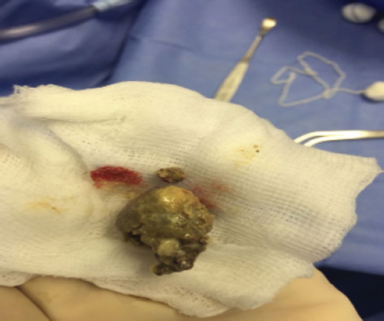
A giant tonsillolith extracted of left tonsil.
Therapeutic intervention
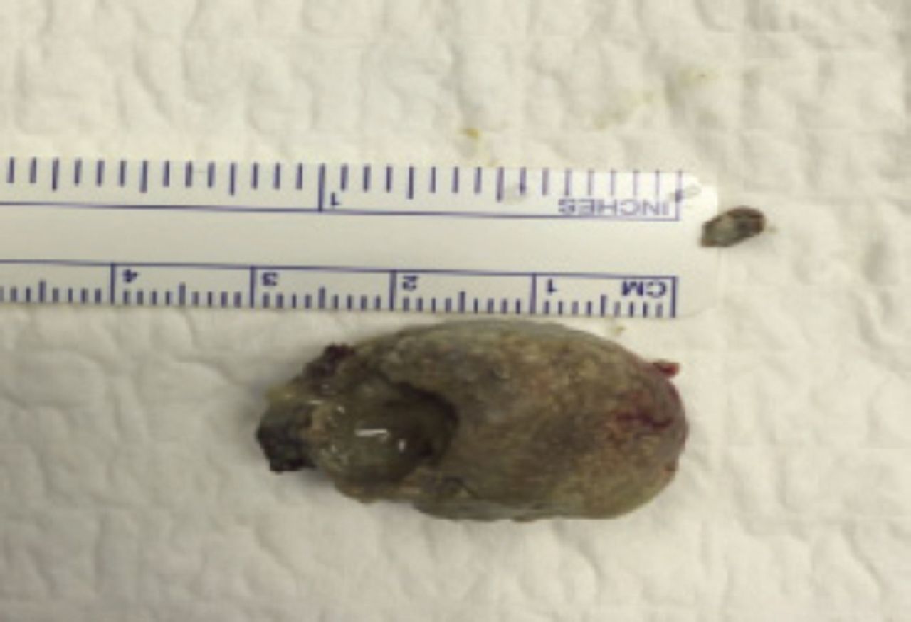
The patient was admitted for elective surgery for removal of the tonsillolith and bilateral tonsillectomy. A large-sized tonsillolith observed during the surgery, had led to the expansion of the entire left upper pole. It was dissected gently using a freer dissector and the stone was removed prior to tonsillar excision. The tonsillolith measured 3.1 × 2.3 cm, right tonsil 3 × 2 × 0.5 cm and the left tonsil 4 × 1.5 × 0.5 cm (Figures F3 & F4). The pathology report for the right and left tonsils revealed a reactive squamous epithelium and reactive lymphoid hyperplasia as well as actinomyces and focal acute inflammation. The occurrence of stone formation in the left tonsil is possibly secondary to the recurrent chronic tonsillitis process. There was no evident malignancy identified.

Tonsillolith measured 3.1 × 2.3 cm.

Tonsillolith measured 3.1 × 2.3 cm.
Follow-up and Outcomes
Two weeks later, the patient presented with secondary hemorrhage post-tonsillectomy, which was controlled under general anesthesia with no complications thereafter. Patient followed up post-operatively, recurrent sore throat and snoring improved. No complications afterward.
Discussion
Tonsillar stones (tonsilloliths) are products of calcified accumulates of food, cellular debris, and microorganism aggregates in the crypts of palatine tonsils.3 Tonsillar crypts are fissure-like invaginations in the medial side of the palatine tonsils and serve to increase the surface area of the tonsils with more than 10 crypts in each tonsil. Small tonsilloliths are common findings associated with recurrent sore throat and if symptomatic, usually present with a chief complaint of halitosis or bad breath.3 However, large tonsilloliths are rare entities and a few cases are reported in literature.4 The exact mechanism of development of tonsillar stones is not well understood. However, several hypotheses exist to explain formation of these calculi. One hypothesis attributes stone formation to recurrent tonsillitis and subsequent crypt fibrosis that lead to epithelial cell retention, which creates an environment for microorganism overgrowth and calcification due to deposition of inorganic salts from salivary gland secretions.5 Stoodley et al6 concluded that tonsilloliths share common characteristics with biofilms. Biofilms are aggregates of aerobic and non-aerobic microorganisms enclosed in an extracellular matrix. Due to the nature of biofilms, it is difficult to treat the microorganisms with antibiotics. In fact, a biofilm serves as a nidus for acute infections rendering it a source of recurrent ENT infections.7 Cases of giant tonsillolith have not been reported in Saudi Arabia. Literature review shows that the largest tonsillolith was reported in a 12-year-old female child in the left tonsil and measured 4.2 × 3.6 × 2.1 cm. A majority of the cases of giant tonsilloliths reported a left tonsil stone rather than the right. Two reported cases showed bilateral giant tonsilloliths.8,9 A patient with tonsillar stone is approached through history taking, physical examination, and imaging studies. A giant tonsillolith may be an incidental finding in asymptomatic patients or maybe associated with common symptoms and signs of tonsilloliths.9 Reported clinical presentations of giant tonsilloliths include recurrent sore throat, halitosis, foreign body sensation, painful swallowing (odynophagia), hoarseness, and rarely, upper airway obstruction. Differential diagnosis of tonsillar stones include foreign body, calcified granulomas, tonsillar malignancy, enlarged styloid processes, embryonic rests originating from the branchial arches.10 Imaging studies may be helpful to identify the size, extension, and location of the stone. Symptomatic giant tonsilloliths are usually treated with tonsillectomy with the pathology report on type of the stone and disease process.
In conclusion, Tonsilloliths are common findings associated mainly with halitosis. However, giant tonsilloliths are rare with few cases reported in the literature. This case supports the hypothesis of tonsillollith formation secondary to recurrent tonsillitis and subsequent crypt fibrosis that lead to epithelial cell retention in the crypts of palatine tonsils. Differential diagnosis of tonsillar stones include foreign body, calcified granulomas, tonsillar malignancy, enlarged styloid processes, embryonic rests originating from the branchial arches. Imaging studies might be necessary to identify the size and extension of the stone beyond the palatine tonsils. This case was managed with tonsillectomy and pathology report on disease process.
Footnotes
Disclosure. Authors have no conflict of interests, and the work was not supported or funded by any drug company.
- Received December 11, 2017.
- Accepted January 25, 2018.
- Copyright: © Saudi Medical Journal
This is an open-access article distributed under the terms of the Creative Commons Attribution-Noncommercial-Share Alike 3.0 Unported, which permits unrestricted use, distribution, and reproduction in any medium, provided the original work is properly cited.
In this issue


Jump to section
Related Articles
Cited By...
- No citing articles found.